TV4101 - Equine MSK 3 - Proximal Limb, Trauma and Joint Sepsis
 Call Kai
 Learn
Learn Practice Test
 Spaced Repetition
 Match
Match Flashcards
 Knowt Play
1/103

There's no tags or description
Looks like no tags are added yet.
Name | Mastery | Learn | Test | Matching | Spaced | Call with Kai | Chat |
|---|
No analytics yet
Send a link to your students to track their progress
104 Terms
Proximal limb lameness
What is a Capped elbow ?
Tx?
Inflammatory swelling of the subcut bursa located over the olecranon process.
Tx
- Topical DMSO +/- aseptic drainage + CCs injection; but beware may lead to abscessation
- Surgical option: cuvette out and pack with iodine gauze
List 3 types of neutrogenic injury/traum
Radial n injury
Brachial plexus - severe gait abnormality
Suprascaular n injury
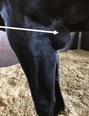
Which neurogenic injury causes lateral subluxation of scapular during weight bearing
Suprascapular n. (sweeney) damage.
*photo shows scapular muscle atrophy due toneurpgenic damge

ddx dropped elbow apperance
- fracuter
- nerve injury
olecranon fracture - more swelling seen
radial nerve injury

Stress fracture of proximal limb radiographs
Initially - may not find anything aside from maybe some mild
If allowed to run around prematurely - fractures can propogate and become massive and obvious

2 common sites of humerus fracture
Greater tubercle
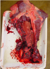
Diaphyseal fracture - often marked local inflammation and massive haematoma (Photo)
Characteristic crepitus on humeral movement and palpation.

Inability to advance limb due to bicep attachment at this location:
Supraglenoid tubercle fracture
Stifle anatomy- 3 joints
femeropatella joint
medial femerotibia
lateral femerotibial

which of the stifle joints communicate
medial femerotibial joint communicates with femoropatella joint in 60% horses
Clinical syndromes stifle Dz
Osteochondrosis
- Subchondral cystic lesions
- Lateral trochlear ridge OCD
Upward fixation patella
Meniscus / Cruciate / Collateral ligament injury
Patella Fractures
Patella luxation
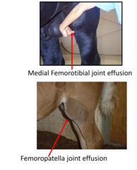
what are the 2 main clinical signs of stifle joint disease
Medial femorotibial joint effusion
Femoropatella jointe ffusion
What is this?
Subchondral cystic lesions secondary to osteochondosis.
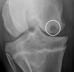
With OC, what determines degree of lameness?
Degree of lameness depends on whether cyst communcates with articular surface. Cyst exposed, engress of joint fluid --> significant lameness.
2 types of OC in stifle
subchondral cystic lesions in medial condyle
OCD (fragment) in lateral condyle
where is OC most common in the stifle
medial femoral condyle
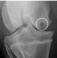
What is the aim for tx of cysts re OC?
to fill the cyst in with bone (debride, CCs and Platelet rich plasma infusions)
Where is OCD of the stifle most common?
lateral trochelar ridge of femur
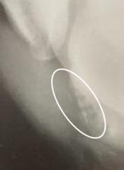
In general, describe clinical signs/findings on lameness exam with stifle soft tissue injuries
Blocks to stifle joint blocks
Positive spavin flexion test
Femorpatella or medial femero tibial joint effusion
3/5 lame
ultrasonogrpahic diagnosis
What does limb locked in rigid extension with toe drag a sign of?
What is getting caught over wht?
Upper fixation patella/patella locking.
- the medial patella ligament hooked over medial femoral trochler
(nb typically straight legged, young horses and occurs first thing in the morning
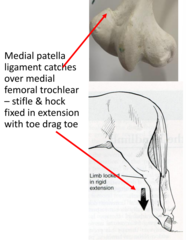
Clinical signifncane of UFP
Common as part of stay apparatus - most horses grow out of it.
Tx of upward fixation patella is conservative at first for young horses. Explain the reasoning behin this and some examples of what to do
Young horse: aim to build up muscle tone so they can 'grow out of it'
- avoid confinement/stables, allow paddock where possible
- graded exercise (straight trotting) but avoiding circles/hills
- PBZ as required
What is the recommended surgical procedure if UFP persists past conservative management (and why)?
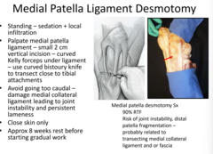
Medial patella ligament desmotomy (transection) - ligament will heal by gap repair = lengthened ligament that is less likely to catch over the medial femoral trochlear
whats another method other than medial patella ligament transection
medial patella ligament fenestraiton with large needle -> inflammation -> ligament repair that may help stabalise and stop process from happened
Explain how to do medial patella ligmanet desmotomy including
- approach
- what structure to differentiate it from
- type of knife/scalpel
- cut close to which landmark?
Standing - sedation and local infiltration
Differentiate from medial collateral ligament!
Curved bistoury knife.
Cut close to tibial insertion - under tension dt weight bearing. Will snap / separate quickly.
8 weeks rest before gradual work.
patella luxation
- how does this occur
congenital - Affected foals may be unable to stand at birth if bilateral
what type of luxation
lateral luxation
ruptured peroneus tertius chracteristic finding?
tx?
Dx: Extension of the hock and simultaneous flexion of the stifle (this is impossible to demonstrate in the normal horse)
Tx: rest 2-3 months

Hip luxation chracteristic finding?
Severe lameness
As in all species: uneven hindlimb lengths - effected will be turned laterally

back pain
- what is the common pitfall surrounding diagnosis of back pain
Common - but also commonly over diagnosed
60% soft tissue pain vs 40% true back pain.
Require through exam to rule out other causes of lameness - needs to be repeatable
What are the msot common causes of back pain
1) secondary to?
2) 2 examples of primary causes
Secondary to
- hind limb lameness
Primary
- impinging dorsal spinous prcoess in performance horses
- stress fractures in young horses
Most common primary cause of back pain
+ radiograph
impingement of dorsal spinour pressess - small gap between processes +/- boney changes

pelvic assymeytry =
pelvic fracture or sacroiliac subluxation

stiff guarded gait all limbs stiff ddx x3
fractured withers
pleuropneumonia
laminit
stringhalt
- what is it
-2 causes
Hypermetra / hyperflexion of hindlimb
Toxic cause - hypochoeris radicata dandelion
Trauma cause - transection of extensor tendons which scars and attaches to cannon bone -> reflexive hyperflexion

tx traumatic stringhalt (not toxic)
Lateral digital extensor tendon transection
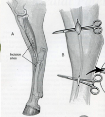
DDX Goose-stepping gait?
(-> functional shortening of hamstring muscle group )
Fibrotic myopathy of hamstring tendons
2ndary to trauma to hamstring mm group due to sliding stops or direct trauma - pulling /falling backwards • Injured site develops progressive fibrosis / ossification muscle bellies of semimembranosis / tendonosis (hamstring mm.)

tx fibrotic myopathy
tx- transection of the fibrotic mass
What is the following
- hindlimb locked in extension with toe drag
- hyper flexion of hindlimb when walking
- flexion of stifle whilst extension of hock
- goose stepping gait
Hindlimb locked in extension with toe drag: Upper patella fixation '
Hyper flexion of hindlimb when walking: Stringhalt (traumatic or toxic)
Flexion of stifle whilst extension of hock: Peroneous tertius rupture
Goose stepping gait: fibrotic myopathy
What is the following
- haematoma around shoulder region
- dropped elbow
Haematoma around shoulder region - diaphyseal fracture of femur
Dropped elbow - radial nerve denervation or olecranon fracture
2 main sites of OCD in stifle
medial femoral cyst
lateral trochlear ridge OCD
Equine MSK Trauma
Understand and recall key principles in history, triage and management of equine musculoskeletal trauma
Apply pre-requisite knowledge of musculoskeletal anatomy and function when diagnosing and managing common equine musculoskeletal traumatic conditions
Recognize, describe and recall clinical features of commonly occurring equine musculoskeletal traumatic conditions
Understand, describe and recall the principles of equine limb immobilization
Understand, describe and recall the 4 regions of equine limb immobilization
Understand, describe and recall methods of equine limb bandaging, splinting or immobilization
Formulate a management and or treatment plan for traumatic equine musculoskeletal conditions and demonstrate an understanding of complications, prognosis and return to function
Summary key principles in history, triage and management of equine musculoskeletal trauma
On the phone to the owner
- Ascertain type of injury
- Specific questions usrrounding type of injury: weight bearing? flail limb? recumbent? haemorrhaage
Check your truck has all things - xray, lethobarb, bandaging materials, splint materials, anaesthesia. analgesics, suture kit etc
First aid management
- mucous membranes, hydration status, pulse, resp
- stabalise horse/limb
Discuss options with owner
- on site management
- referral
- euthenasia
Stabalising the traumatised horse
- 2 main components?
Sedation
Analgesia
Sedative options
- judicious use to reduce stress without increasing ataxia
Low dose xylazine (0.4 mg/kg IV) shorter acting
Detomidine (0.005 mg/kg IV) longer acting
Butorphanol 0.01 mg/kg IV/IM combined with alpha-2; IM morphine
ACP - lasts longer but potential hypotension and sedation not as profound in excited horse
nb polypharmacy can be useful
What is the aim of limb immobilisation?
Which joints to immobolise?
Reduce mechanical forces on limb, allow callus formation and faster healing.
Immobilize joint proximal and distal to injury site
3 Immobilisation options
Kimzey Splint (raised toe, strapped over RJ bandage)
Compression/ski boot (not raised toe but doesnt require RJ bandage)
DIY splints - robert jones, cotton wool, elastoplast, duct tape, PVC pipe

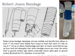
DIY robert jones splint
5th year.
Elastoplast anchor stirrup + proximal to distal cotton wool (at least 3 rolls) + elastoplast outside + splint ie PCV pipe duct taped on top
4 regions of immobolisation
1. Phalanges / distal limb
2. Metacarpus/Metatarsus
3. Radius/Forearm & Hock/Tibia
4.Elbow/Olecranon/Shoulder/Humerus/distal scapula/Stifle/Femur
(I would personally put elbow/olecranon is a separate category)
Where is it impossible to immobilise using a splint/RJ
shoulder and hip joints - hence femoral and humeral fractures cannot be fixed by immobilisation. .
1. What is the most common type of splints for phalangeal/distal limb fractures?
RJ with dorsal splint + heel wedge.
Ending just below the carpus/tarsus joint.
Splints options kimzey or PCV or complression boot

Why do a raised heel wedge for distal limb splints ? *red*
Heel wedge in order to align dorsal cortices of phalances with dorsal aspect of cannon bone - prevents weight bearing on these joints

2. Metacarpus/metatarsus injury splints?
Same as distal limb splints RJ, dorsal splint, but extending proximally to include the carpus/tarsus in the bandage +/- the raised toe
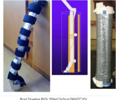
3 and 4. Carpal, radius, tibia/fibula splints?
From the carpus/tarsus joint and up, Brad seems to just suggest full limb casts via RJ bandages.
Options of adding a lateral splint to the heel

5. Olecranon fractures?
Full limb RJ bandage + caudal splint from elbow to heel
This allows even weight sharing between front limbs when it overwise wouldnt be possible due to disruption to triceps apparatus so horse unable to fix carpus (reduces chance of contralateral limb laminitis*).
Additionally forces olecranon fracture into better position for repair.

Rupture of which ligamnets has the following presentations:
a) Horse has dropped fetlock
b) Horse has toe raised and dropped fetlock
c) Horses fetock is on the floor
Rupture of
a) SDFT
b) DDF + SDFT
c) SL + DDFT + SDFT

DDF rupture pathogneumonic look
- fetlock Drops & toe flips up

Outline the generalised treatment protocol for flexor tendon laceration
- conservative vs surgical
Conservative
- Splint for 4-6 weeks
- but cannot immobilize origin of the tendons/will always have gap heeling
+ 12 month rehab and staged weaning off support.
Surgical repair. Suture the ends together via 6 strand savage or 3 pulley system.
- This orientates the tendon ends better and gets them closed, however there will always be some gap healing involved
Extensor tendon laceration presentation
Knuckling gait
*extremely common
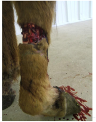
Prognosis of extensor tendon lacerations wihtout tx
heal pretty well by themselves - 60-80% RTF (compared to SFT/DDFT which is 50-75%)
Tx options extensor tendon lacerations
Debride tendon ends if exposedd
+
RJ bandage +/- dorsal splint initially until horse learns how to place the limb (encouraged through controlled walking rehab)
Whats this indicative of/

suspensory ligment disruption *looks same as SFFT rupture?
stretching of suspensory ligament
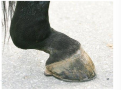
suspensory ligament laceration?
toe up ,entire fetlock on floor

Summary 1

summary 2

MSK sepsis
• Recognize, describe and recall the clinical features of equine musculoskeletal sepsis
• Understand, describe and recall the aetiopathogeneses of equine musculoskeletal sepsis
• Understand, describe and recall the diagnostic methods applied to equine musculoskeletal sepsis
• Recognize, describe and recall the common pathogens involved in equine musculoskeletal sepsis
• Recognize, describe and recall the principles of antimicrobial therapy, common antibiotic agents used to treat equine musculoskeletal sepsis including does rates, routes of administration, frequency of administration and side effects and complications
• Understand, describe and recall surgical methods of treating equine musculoskeletal sepsis
• Formulate a management and or treatment plan for equine musculoskeletal sepsis and demonstrate an understanding of complications, prognosis and return to function and outcomes
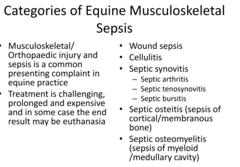
Categories of MSK sepsis
Wound sepsis
Cellulitis
Septic synovitis
Septic osteitis
Septic osteomyelitities
Clinical signs of sepssi
4-5/5 lameness
Joint synovial effusion, heat, painful
Periarticular swelling too
Often with discharging wound
Often acute onset
Not febrile in adults. Febrile in foals and WCC elevated in foals

list 4 DDX for sepsis?
Hoof abscess
Fracture
Cellulitis
Neuropathy
What structures may be involved here?
Digital tendon sheath
Coffin / DIP joint
Collateral cartilages
Pastern / PIP joint

A practical approach to assessing sepsis in open wounds
Nerve block + aseptic prep + digital palpation ti assess depth and see if you can feel the structures
Then
- Joint tap (neutrophils)
- U/s
- radiography

How to classify a wound as infected vs normal healing process based on palpation
Heat, pain, swelling, discharge - indications of infection due to replicating microrganimals.
If just mucopurulent dischrage, probably not infected.
What is the disease in foals, characterisedby multiple joint swellings and owners tend to misinterpret as foals stood on by mare.
neonatal septic arthritis
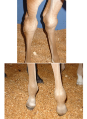
Foals, unlike adults, will have _____ as signs fo sepsis
increased WCC and fever
What are 2 forms of bone sepsis?
osteomyelitis or septic osteitis +/- sequestration
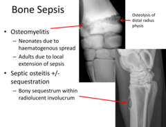
Diagnostic methods
- when would radiography be practical
Osteomyelitis in foals especially
Metal FB or in chronic or non-responsive cases - signs of OA or osteomyelitis
Diagnostic methods
- when would u/s be pracatical
Ass effusions, fibrin and wood foreign bodies
Diagnosti methods
- when would synoviocentesis be useful and
All cases
Use of MC&S in sepsis
Is a good and ncie thing to do, but may be difficult.
Sample the primary area of sepsis.
Allows targetting of primary organism. Lots of the time get mixed cultures due to contamination
Main organsims isolated in septic arthritis?
Gram negatives - e coli, enterobacteria, pseudomonas
Sometimes gram positive, more commonly if iatrogenic cause ie arthroscopy - staph and strep
What 4 things can you assess on synoviocentesis?
TP
TNCC
Cell populations on cytology
Gross Apperance
Compare normal vs Septic synovial fluid characteristics *in red*
- TCC
- g/L TP
- % PMN
TCC
- normal:
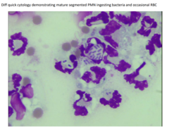
What gross mirpholog of synovial fluid would you see with septic synovitis
Flocculent and turbid fluid

How much synovial fluid to collect?
5ml
MC&S takes how long? what to do in meantime
7-10 days.
Clinicians must commence antibiotic therapy before receiving culture and sensitivity results
Principles of antibiotic therapy for the treatment of MSK sepsis
- List 3 options for routes of delivery and when they would be indicated
IV and IM most common in acute cases
PO in prolonged therapy
Regional anaesthesia in conjunction with systemic methods (IV/IM/PO) to increase efficacy of antimicrobials
When to reassess antibiotic selection, dose and/or route ?
If no improvement in CS within 48-72 hrs
What should you use as first principle antibiotics
Beta lactam (gram + and anaerobe)
PLUS
Aminoglycoside (gram neg activity)
why both? synergistic and provide good broad specrtum coverage
IE PENICILLIN + GENTAMYCIN
How long to administer Abs for
required for 2-4 weeks in total and usually for at least 48-72 hrs after resolution of CS
List 3 examples of local AB administration
- Intrasynovial injection
- Pumps
- Depot methods
Whats the main negative of intra-synovial antibiotic injections as a treatment option for septic arthritis?
Repeated injections required into synovial space = increased risk of iatrogenic infections
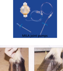
Local antibiotic depot methods
Less so the rage now adays. Called PMMA beads in wounds or in joint but will need to be removed

Regional antibiotic delivery - 2 methods of delivery
Intra-osseous
Regional IV perfusion
Intravenous regional limb perfusion method - explain this method of antimicrobial deposition
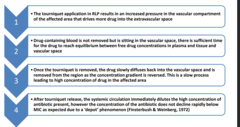
Torniquetto to occlude venous return/keep antibiotic localsied.
Place catheter and inject into vein of choice (saphenous, cephalic, digital).
Leave tornqiuet on for 20-30 minutes leading to leakage of antimibrial from vessel into tissue due to high hydrostaic pressures --> Achieves antimicrobial concentrations in tissue that are significantly above the minimum inhibitory concentration
Relatively small doses but still reach significantly above MIC
Take torniquet off. Will take a long time for abs to diffuse back out thus sustained concentrations in tissue for 24-48 hours, then repeat.
*always in conjunction with systemic antibiotics
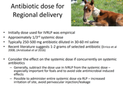
What dose of regional IV ?
1/3 systemic dose
If on concurrent systemic dose, reduce systemic dose (minus the concnetraiton you put in the regional vein)
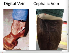
Which vessel for injection?
- carpus, tarsus
- fetlock region
- distal limb region
Veins >>> arteries
Cephalic (medial) and saphenous (lateral) veins for carpus, tarsus and fetlock
Digital vein for distal limb
What are the options for Oral antibiotics in horses?
TMS +/- rifampicin
Enrofloxacin (staph septic arthritis)
risks of oral antibiotics?
diarrhoea / colitis.
List 3 Surgical methods of treating sepsis (an adjunct to systemic and regional antibiotics)
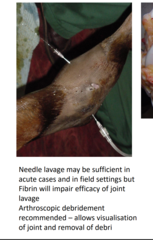
Needle joint lavage
- 14G needles ingress + egress 1-3L Ringers
Arthroscopic lavage
Arthrotomy