Unit 2 Skull, Scalp, Meninges, and Dural Venous Sinuses
 Call Kai
 Learn
Learn Practice Test
 Spaced Repetition
 Match
Match Flashcards
 Knowt Play
1/51

There's no tags or description
Looks like no tags are added yet.
Name | Mastery | Learn | Test | Matching | Spaced | Call with Kai | Chat |
|---|
No analytics yet
Send a link to your students to track their progress
52 Terms
neurocranium
- consists of calvaria and cranial base

calvaria
- "skull cap"
- surrounds the brain superiorly, anteriorly, posteriorly, and laterally
- comprised of the parietal bones and parts of the occipital, temoporal, frontal, and sphenoid

cranial base
- supports the brain inferiorly
- contains foramina for the exit of CNs
- comprised of ethmoid and parts of frontal, sphenoid, temporal, and occipital bones
- includes and forms the anterior, posterior, and middle cranial fossae

viscerocranium
- facial skeleton
- comprised of 14 bones - lacrimals, nasals, zygomatics, palatines, inferior nasal conchae, maxillae, mandible (1), and vomer (1)

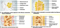
intramembraneous ossification
- calvaria and facial skeleton
- most skull bones form from dense CT membranes
- ossification centers develop and edges of bony plates grow toward each other
fontanels
- six non-ossified areas of membrane that connect adjacent cranial bones at birth
- anterior, sphenoidal, mastoid, and posterior fontanels
- normally close between 3 and 18 months post-natal

Wormian bones
- extra ossification centers form sutural bones in sutures (especially lamboid)

endochondral ossification
- some skull bones, or parts of bones, form from hyaline cartilage
- occurs in the four bones forming the cranial base, entire ethmoid, most of the sphenoid, occipital bone around the foramen magnum, and the mastoid process and petrous portion of the temporal bones
joints of the skull
- skull bones articulate via sutures (fibrous joints - sutural ligament inside)
- become synostoses gradually starting at ~25-30 years
- few exceptions
temporomandibular joints
- bilateral synovial joints between the mandibular fossa and the condyle of the mandible

auditory ossicles joints
- malleus/incus and incus/stapes articulate via synovial joints

mandible joint
- right/left halves joined by mandibular symphysis, synostosed by 24 months
frontal bone joint
- paired, united by metopic suture that usually synostoses by 8 years old, but can persist into adulthood
gomphosis
- peg and socket joint between teeth and mandible/maxilla

scalp 5 layers
- consists of 5 layers of tissue, three superficial are fused to form a single unit
- skin, connective tissue, aponeurosis, loose CT, pericranium

S of scalp
- skin
- thin except for in the occipital region
C of scalp
- connective tissue
- thin superficial fascia with abundant blood supply, mainly from the external carotid a. and some from internal carotid a. via the ophthlamic a.
A of scalp
- aponeurosis
- epicranial aponeurosis which is dense CT connection between frontalis and occipitalis muscles
L of scalp
- loose CT
- loosely connects the aponeurosis to the underlying periosteum
- provides mobility for the three fused superficial layers
- transversed by emissary veins
P of scalp
- pericranium
- external periosteum of calvaria
frontalis
- paired
- each muscle belly origininates from skin and superficial fascia of eyebrow and inserts into epicranial aponeurosis

occipitalis
- paired
- each muscle belly originates from superior nuchal line and inserts into epicranial aponeurosis
- some reference as single muscle with frontalis : occipitofrontalis innervated by CNVII

sensory innervation of scalp
- anterior 2/3 of scalp innervated by branches of trigeminal n.
- posterior 1/3 of scalp innervated by C2-3 rami
trigeminal nerve innervation of scalp
- supratrochlear n. (more medial) and supraorbital n. (more lateral) : both branches of ophthalmic n.
- zygomaticotemporal n. : branch of maxillary n.
- auriculotemoporal n. : branch of mandibular n.
C2-C3 rami innervation of scalp
- lesser occipital n. : primarily fibers from anterior rami of C2, but can be from C3
- greater occipital n. : posterior rami of C2
- third occipital n. : posterior rami of C3
blood vessels of scalp
- supratrochlear a. (more medial) and supraorbital (more lateral) : both branches from the ophthalmic a.
- superficial temporal, posterior auricular, and occipital a. : branches of the external carotid a.
emissary veins
- connect veins in the scalp to the dural venous sinuses via emissary foramina
- venous flow is normally away from the dural sinuses (this is opposite)
- function in selective brain cooling
- can create a route for extracranial infections to spread into cranium

dura mater in head
- cranial dura is formed from two layers that are fused in most places
- forms dural septa that support cerebral hemispheres and cerebellum and prevents displacement of these structures
- major blood supply is the middle meningeal a., which lies in the epidural space
periosteal (endosteal) dura
- made from the internal periosteum of the calvaria
true (meningeal) dura
- dura mater 'proper'
- continuous at foramen magnum with the spinal dura
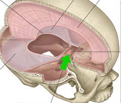
falx cerebri
- midsagittal double layer of truue dura in longitudinal fissure of the cerebral hemispheres
- attaches anteriorly to crista galli and continuous posterior with tentorium cerebelli

tentorium cerebelli
- double layer of true dura in the transverse fissure between the occipital lobes and cerebellum
- attaches to both of clinoid processes, superior border of the petrous portion of temporal bones (forming the superior petrosal sinus) and the lips of the grooves for the transverse sinus (forming the transverse sinuses)

tentorial notch/incisures
- central opening for midbrain within the margin of the tentorium attaching to the anterior clinoid processes

falx cerebelli
- small, double layer of true dura between the cerebellar hemispheres
diaphragma sellae
- single layer of dura over the sella turcica with an opening acccomodating the infundibulum of the pituitary gland
arachnoid mater in head
- loosely adheres to dura
- forms arachnoid granulations which project into the superior sagittal sinus
pia mater in head
- covers the brain forming a "pial coat"
- extends into the brain for a short distance to cover the cerebral vessels
dural venous sinuses
- spaces between the periosteal and true dural
- receive cerebral veins and veins draining the calvaria, orbit, infratemporal fossa, inner ear, and scalp
arachnoid villi
- minute projections of arachnoid mater through the true dura into the sinuses for return of cerebrospinal fluid from subarachnoid space to systemic venous return
- groups of villi are known as arachnoid granulations
superior sagittal sinus
- in the fixed margin of the falx cerebri
- receives cerebral veins
- receives most arachnoid villi
- empties into confluence of sinuses

inferior sagittal sinus
- along the inferior margin of the falx cerebri
- joints the great cerebral vein posteriorly to form the straight sinus

straight sinus
- lies at the junction of falx cerebri and tentorium cerebelli
- empties into the confluence of sinuses

confluence of sinuses
- located at the internal occipital protuberance
- receives blood from superior sagittal and straight sinuses
- drains into right and left transverse sinuses

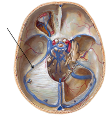
transverse sinuses
- lie in fixed border of tentorium cerebellum in grooves of occipital bone
- drains blood from the confluence of sinuses
- leaves tentorium laterally to drain into sigmoid sinuses

sigmoid sinuses
- curve inferiorly and anteriorly to become internal jugular veins at jugular foramen

cavernous sinuses
- lie between the two dural layers on either side of the body of the sphenoid bone and communicate across midline
- receives blood from ophthalmic and cerebral veins
- several structures pass through this sinus

structures that pass through the cavernous sinuses
- internal carotid arteries and sympathetic nerves that run along the surface of these arteries
- CN III, IV, V1, V2 in the lateral wall of sinus
CN VI passes through the sinus more medially
superior petrosal sinus
- along superior edge of petrous portion of temporal bone
- transmits blood from the cavernous sinus, joins the terminal end of transverse sinus to drain into sigmoid sinus
inferior petrosal sinus
- transmits blood from cavernous sinus into the internal jugular vein at the jugular foramen

epidural space
- potential space between the periosteal dural and cranial bones
- skull fracture, especially in the temple can tear the middle meningeal a. and result in an epidural hematoma
subdural space
- potential space between arachnoid mater and true dura
- tearing of cerebral veins as they enter the superior sagittal sinus results in a subdural hematoma
subarachnoid space
- space between pia and arachnoid mater containing cerebrospinal fluid and cerebral arteries/veins
- ruptured cerebral arteries (often aneurysms) result in a cerebral hemorrhage and blood in the CSF