Bones
1/14
There's no tags or description
Looks like no tags are added yet.
Name | Mastery | Learn | Test | Matching | Spaced | Call with Kai |
|---|
No analytics yet
Send a link to your students to track their progress
15 Terms
Bone
living tissue capable of changing its structure as the result of stresses to which it is subjected
consists of cells, fibers and matrix
calcification hardens surface
protective function
lever storage and protection
forms
compact
cancellous
Compact bone
solid mass
Cancellous bone
branching network of trabeculae
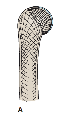
Long bones
found in limbs (humerus, femur, metacarpals/tarsals. phalanges)
tubular shaft, diaphysis, and ephyisis
growth
epiphysis separates by epiphyseal cartilage
metaphysis = lies between diaphysis
central marrow cavity with bone marrow
periosteum = connective tissue sheath covering the outer compact bone
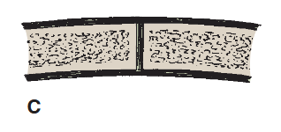
Short bones
found in hand and foot
cuboidal in shape
composed
cancellous bone surrounded by thin layer of compact bone
periosteum and hyaline cartilage
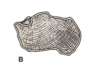
Flat bone
found in vault of skull
thin inner and outer layers of compact bone,
tables separated by cancellous bone (diploe)
Irregular bones
unassigned groups
thin shell of compact bone with interior made of cancellous bone
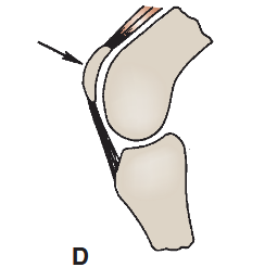
Sesamoid bone
small nodules of bones found on certain tendons
EX: Patella
Surface markings of bones
irregularities in bones that first appear at puberty
some are large with special names
Bone marrow
occupies marrow cavity in long and short bones and cancellous tissue in flat and irregular bones
starts as red at birth = becomes yellow at the age of 7
periosteum
thick layer of fibrous tissue has abundant vascular supply and osteogenic deep cells
provides rich nerve supply = highly senstiive
Bone development
menbranous and endochondral
developed directly from connective tissue membrane
cartilaginous model is laid down and later replaced by bone.
Membranous method
bones of the vault of skull
serves to protect underlying developing brain
Endochondral method
long bones of limbs are developed in a slow process until 18-20th year
diaphysis (found in shaft of bone)
epiphysis (ends of bone)