Looks like no one added any tags here yet for you.
receptive field
refers to the specific receptors that feed into a given cell in the nervous system; region of space in which the presence of a stimulus will alter the firing of a neuron
excitation, inhibition
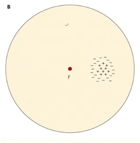
Receptive field recordings can be determined by inserting a microelectrode into an anesthetized animal and directing the fovea of that animal to a point on a screen. Spots of light are directed onto various areas the screen. The microelectrode then records activity of a neuron on the retina.
When the spotlight is within a neuron's receptive field, what 2 things can happen?
excitation
(higher rate of action potential)
What happens if stimulation occurs in the center part of a neuron's receptive field?
inhibition
(lower rate of action potential)
What happens if stimulation occurs in the peripheral part (outer ring) of a neuron's receptive field?
true
True or false: Receptor field properties are different for different types of cells.
hyperpolarized
If light falls in a photoreceptor's receptive field, is the photoreceptor depolarized or hyperpolarized?
more
A larger/brighter light stimulus results in [more/less] hyperpolarization in photoreceptors.
nothing
(no activity)
What happens when light falls outside of a photoreceptor's receptive field?
horizontal cells
(due to spatial summation)
Which has a larger receptive field: photoreceptors or horizontal cells?
light falling anywhere in receptive field can initiate any cell
How does spatial summation occur in horizontal cells?
hyperpolarized
If light falls in a horizontal cell's receptive field, is the horizontal cell depolarized or hyperpolarized?
center surround organization
organization of bipolar & ganglion cell receptive fields in which light falling on the center of the RF has the opposite effect of light falling on the surrounding area of the receptive field
bipolar cells, ganglion cells, amacrine cells
What kind of cells in the retina have center-surround organization?
off center, on surround
bipolar cell receptive field in which light striking the center field causes an inhibitory response (hyperpolarization)
off center, on surround
bipolar cell receptive field in which light striking the surrounding field causes an excitatory response (depolarization)
on center, off surround
bipolar cell receptive field in which light striking the center field causes an excitatory response (depolarization)
on center, off surround
bipolar cell receptive field in which light striking the surrounding field causes an inhibitory response (hyperpolarization)
invaginating
What kind of synapses do on-center bipolar cells have?
flat
What kind of synapses do off-center bipolar cells have?
midget
bipolar cells that have a smaller receptive field but high spatial resolution; they are also color-opponent
they connect to either one M cone or one L cone
(center has a narrow range of wavelength that it'll respond to, whereas nearby horizontal cells connect to multiple cones)
What does it mean that midget bipolar cells are "color-opponent"?
diffuse
bipolar cells that have a larger receptive field but they are not color-opponent
can process different wavelengths
What does it mean that diffuse bipolar cells are NOT "color-opponent"?
S-cone bipolar cells
bipolar cells that synapse only with one specific type of cones and are color-opponent
midget
Which bipolar cells are responsible for high spatial resolution, especially at the fovea?
midget, S cone
Which 2 bipolar cells are color-opponent?
action potential
Like bipolar cells, amacrine cells have a center-surround organization. However, they are the FIRST retinal neurons to display what kind of electrical response?
graded potentials
(amacrine = first to have action potential)
Up until amacrine cells, what kind of electrical responses did we get in photoreceptors, horizontal cells, and bipolar cells?
photoreceptors, horizontal cells, bipolar cells
Which retinal cells have graded potentials?
amacrine cells, ganglion cells
Which retinal cells have action potentials?
can be both
Are ganglion cells on-center or off-center?
faster
If light stimulates the center of an on-center ganglion cell, is the rate of an action potential faster or slower?
slower
If light stimulates the center of an off-center ganglion cell, is the rate of an action potential faster or slower?
on center
Do on-center midget ganglion cells synapse with on or off center midget bipolar cells?
off center
Do off-center midget ganglion cells synapse with on or off center midget bipolar cells?
retinal parvo cells
(part of parvocellular visual pathway)
other term for midget ganglion cells - over 70% of ganglion cell population
parvocellular
What visual pathway are midget ganglion cells involved in?
midget bipolar cells
What other kind of cells do retinal parvo cells synapse with?
midget ganglion cells
(retinal parvo cells)
70% of retinal ganglion cells are what specific type?
diffuse bipolar cells
What other kind of cells do retinal magno cells synapse with?
parasol ganglion cells, retinal magno cells
2 names for type of ganglion cells that synapse with diffuse bipolar cells
magnocellular
What visual pathway are parasol ganglion cells involved in?
small bistratified ganglion cells
type of ganglion cells that synapse with S-cone bipolar cells
S-cone bipolar cells
What other kind of cells do small bistratified ganglion cells synapse with?
koniocellular visual pathway
What visual pathway are small bistratified ganglion cells involved in?
midget, parasol, small bistratified
What are the main 3 types of ganglion cells in order from most to least numerous?
parasol ganglion cells
(since these are involved in the magnocellular visual pathway)
Which ganglion cells are involved in the MAIN visual pathway: midget ganglion cells, parasol ganglion cells, or small bistratified ganglion cells?
ipRGCs
(intrinsically photosensitive retinal ganglion cells, or melanopsin containing ganglion cells)
other kind of ganglion cells that contain melanopsin and therefore can absorb photons directly; peak sensitivity at 483 nm, related to circadian rhythms
ipRGCs
(since these contain melanopsin)
Which type of ganglion cells are involved in seasonal affective disorder?
**may be treated with standardized broad-spectrum light to help modulate circadian rhythms better
C, D, E
(other two are just regular circular receptive fields, no distinct areas)
Which of the following cells have a center-surround organization? Select all that apply.
A. photoreceptors
B. horizontal cells
C. bipolar cells
D. amacrine cells
E. ganglion cells
D, E
Which of the following cells have action potentials? Select all that apply.
A. photoreceptors
B. horizontal cells
C. bipolar cells
D. amacrine cells
E. ganglion cells
A, B, C
Which of the following cells have graded potentials? Select all that apply.
A. photoreceptors
B. horizontal cells
C. bipolar cells
D. amacrine cells
E. ganglion cells
maintained discharge
(or simply just spontaneous activity)
In the absence of a stimulus, cells with a center-surround receptive field can spontaneously generate action potentials in what process?
true
True or false: When there is no light stimulation, action potentials still occur.
decreases
If light falls on the surround of an on-center cell, what happens to the frequency of action potentials?
increases
If light falls on the center of an on-center cell, what happens to the frequency of action potentials?
false
(always occurring, but light = higher rate of action potentials)
True or false: When there is no light stimulation with center-surround receptive fields, action potentials will not occur. They require light exposure.
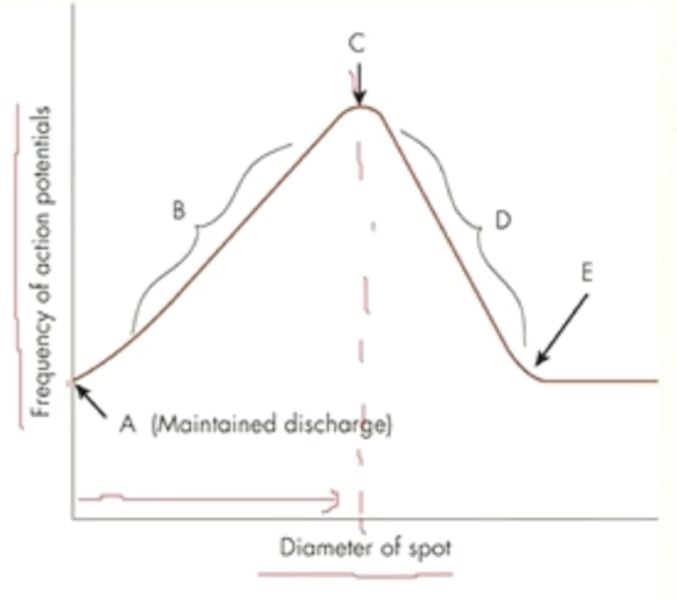
increases up until a certain point, then decreases
(this is because the stimulus size ends up surpassing the center field, now going into the inhibitory surround field)
When measuring receptive field in an animal (such as a monkey or a cat), how does the action potential rate change with an increasing stimulus size?
maintained discharge
(none of the receptive field is being stimulated)
What does A represent in an on-center/off-surround cell?
stimulus size is increasing but staying within the center field only
What does B represent in an on-center/off-surround cell?
stimulus size = size of center field only
What does C represent in an on-center/off-surround cell?
stimulus size is increasing and spilling over more into the surround field
What does D represent in an on-center/off-surround cell?
stimulus size = size of entire receptive field (center and surround)
What does E represent in an on-center/off-surround cell?
it doesn't
(the center and surround fields fully cancel each other out, so the frequency of action potentials is the same as the baseline maintained discharge)
If a light stimulus is larger than the entire receptive field, how does that influence the activity of the neuron?
after our eyes have remained open and acclimated to the current light level, they go back down to baseline
(after having a very brief period of increased activity)
When we close our eyes, the cells receive no stimulation and are at baseline level.
However, the ability of our visual system to adapt to different light levels is very quick. So how is it possible to be at baseline level when our eyes are open?
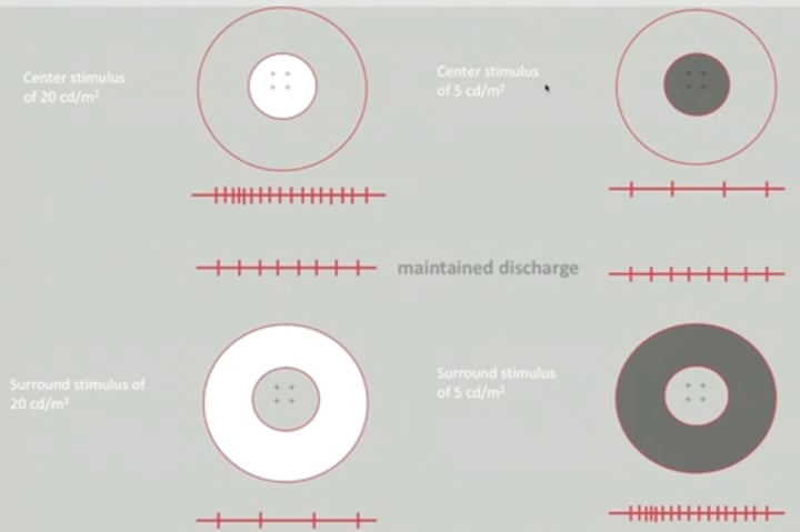
increases
(due to excitation of center field)
If our eyes are adapted to a background light level and therefore have baseline activity, but we have a light stimulus presented to the center field that is brighter than the background light level, what happens to the rate of action potentials?
decreases
(due to inhibition of center field)
If our eyes are adapted to a background light level and therefore have baseline activity, but we have a light stimulus presented to the center field that is darker than the background light level, what happens to the rate of action potentials?
decreases
(since we are exciting the inhibitory surround field)
If our eyes are adapted to a background light level and therefore have baseline activity, but we have a light stimulus presented to the surround field that is brighter than the background light level, what happens to the rate of action potentials?
increases
(since we are inhibiting the inhibitory surround field)
If our eyes are adapted to a background light level and therefore have baseline activity, but we have a light stimulus presented to the surround field that is darker than the background light level, what happens to the rate of action potentials?
A, D
What 2 choices together would cause the greatest increase in frequency of action potentials?
A. stimulus brighter than background presented to center field
B. stimulus brighter than background presented to surround field
C. stimulus dimmer than background presented to center field
D. stimulus dimmer than background presented to surround field
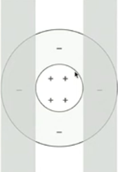
spatial grating pattern
What is a strong stimulus for a cell with center-surround organization?
increase
(dark bars inhibit surround, center bright bar stimulates center)
If you present a vertical spatial grating pattern that has two darker bars and a bright bar in the middle to a cell with a center-surround receptive field, will the rate of action potentials increase or decrease?
spatial contrast
(alternating dark and light bars)
Cells with center-surround organization are selective for what kind of contrast?
ganglion cells
Cell with center-surround organization are selective for spatial contrast, especially what cells in particular?
contrast information
(since cells with center-surround receptive field are selective for spatial contrast - our visual system is very well built for contrast!)
What kind of visual information is extracted from the visual stimulus usually very early in the process?
lateral inhibition
process in which the activity of one cell laterally inhibits the activity of its neighboring cells
horizontal cells
Which retinal cells are mostly involved in mediating lateral inhibition?
horizontal cells
In addition to sending signals to the bipolar cells, photoreceptors also send signals to what other kind of cells, which then laterally connect to surround ones to mediate their input/output?
limulus
(scientists placed electrodes into 2 separate large photoreceptors in the crab, saw how each affected the other)
In which animal was lateral inhibition first discovered?
edge detection, contrast enhancement
Lateral inhibition is important for neurons with center-surround receptive fields because it plays a role in which two perceptual processes?
luminance
(the abrupt changes in luminance that occur at the border of objects create a contrast that could be detected by cells with center-surround receptive fields)
Lateral inhibition plays a role in detecting abrupt changes in what physical property of objects?
Mach bands, simultaneous contrast, Hermann grid
3 visual phenomena that can be explained by the interaction between center-surround receptive fields and lateral inhibition
simultaneous contrast illusion
illusion in which 2 objects of equal luminance will change brightness depending on the background
background
What does the perceived brightness of the 2 objects depend on in a simultaneous contrast illusion?
square with lighter background
You have two squares of the same luminance - one surrounded by a darker background and one surrounded by a lighter background. Which one has more lateral inhibition?
darker
If there is a square surrounded by a certain background, if there is more lateral inhibition, will this make the square appear lighter or darker compared to the background?
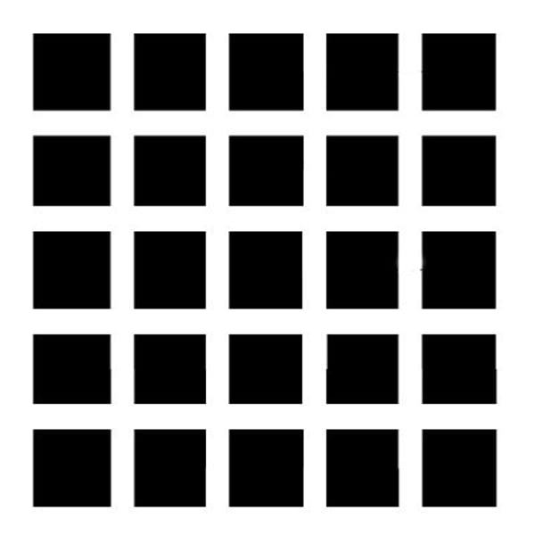
Hermann grid
geometrical display that results in the illusion of dark areas at the intersection of two white "corridors"
fovea
With the Hermann grid, if you are fixating a certain "intersection" point, what part of your eye is specifically fixating on that part?
also stimulates the fovea and is included in the receptive field
(more light stimulation where we are directing our gaze)
With the Hermann grid, if you are fixating a certain "intersection" point, why does the immediate surrounding area also appear white?
darker
If you have a Hermann grid with equally spaced black squares, if there is more lateral inhibition, will looking at a certain area appear lighter or darker?
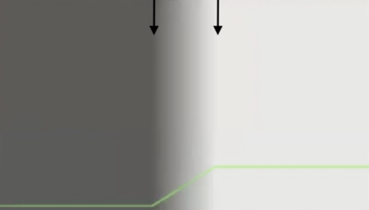
Mach bands
perception of a thin dark band on the dark side of a gradual light-dark border and a thin light band on the light side of the border
These bands are an illusion because they occur even though corresponding intensity changes do not exist.
more LI at dark band, less LI at light band
At the points where we see the dark/light Mach bands, do those represent an area of more or less lateral inhibition?
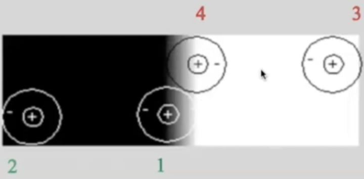
1
(which is why 1 appears slightly darker)
Which area has more lateral inhibition: 1 or 2?
3
(which is why 4 appears slightly brighter)
Which area has more lateral inhibition: 3 or 4?
B
Which of the following phenomenon illustrates a contrast enhancement at the edge between two areas?
A. simultaneous contrast
B. Mach bands
C. Hermann grid
D. none of above
C, D
Why is center-surround receptive field/lateral inhibition important? (2)
A. to control light and dark adaptation in the retina
B. to process the beginning stages of color vision
C. to enable and enhance the process of edge detection
D. to enhance contrast processing between two areas of different luminance
B
Which of the following correctly point out a reason why a dark spot is not perceived at the central fixated intersection in a Hermann grid?
A. foveal ganglion cells have large receptive field
B. foveal ganglion cells have small receptive field
C. peripheral ganglion cells have large receptive field
D. peripheral ganglion cells have small receptive field
B, C
Dark spots are perceived at the peripheral intersections in Hermann Grid because: (2)
A. peripheral ganglion cells have small receptive field
B. peripheral ganglion cells have large receptive field
C. there is more lateral inhibition for cells at the intersections
D. there is more lateral inhibition for cells at the streets
B
How can lateral inhibition explain the phenomenon of simultaneous contrast illusion?
A. there is more lateral inhibition from the darker surround to the center small square than from the lighter surround
B. there is less lateral inhibition from the darker surround to the center small square than from the lighter surround
B
Lateral inhibition in the retina is via the connection between cones with which type of retinal cells?
A. bipolar cell
B. horizontal cell
C. amacrine cell
D. RGC