Microbiology Lab Exam 1
 Call Kai
 Learn
Learn Practice Test
 Spaced Repetition
 Match
Match Flashcards
 Knowt Play
1/148

There's no tags or description
Looks like no tags are added yet.
Name | Mastery | Learn | Test | Matching | Spaced | Call with Kai | Chat |
|---|
No analytics yet
Send a link to your students to track their progress
149 Terms
Occupational Health Care provider
call if you have health concerns or questions
Questions about chemicals can be answered by referring to the
appropriate Material Safety Data Sheet (MSDS)
first unexcused absense deduction
5 lab points
second unexcuse absense deduction
10 lab points
3rd unexcused absense deduction
zero for your lab
receive an unexcused absense for reasons including, but not limited to:
3 Tardies (arriving within 10 minutes of the start of lab according to the lab clock)
arriving more than 10 minutes after the lab period has begun (you will not be allowed to enter the lab at this time)
Leaving before all experiments are complete
must schedule a make-up at least how many hours before leaving on a University excused field trip or interview
72 hours
all other excused absences should be schedules within 48 hours of the absence and completed within 5 working days
Total absences that exceed 20% of scheduled lab meetings may result in an
F in the course
For excused absences you must get an excuse from
Lacy Basile
complete the make-up request form, link on canvas, include course and section number
Glass microscope slides 🡪
Glass disposal
Glass tubes with media (sterile or not/broken or not) 🡪
Biohazard bin
Pipette tips 🡪
Biohazard waste
Paper towels 🡪
Regular trash
Microorganisms are
“ubiquitous in nature”
Found everywhere that other lifeforms exist
Have a long, rich history on Earth
And have successfully adapted to a wide range of habitats
Microorganisms can be isolated from
Soil (all kinds!)
Water (over a large range of salinity)
Plants
Animals (even humans!)
Even seemingly uninhabitable places such as the hot acid pools in Yellowstone (approx. 85ᵒC with a pH near 1)
Does NOT apply to EVERY SPECIES, but certainly applies to microorganisms as a group
Classification of microorganisms
Many microorganisms do not reside on or in a specific plant or animal host and are not known to cause disease
Free-living and nonpathogenic
Often saprophytes that perform important role of decomposition in the ecosystem
Other microorganisms reside on or in a host organism
pathogens
mututalism
commensals
pathogens
cause damage to host
Mutualism
both host and microbe benefit
Commensals
microbe benefits but no effect on host
Many of the commensal /mutualistic strains are
opportunistic pathogens
Inhabit our bodies and are capable of producing a disease state if introduced into a suitable part of the body
reservoir
reservoir
any area where a microbe with the potential to cause infection resides
Purpose of blood agar
To differentiate bacteria based on their hemolytic characteristics
Blood agar
Includes 5% sheep blood in a Tryptic Soy Agar base
Several species of Gram-positive cocci produce exotoxins called
hemolysins, (streptococci produce streptolysins) which are able to destroy RBCs and hemoglobin
Three major types of hemolysis:
β – hemolysis
α – hemolysis
γ – hemolysis

β – hemolysis
complete destruction; results in clearing of the medium
yellow glow

α – hemolysis
partial destruction; results in greenish discoloration of the medium
brownish/green

γ – hemolysis
no hemolysis
clear/blue

Germicides
Refers to substances or systems, both chemical and physical, that prevent the spread of pathogens
Some are specific but most target a broad-spectrum
Three categories of Germicides
Decontamination
Disinfection
Sterilization
Decontamination
Lowest level of control
Defined as “reduction of pathogenic microorganisms to a level at which items are safe to handle without protective attire”
Usually includes physical cleaning with soaps or detergents, and removal of all or most organic and inorganic matter
Disinfection
Next level of control
Divided into three sublevels – low, medium and high – based on effectiveness against specific control pathogens or their surrogates
All kill most, if not all, of the targeted pathogens but typically do not kill large numbers of spore
Typically liquid chemical agents but can be solid of gaseous
Other methods include dry heat, moist heat, and ultraviolet light
Types of disinfectants
Chemical sterilants
Antiseptics
Chemical sterilants
High-level disinfectants that have the ability to kill all vegetative cell and some spores
Antiseptics
Disinfectants designed to reduce or eliminate pathogens on or in living tissue
Sterilization
Highest level of pathogen control
Complete elimination of viable organisms including spores
Can be achieved by some chemicals and gases, incineration, dry heat, moist heat, ethylene oxide gas, ionizing radiation, low-temperature plasma, or low-temperature ozone
Steam Sterilization
Most effective and most common method
Autoclave
Autoclave
Device most commonly used for steam sterilization
Use superheated steam under pressure to kill heat-resistant organisms
In the microbiology laboratory:
Sterilizing temperature is set between 121°C and 127°C
Must reach optimum temperature for a least 15 minutes
Time varies according to the size and consistency of the material
Color-coded autoclave tape
Biological indicators
Biological Indicator
Indicator vial includes:
Small ampule containing fermentation broth with pH indicator
Strip of filter paper containing bacterial spores
Test:
Vial is autoclaved at 121°C for 15 minutes
Ampule is crushed
Allows fermentation broth to come into contact with bacterial spores
Vials are incubated at 55°C for 48 hours
Biological indicators are the only way, with certainty, to determine that
sterilization has been achieved
Acidic condition from fermentation
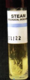
right colored indicator

Negative Control(not crushed)
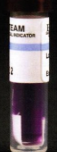
Ubiquity of Microorganisms - Procedure
Day 1
You will transfer microorganisms from seemingly uninhabited sources and grow them on agar plates
When you inoculate the plates, you will transfer an unknown number of unknown cells
All cells that are able to grow on the plate will divide and produce visible colonies
Each source will likely have multiple species present representing a mixed culture
Day 2
You will examine various growth characteristics produced by these “invisible” cohabitants
If a colony is not contacting any other colonies, it is said to be isolated and can be transferred to a sterile medium to start a pure culture
Consider mixed vs. pure culture
isolated
If a colony is not contacting any other colonies
can be transferred to a sterile medium to start a pure culture
writing vs typing organism names
typing will be italisized and writing will be underlined
What does aseptic transfer mean?
Transfer of living microbes from one place to another without contamination of:
The culture
The sterile medium
Or the surroundings
To prevent contamination
Inoculating instruments must be sterilized prior to use
Wire loops are sterilized immediately before use in an incinerator
Cool loop in agar before transfer
Mouths of tubes containing culture or media are also incinerated at the time of transfer
Instruments that can not be incinerated are sterilized inside wrappers or containers by autoclaving prior to use
What is a culture?
A medium that contains living microbes
A pure culture is one that contains a single species
A mixed culture is one that contains multiple species
All cultures are assumed to be mixed prior to obtaining isolation!
Microbial cultures are grown and maintained on or in solid and liquid substances called
media
Media come in many forms, each with a specific application
Broths are used to grow microbes when fresh cultures or large number of cells are required
Agar slants are generally used to grow stock cultures that can be refrigerated after incubation and maintained for several weeks
Plated media are typically used for obtaining isolation of species, differential testing, and quantifying bacterial densities
A bacterial sample is ALWAYS assumed to be a
mixed culture
Mixed culture
microbial culture consisting of two or more species
Pure culture
microbial culture containing only a single species
Methods of isolation
Streak Plate
Pour plate (Lecture Only)
Spread plate
In the 1850’s, Louis Pasteur developed concept of the
pure culture
Allowed microbiologists to study individual species
His methods of isolation were terribly inefficient
Tried to dilute cultures until he thought he had a single cel
In the 1880’s Robert Koch developed a procedure for
“streaking for isolation”
The primary purpose of streaking for isolation is to
obtain isolated colonies
Can then be used to obtain a pure culture
A single colony (composed of identical cells) is then used to inoculate another culture
An isolated colony is composed of
millions of cells – all identical and all descendants from a single cell or group of cells
In the quadrant streak method:
A bacterial sample is streaked over the surface of a plated agar medium.
During streaking, the cell density decreases, eventually leading to individual cells being deposited separately on the agar surface
This results into a colony consisting only of the original cell type
COMMONLY USED TECHNIQUE: QUADRANT STREAK
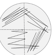
Quadrant Streak
Used with samples suspected of high cell density
Quadrant Streak Method 2
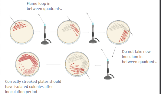
Quadrant Streak Method 3
Properly streaked plate with isolated colonies in the 4th quadrant
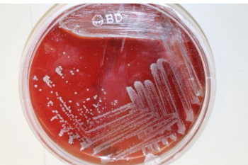
Result of not sterilizing loop between quadrants

Improperly streaked quadrants
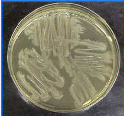
Result of not rotating plate properly

Either loop was not cooled or loop did not pass through previous quadrant
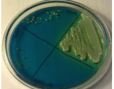
Colony Morphology
One cell makes two, two make four, four make eight…one million make two million
Eventually, a visible mass of cells known as a colony appears
Color, size, shape, and texture are determined by the genetic makeup of the organism

Growth characteristics of colony morphology are influenced by:
Nutrient availability
Temperature
Incubation time
colony morphology shapes
Round
Irregular
Punctiform (tiny pinpoint dots)
colony morphology Margin
Entire (smooth, with no irregularities)
Undulate (wavy)
Lobate (lobed)
Filamentous (unbranched strands)
Rhizoid (branched like roots)
colony morphology Elevation
Flat
Raised
Convex
Pulvinate
Umbonate
colony morphology Texture
Moist
Mucoid
Butyrous
Dry
Shiny
Dull
Other properties include color (pigment production) and optical properties (opaque and translucent)
Agar slants are primarily used for
cultivation, maintenance, and storage of stock cultures
Organisms cultured on slants display a variety of growth characteristics:
Filiform
Friable
Spreading edge
Pigmented or translucent/transparent
Filiform
dense and opaque with a smooth edge
Friable
crusty
Spreading edge
produced by motile organisms
Microorganisms culture in a broth also display
characteristic growth patterns
Pellicle
Sediment
Uniform fine turbidity
Flocculent
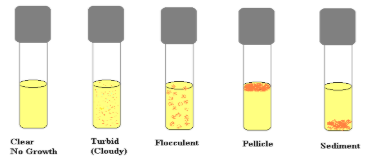
Pellicle
Growth floats on top of the medium

Sediment
Growth sinks to the bottom

Flocculent
Clumped growth

What is the Pour Plate Technique
Yields isolated colonies of bacteria and fungi
Original sample volume is diluted several times to reduce microbial population and to calculate original sample concentration
Small volumes from each dilution are mixed with molten Tryptic Soy Agar (TSA) (48-50C) in sterile petri dishes
After the agar has solidified, each cell will be fixed in place and form a colony
The total number of colonies can then be used to calculate the number of viable microorganisms in the sample
Calculated as CFU/mL (colony forming units/mL of sample)

What is the spread plate technique?
A method of isolation in which a diluted microbial sample is deposited on an agar plate and spread uniformly across the surface
If properly diluted, cells (colony forming units/CFUs) will be deposited far enough apart on the agar surface to grow into individual colonies
After incubation, a portion of an isolated colony can be transferred to a sterile medium to begin a
pure culture
Also can be used to quantify cell density of a broth culture
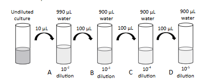
Perform a serial dilution of a broth culture
Take 10 μL of undiluted culture, add that to a tube containing 990 μL of sterile H20, and gently mix/vortex. (Tube A)
Then take 100 μL from tube A and transfer to another tube containing 900 μL of sterile H20, and gently mix/vortex. (Tube B)
Continue this process of transferring samples from higher dilutions to sterile water to create more and more diluted samples, hoping to get a plate that will have the right number of colonies to be considered “countable” (30-300)
Transfer and spread 100 μL of each dilution on a TSA plate.
Lastly, incubate and count colonies
How is the spread plate technique useful?
Achieve isolation
May be used to start a pure culture
Estimate cell density
CFU/mL = # colonies .
volume plated x dilution

Calculating Dilutions for Spread Plate
1st tube - Dilution = Volume transferred/total volume of tube
10μL/(10μL+990μL) = 10/1000 = 0.01 = 10-2 (Tube A)
2nd tube – Dilution = concentration from the previous tube x (volume transferred/total volume)
10-2 x (100μL/100μL+900μL) = 0.001 = 10-3
3rd tube – Same as the second tube calculation
10-3 x (100μL/100μL+900μL) = 0.0001 = 10-4

Bright-field microscopy
Produces an image made from light transmitted through a specimen
Specimen restricts light transmission and appears dark against a light background
Because most biological specimens are transparent, contrast is improved with the application of stains to the specimen
Staining process usually kills cells
Image formation of microscope
Begins with light from an internal or external light source
Light passes through the condenser, which concentrates the light, then through the specimen and the objective lens
As the light passes through the objective lens it is refracted, or bent, and produces a magnified “real image”
The image is magnified again as it passes through the ocular lens to produce a “virtual image”
Total Magnification =
Magnification of ocular lens X Magnification of objective lens
Immersion oil will ONLY be used on the
100x objective
Other types of microscopy
Dark-field microscopy
Phase contrast microscopy
Fluorescence microscopy
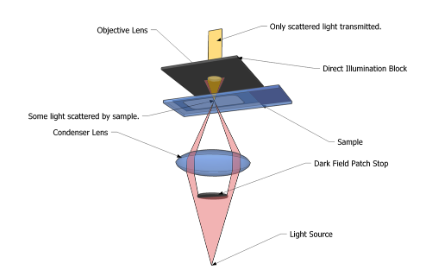
Dark Field
Light travels in a path past the objective unless scattered into it
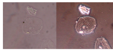
Phase Contrast
•Scattering produces 2 changes in light: change in brightness and change in phase.
•Human eye is only sensitive to changes in brightness
•Phase contrast microscopes make phase shifts visible as differences in brightness
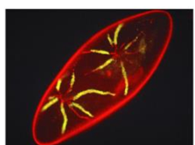
Fluorescence
•High energy light (usually a laser) outside the visible spectrum strikes the sample
•Any fluorescent molecules absorb that light and emit lower energy, visible light back to the viewer
How are bacterial stains useful?
They allow us to see cellular morphology which include:
Size (length, width, diameter)
Shape
Arrangement
Other structures or differentiating features
Determining cellular morphology is an important step in identifying a bacterial species