Paediatric limb injuries
 Call Kai
 Learn
Learn Practice Test
 Spaced Repetition
 Match
Match Flashcards
 Knowt Play
1/21

There's no tags or description
Looks like no tags are added yet.
Name | Mastery | Learn | Test | Matching | Spaced | Call with Kai | Chat |
|---|
No analytics yet
Send a link to your students to track their progress
22 Terms
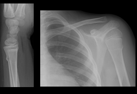
Torus or buckle fractures
•Incomplete fracture of the diaphysis of a long bone.
•Caused by axial loading along the long axis of the bone.
•Trabecular compression results in bulging of the cortex.
•Frequently involves the distal radial metaphysis.
•Very common in children aged 5 -10 years.
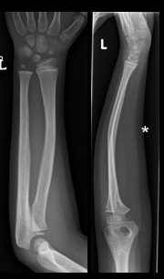
Bowing fracture -
•Incomplete long bone fracture.
•Commonly found in the radius and ulna.
•Usually in younger children but can occur in adolescents.
•Caused by an angulated longitudinal force which causes the bone to bend.
•May return to a normal appearances once the force is removed – no radiological evidence of injury.
Often only visible on
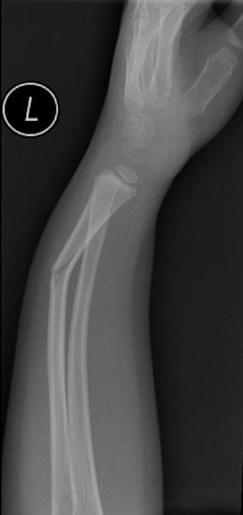
Greenstick fracture-
•Incomplete fracture of the diaphysis of a long bone.
•Usually seen in children under 10 years.
•Commonly seen in the forearm and tibia.
•Caused by angulated axial loading along the bone or by a force perpendicular to the bone.
•Much less common than a torus fracture.
•Usually mid-diaphyseal and obviously angulated.
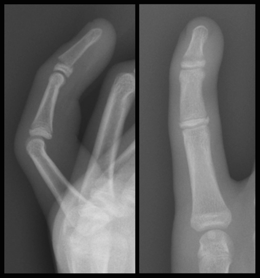
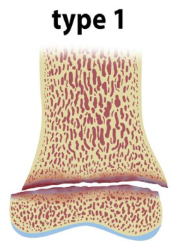
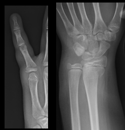
Salter harris type 1 fracture
•Relatively uncommon – 5-7%.
•The fracture passes all the way through the physis and does not extend into the bone.
•No epiphyseal or metaphyseal fragments.
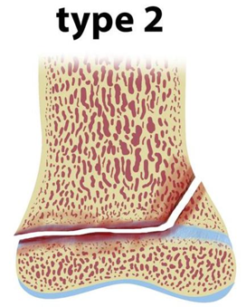
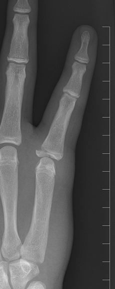
Salter harris type 2
•~ 75% of injuries – by far the most common type.
•The fracture passes through the physis and through part of the metaphysis.
•Triangular metaphyseal fragment.
•Common in the distal radius, phalanges and the distal tibia and fibula.
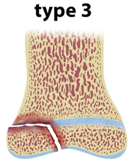
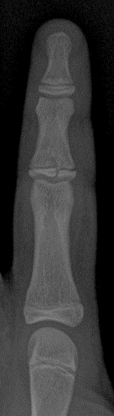
Salter harris type 3
•7-10% of injuries – uncommon.
•Fracture passes through the physis and then down through the epiphysis.
•Oblique or vertical fracture through epiphysis with a horizontal component through the physis.
•No fracture of the metaphysis.
•Commonest in the distal phalanges and the distal tibia.
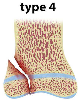
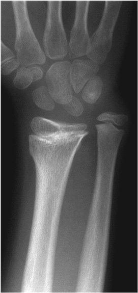
Salter harris type 4
•10% of injuries – uncommon.
•Fracture passes through the metaphysis, physis and epiphysis.
•Intra-articular injury – possible joint effusion.
•Common in the distal radius, phalanges and distal tibia.
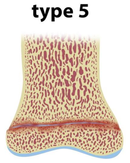
Salter harris type 5
•Rare - < 1% of injuries.
•Crush injury which does not displace the physis but causes damage by direct compression.
•Potential growth arrest, impaired joint mechanics and functional impairment.
•Often not visible – may see narrowing of the physis and/or sclerosis close to the physis.
•Soft tissue signs very important.
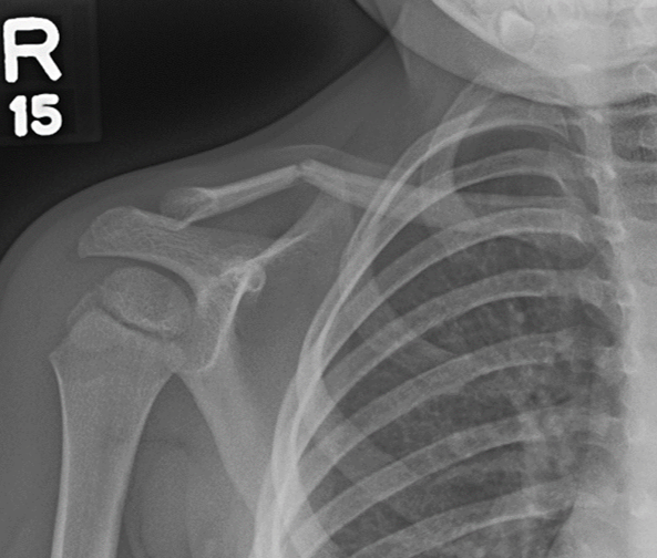
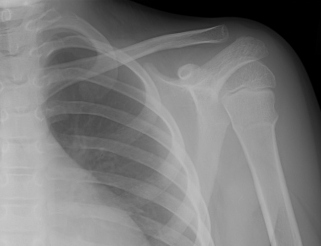
Clavicle fractures
•Mid-shaft fractures common in all age-groups.
•90% are greenstick fractures.
•Uncommon as a birth injury – shoulder dystocia.
•Uncommon below 2 years – suspicion of NAI.
•Can be very subtle.
•Often leave residual deformity.
Proximal humerus fractures
•Metaphysis common – below the surgical neck.
•Medial buckle fractures and greenstick fractures.
•5 – 12 years.
•Uncommon below 5 years.
•Can be sport-related injuries – little leaguers' shoulder.
•GH dislocation rare prior to fusion of the proximal humeral epiphysis.
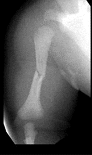
Humeral diaphysis fractures
•Only 2% of paediatric fractures – more common in adults.
•Most common in the over 10’s.
•Suspicious of NAI in the under 3’s.
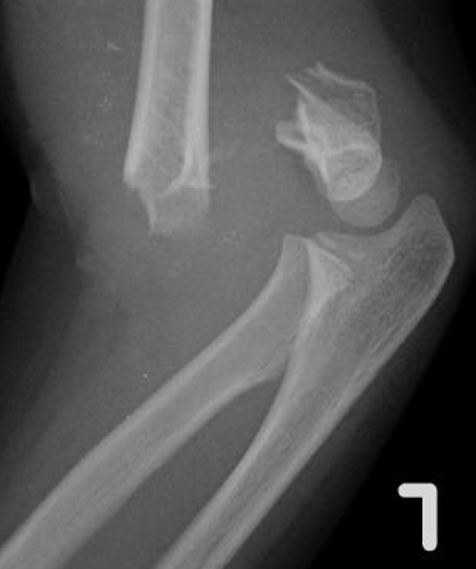
Elbow fractures
•Supracondylar fracture – 5-8 years can be very subtle, uncommon (5-7%).
•Lateral epicondyle – 5-10 years, relatively common (15-17%).
•Medial epicondyle – 8-14 years, relatively common (12%), often related to a dislocation.
•Radial head and neck – 9-10 years, uncommon (5%).
•Pulled elbow – 2-5 years, frequently not visible radiologically.
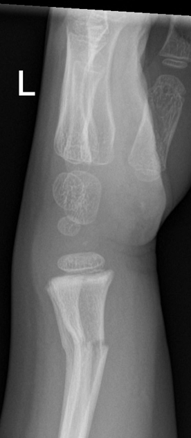
Distal forearm and wrist fractures
•Distal radial fractures are the most common injuries.
•Physeal injuries are usually Salter-Harris I or II.
•Carpal bone fractures are rare until adolescence due to the protective effect of cartilage during ossification.
•Scaphoid is the most commonly fractured carpal bone.
•Carpal fractures may be underestimated due to incomplete ossification.
•Overuse injuries are common in high impact sports with repetitive loading such as gymnastics.
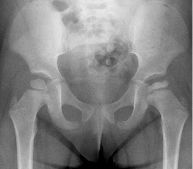
Perthe’s disease
•Asymmetric capital femoral epiphyses – smaller on the affected side.
•Sclerosis of the affected epiphysis.
•Fragmentation of the capital femoral epiphysis.
•Deformity of the femoral neck – widening.
•Idiopathic osteonecrosis of the proximal femoral epiphysis.
•No clear disposing factors.
•Relatively uncommon - 5-15 cases per 100,000.
•Boys are five times more likely to be affected than girls.
•Peak presentation is younger than SUFE – 5-6 years.
•Bilateral in 15% of cases.
•Children present with atraumatic hip pain or limp.
•Secondary to an abnormal/damaged blood supply leading to fragmentation, bone loss and eventual collapse.
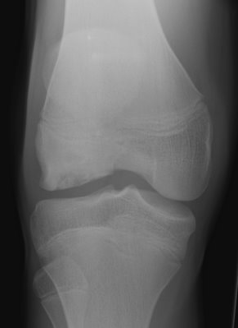
Osteochondral defects
•Indistinct lucency in the articular surface.
•As the disease progresses there is increasing deformity, fragmentation and patchy lucency/sclerosis.
•If an osteochondral fragment becomes unstable and displaced a donor site and intra-articular fragment may be seen.
•Can result in early degenerative change.
•Usually heal spontaneously unless there is an unstable fragment - surgical intervention may be needed.
•Due to aseptic separation of an osteochondral fragment with gradual fragmentation of the articular surface resulting in an osteochondral defect. Often associated with intra-articular loose bodies.
•Majority of patients are aged 10-40 years. More common in males.
•Most cases are thought to be trauma related, but may also be related to AVN, fat embolism, repeated microtrauma (osteochondritis dessicans) or familial dysplasia.
•May be asymptomatic or present with significant pain and locking. Joint effusions and synovitis are common.
•Most common sites are the femoral condyles, but the talus, capitellum and glenoid can also be affected.
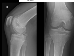
Tibial spine avulsion fracture
•Peak incidence 8-17 years.
•Avulsion of the tibial attachment of the ACL.
•The ACL is stronger than the bone in a child.
•Results from hyperextension and rotation - kicking a football.
•A related meniscal tear is common - MRI needed.
•Surgical management needed if the fragment is significantly displaced or inverted.
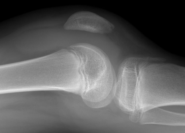
Patellar sleeve fractures
•Uncommon – less than 1% of paediatric fractures.
•Caused by sudden, forceful contraction of the quadriceps muscle.
•Chondral/osteochondral avulsion injury to the apex of the patella but may occasionally affect the proximal pole.
•Most common in boys between 8 and 16 years.
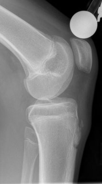
Osgood-Schlatter disease
•It occurs 3 times more frequently in boys than girls and is characterised by fragmentation of the tibial tuberosity, soft tissue swelling, and thickening at the insertion of the patellar tendon.
•In 25% of cases, it is bilateral is best seen on lateral projection.
•This should be a clinical finding not radiographic.
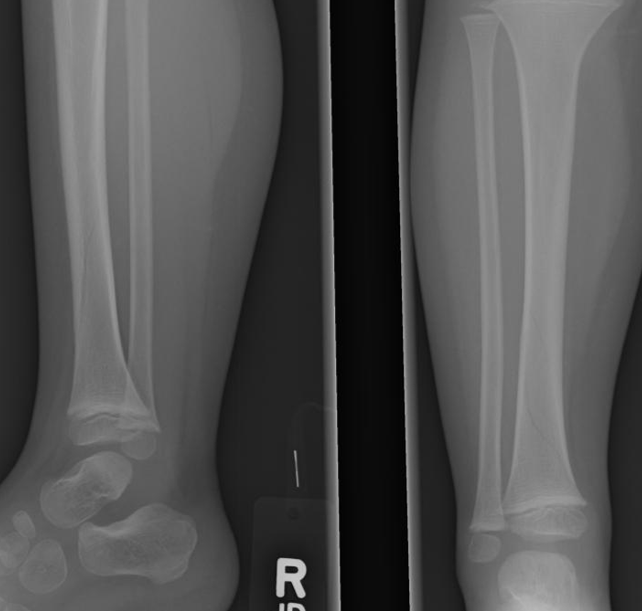
•Fracture is likely to be visible on only one projection.
•Oblique projections sometimes needed.
•Most common in the middle and distal thirds of the tibial shaft.
•Sometimes only visible on follow-up imaging with slight sclerosis and periosteal reaction.
Toddlers fracture/ Childhood accidental spiral tibial fracture
Toddler fractures, also known as childhood accidental spiral tibial (CAST) fractures, are minimally displaced or undisplaced spiral fractures, usually of the distal tibia, commonly encountered in toddlers. It is potentially difficult to diagnose because the symptoms and imaging findings can both be subtle
Follow-up radiographs usually demonstrate slight sclerosis and periosteal reaction
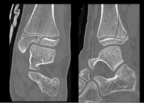
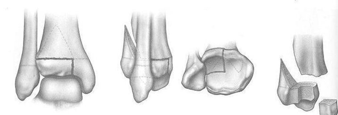
Triplanar fractures
•Usually seen in adolescents because of the growth plate fusing medial to lateral.
•Involves all three planes. Vertical fracture through epiphysis (SH III), horizontal fracture through lateral aspect of the growth plate and a coronal oblique fracture extending superiorly (SH 2).
•CT is essential for confirmation and/or surgical planning.
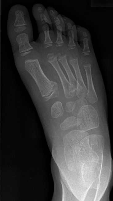
Paediatric foot fractures
•Fractures of the metatarsals and phalanges tend to be greenstick or torus fractures – especially in the under 5's.
•Salter-Harris I and II fractures are common at the base of the 1st MT (in this image)– often caused by a fall from height.