BIOC221 Whole course flashcards
 Call Kai
 Learn
Learn Practice Test
 Spaced Repetition
 Match
Match Flashcards
 Knowt Play
1/198

There's no tags or description
Looks like no tags are added yet.
Name | Mastery | Learn | Test | Matching | Spaced | Call with Kai | Chat |
|---|
No analytics yet
Send a link to your students to track their progress
199 Terms
Define CTD
C-terminal Domain. It’s super important in RNAP II as it is the first part of RNAP that is recruited for intiation of transcription.
What does TSS, CP and CRE stand for
Transcription start site, Core Promoter and Cis-regulatory element.
What are the main types of non-coding genes in eukaryotes
Non-coding genes generally produce functional RNA products that can do:
protein translation: ribosomal (rRNA), transfer RNA (tRNA)
RNA-processing: small nuclear RNA (snRNA), small nucleolar RNA (snoRNA)*
regulation of gene expression: microRNAs (miRNA) and long non-coding RNA (lncRNA)
Describe the stages at which gene expression can be controlled.
• Transcription: whether a gene is copied into mRNA (or ncRNA)
• Co-transcription/mRNA processing – E.g. alternative splicing giving different forms of a protein
• Post-transcription: mRNA stability and translation efficiency determines how much protein is made from each mRNA – E.g. miRNAs (microRNAs)
• Post-translational (protein-coding genes): covalent modifications, localization and degradation alter protein function or abundance (Liz Ledgerwood)
lnc-RNAs can affect gene expression at multiple stages
What are the products of the different nuclear RNA polymerases in eukaryotes?
RNA Polymerase I makes: Most rRNA, located in the Nucleolus
RNA Polymerase II makes: mRNA, snoRNA, some snRNA, miRNAs, and lncRNA, located in the Nucleoplasm
RNA Polymerase III makes: tRNA, 5s rRNA, some snRNA, and other small ncRNAs. Located in the Nucleoplasm
What are the different subunits of a DNA-based RNAP called?
(called Rpb#), e.g. Rpb3 or Rpb11
Describe the CTD of the RBP1 subunit of the eukaryotic RNA Polymerase II and how it changes during transcription.
Not structured, not catalytic. (multiple copies of the same seven amino acid sequence).
The CTD is reversibly phosphorylated during transcription - Ser 2 and Ser 5 are the main residues that get phosphorylated - Kinases add phosphate groups - Phosphatases remove phosphate groups - Different phosphorylation patterns alters RNA Pol II interactions with other proteins/factors
What part of Transcription Initiation does RNAP need help with?
RNAP requires additional assistance to find the correct start codon to start transcribing the reading frame. It is very important that it finds the correct AUG start codon as incorrect starts will cause a frameshift.
How are histone tails modifed and how does this affect gene expression
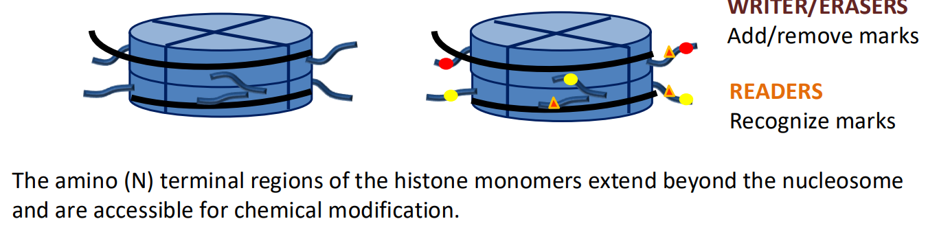
The N-terminal of histone monomers. They are modified by HAT’s and HMT’s to add acetyl and methyl groups respectively. These HTM’s can affect RNAP binding affinity, signalling when or if the gene should be expressed
Break down the events preceding RNAP II binding to a gene.
Transcription Factors bind to the cis-regulatory elements (CRE’s) of a specific gene in an enhancer region next to a promoter, which is upstream of its exxons and the core promoter. These transcription factors are transcription activators, after the activators bind they recuits co-activators.
Co-activators acetylate histone H3 and histone H4 Co-activators may also make other histone-tail modifications (e.g. methylation)
Nucleosome remodelers - Part of, or recruited by, co-activators - Reads histone tail modifications and slides or evict nucleosomes - often partially by removing H2A/H2B dimers, leaves H3/H4 dimers with tail modifications - helped by DNA-histone interactions being a little bit looser
What are the two domains of a transcription factor?
Typical activators consist of two domains
Activation domain (AD): Protein-protein interactions - Other activators (complex) - Co-activators
DNA binding domain (DBD): Binds an enhancer Hold the activation domain in the vicinity of the promoter
After activator/co-activator recruitment, how is RNAP guided to the TSS?
Activators/co-activators recruit RNAPII and form to pre-initiation complex (PIC). This contains many general transcription factors (GTF), TFIIA to TFIIH, which have different roles in helping RNAPII reach the TSS, by the ordering of their recruitment, the GTF’s can help recruit RNAPII to the TSS.
Which GTF is recuited first to form the pre-initiation complex and how is this done?
TFIID is a protein complex.
TFIID is recruited to the core promoter by a combination of:
1. TFIID binding to DNA sequences (motifs) in the core promoter e.g. TATA box
2. Interactions with co-activators (multiple GTFs)
3. Histone-tail modifications - TFIID has reader proteins - H3 and H4 acetylation, methylation of specific residues
Describe the events associated with the initiation of transcription, getting to the unstable, abortive transcription stage
Once the PIC has been formed, RNAP has been recruited to the TSS to start transcription. GTR TFIIH has helicase activity so can unwind the DNA, forming a transcription buble inside the PIC. The GTF’s help load the template strand onto the cleft of RNA polymerase II
Once RNAP has bound to the TSS, what steps must the GTF’s take to stabilise RNAP so it can begin transcription
To become more stable RNA polymerase II must adapt an active conformation with the clamp close to hold onto the DNA and form the stabilising DNA-RNA hybrid helix. RNAPII also needs to break contacts with the GTFs that are anchored to the core promoter so RNAPII can move along the template strand. The contacts are broken by TFIIH kinase activity, it adds phospates to serines at position 5 (Ser 5) of the heptapepetide repeats of the CTD, changing the proteins the CTD interacts with. The DNA/RNA hybrid helix helps remove the stabilising GTFs (mainly TFIIB) that are attached to the PIC from the RNAPII, so RNAPII can move forward and transcribe the whole gene.
Describe the addition of the 5’cap, including when capping occurs and how this relates to the CTD
The addition of the 5’ Cap is a PTM that happens while transcription is taking place.
The mRNA Capping enzyme (CE) is recruited by Ser5 P of CTD but performs its job on the 5’ RNA strand being transcribed. Triphosphatase activity cleaves 5’ phosphate - guanylyltransferase activity uses GTP (energy) to add an inverted guanine (G) (GMP added) - stable but poor translational efficiency
RNA methyltransferase (RNMT) recruited by Ser5 P of CTD - methylates nitrogen 7 of the guanine base (m7) - stable and high translation efficiency
What does PTEF-b complex do to promote productive elongation during transcription? (3 main things)
Positive Transcription Elongation Factor b (P-TEFb) complex. It does:
phosphorylation of Ser 2 of the CTD: recruits a range of Elongation Factors (EF) that help RNA Pol II:
Nucleosome writer: elongation factors have similar roles to co-activators at the core promoter - HAT activity: H3/H4 acetylation (loosen/ recruit readers)
Nucleosome remodelers: evict (full or partial) nucleosomes in front of RNA Pol II and replace them after transcription.
What is the basic structure and function of the spliceosome?
• Complex of splicing factors (U#) which consist of snRNAs and proteins
• Some splicing factors recruited by RNA Pol II with Ser2 P on CTD
• Splicing factors are small nuclear ribonucleoproteins; snRNPs or “SNERPS” – RNA molecules form complex shapes – Catalytic site of the spliceosome largely formed by RNA molecules • Ribozyme - ribonucleic acid enzymes
Its function is to remove introns from transcribed RNA via transesterification reactions: - the breaking of one phosphodiester bond is coupled with the creation of another (Requires ATP)
Describe the process of RNA splicing by the spliceosome
Some splicing factors associate with Ser2 P on CTD of RNA Pol II
Recognize consensus sequences (base pairing) in the pre-mRNA as it exits RNA Pol II. Transfer onto pre-mRNA.
There are three sites recognized: 5’ splice site, Branch point/branch site (A), 3’ splice site
First, the Splicing factors bind to an A site, which is just upstream of the 3’ end of the intron to be deleted, and bind to the 5’ end of the intron. Then they catalyse a transesterification to move that bond so the 5’ end bonds to the A site, forming a lariat (lasso shape).
Splicing factors change out
A second transesterification reaction occurs, the 5’ intron site to the A site bond is switched to a bond between the 3’ and 5’ ends of the exons flanking the intron lariat. This removes the intron successfully.

How does the CTD of RNAPII change during productive elongation?
- a phosphatase removes P from Ser 5 in heptapeptide repeats
- P-TEFb continues to add P to Ser2 in heptapeptide repeats
What phosphorylation state of the CTD causes termination and adenylation of transcription
Low Ser 5/ high Ser 2 on the CTD recruits:
CPSF (cleavage and polyadenylation specificity factor)
CstF (cleavage stimulation factor)
Describe the process of termination and adenylation of mRNA transcription
CPSF (cleavage and polyadenylation specificity factor)
CstF (cleavage stimulation factor)
These factors are recruited when the cleavage signal in mRNA exists RNA Pol II, they recruit more CF’s, and they cleave the mRNA
PAP (poly-A polymerase) recruited by CPSF
PAP adds multiple As (polyA) to 3’ end of mRNA (no template, uses ATP)
Poly-A binding proteins (PABP)bind to provide protection and translation efficiency
What happens downstream of the cleavage site after transcriptional termination?
Downstream of cut site:
termination 5’ end of cleaved mRNA no longer protected by 7mG-cap
Exonuclease degrades mRNA that is still associated with Pol II
DNA/RNA hybrid helix is disturbed
Pol II stops transcription and dissociates from the DNA CTD dephosphorylated
Explain what is meant by activator “activity”.
Typical activators consist of two domains:
DNA Binding domain (DBD): Binds an enhancer, holds the activation domain in the vicinity of the promoter. “Activity”: ability to bind an enhancer.
Activation domain (AD) Protein-protein interactions - Other activators (complex) - Co-activators. “Activity” ability to recruit co-activators.
Describe how activator “activity” is regulated
1. Presence/absence Gene expression and degradation
2. Conformation Ligand binding Post-translational modifications
3. Complex that activator binds to changing formation
4. Inhibitors, repressors that bind the promoter or to the activator, changing its form.
5. Nuclear localisation. (if in nucleus to transcribe genes or not)
What techniques would we use to measure the activity of an activator
RT-qPCR to make cDNA from mRNA transcripts in cell conditions. We can use poly-T primers to bind to the poly-A tail of mRNAs so we only read mRNA transcripts, and design more specific primers to measure mRNA expression of one gene.
RNA-Seq can tell us about the expression of all the genes in a cell. These expression measurement techniques allow us to measure the effect of activators on gene expression, or you could directly measure the expression of activator genes.
How can some activator genes end up creating multiple different functional activators?
Activators usually bind their Activator domains to form heterodimers or larger complexes with other proteins, so there may be multiple forms that a single activator monomer could end up in, each with a different function.
How does euchromatin conformation affect transcription?
Even within euchromatin which is most protein-coding genes are, Chromatin can be tightly packed, “closed”, with activators unable to bind to its CRE’s, or it can be open with space for activator binding and then RNAP II binding
What are the two types of proteins that facilitate the histone code?
Writers/ erasers: proteins that add/remove specific modifications, write the code Examples: HATs, HDACs, HMTases, histone kinases
Readers: recognize specific histone modifications and can alter chromatin structure
Examples: nucleosome remodelers, chromatin compaction proteins/ structural proteins, DNA methyltransferase
What can different histone tail modifications be read as?
- acetylated histone tails recruit readers associated with open chromatin
- methylated histones can recruit readers associated with open or closed chromatin depending on the histone, the residue and the number of methyl groups added
Describe how to perform a chromatin immunoprecipitation (ChIP)
Formaldehyde treatment forms covalent bonds between DNA and proteins and between close (interacting) proteins: crosslinks
Purify the chromatin (still cross linked)
Fragment DNA into pieces (200-1000 bp) (sonication)
Incubate cross-linked DNA with antibody that is specific for the protein-of-interest
Antibodies are usually attached to a column or bead so they be isolated from solution with the bound cross-linked DNA.
Stop antibody binding and separate from protein/DNA
Reverse cross link, separate and analyses the DNA

Why would you perform a Chromatin Immunoprecipitation?
Used to detect interactions between proteins and genomic DNA within cells
A. Which genes are currently being transcribed?
B. Where does an activator bind to genomic DNA?
C. Which regions of the genomic DNA have a specific histone-tail modification?
D. Where in the genomic DNA are histone tails being modified?
Once you have isolated DNA and proteins from chromatin immuno precipitation, how do you analyse the DNA?
- Sequence the immunoprecipitated DNA and map reads to the genome (like with RNA-Seq data)
- Find which regions of the genome, and how much of each region, were immunoprecipitated
What is the CRE on mRNA that allows for post-transcriptional modifications?
sequence motifs
- bound by RNA biding factors (proteins, RNPs, non-coding RNAs)
- role in alternative splicing, post-transcriptional regulation
Explain how alternative splicing can produce different functional proteins and how this could alter a protein and protein function.
The spliceosome somewhat randomly splices introns out so can produce a wide range of proteins with slightly different AA patterns and therefore structure.
What is the regulation mechanism to control intron splicing?
The sequence motifs in the mRNA and how well they recruit splicing factors is a big determinant in an introns chance of being spliced from the mRNA.
This is also affected by proteins, ribonuclearproteins, lncRNA’s binding to CRE’s on the mRNA and affected SF binding affinity
How does the length of the poly-A tail of an mRNA affect its transcription?
mRNAs are often circular with the 5’ and 3’ ends held together by interactions between 5’ cap, eukaryotic translation initiation factors and poly-A-binding proteins (PABP) - protected from exonucleases (stable) and efficient translation. If poly-A tail is short (<25 As), PABP stop binding - Not circular - RNA will be degraded and difficult to translate.
Length of the poly-A tail can be used to alter mRNA half-life and translation
How are CRE’s often made of double-stranded RNA?
Complementary base pairs on RNA’s in different parts of the sequence can bond together, forming secondary structures on the RNA and folding the RNA into a certain shape, this forms a CRE which can be bound to to control gene expression.
What is RNAi?
A process in which RNA molecules inhibit gene expression by altering mRNA stability or translation efficiency. Triggered by a double-stranded RNA molecule (dsRNA) in the cytoplasm
How are miRNA produced endogenously?
miRNA production - Transcription from a miRNA gene (non-coding) in the nucleus, usually by RNA Pol II (regulated). Folded into a pri-miRNA (primary miRNA; forms a stem-loop). A ribonuclease (RNase; Drosha) cleaves ssRNA and exports pre-miRNA (preliminary miRNA) cytoplasm (mostly) - RNase Dicer processes it to miRNA duplex (21-23 nts, still dsRNA)
How do miRNA contribute to gene expression?
Regulates gene expression by binding a cis-regulatory element in mRNA, usually in non-coding regions
Once passed into the cytoplasm, what happens to the ds mi/siRNA?
One strand can base pair with the Agonaut protein, which has complementary base pairs with either the strand or its antisense pair. The other strand is released and degraded. The Agonaut protein with the mi/siRNA binds to the RISC, the RNA-Induced Silencing Complex.

What does the RISC do?
RISC binds to mRNA leading to either
- mRNA degradation (RNase/ slicer) or prevents translation - common in plants
- Inhibition of the ribosome (blocks translation) - common in animals

Describe the characteristics of lncRNAs including how they are generated.
RNA transcripts not from protein-coding genes or known noncoding genes (rRNA, tRNA, etc)
LncRNA can be transcribed in many ways from DNA:
- antisense (opposite direction to an mRNA)
- bidirectional (opposite direction from a promoter)
- intergenic (between protein-coding genes)
- intronic (from an intron of a protein-coding gene)
(Mainly) transcribed by RNA Pol II - processed like an mRNA - 5’ 7mG cap, spliced, poly-A tail
What are the mechanisms through which lncRNAs can influence gene expression?
lncRNA can: base pair with complementary DNA and other RNAs and form complex shapes (base pairs within lncRNA): It can do lots of regulatory roles
i) Direct regulator - promote/ inhibit processes
ii) Guide - recruit a protein (proteins) to a nucleic acid sequence or another protein
iii) Scaffold - bind multiple proteins/other RNA to form a complex
iv) Decoy or sponge - binds another molecule (protein or RNA) that prevents the molecule binding elsewhere
v) Source of dsRNA (antisense transcript)
Come up with three examples of how lncRNA can regulate different stages of gene expression
Three of these from different levels:
Transcription
- activators:
- direct regulator: plays the role of an activator (binds Co-activators to core promoter)
- decoy: activator binds lncRNA instead of DNA (sequence matches enhancer sequence)
- guide: helps bring an activator to an enhancer
- scaffold: help bring activators and co-activators together
Transcription – chromatin:
- guide: recruit writers and readers to certain genes to open/close chromatin
- scaffold: help range of proteins interact/ remain associated Co-transcriptional
- direct regulator: help or inhibit recruitment of first splicing factors to RNA consensus sequences
Post-transcriptional
- direct regulator: influence the competition between extension and shortening of the poly-A tail
- direct regulator: inhibit the ribosome or promote translation
- decoy/sponge: provides miRNA binding sites, bind to lncRNA instead of mRNA
- other: source of dsRNA to trigger RNA interference (RNAi)
Draw and annotate the process of Eukaryotic gene expression from DNA to and active protein.


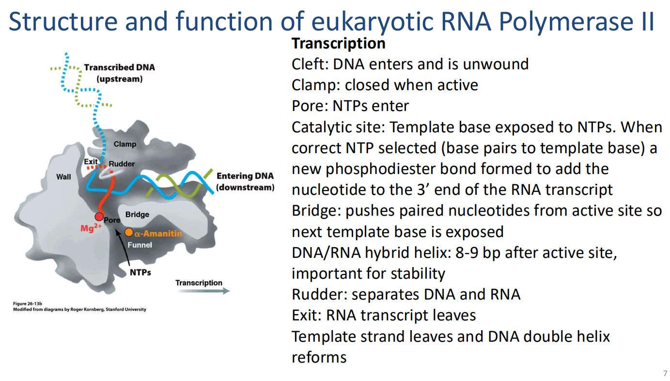
Explain the function of the following parts of this protein-RNA complex.
Transcription
Cleft: DNA enters and is unwound
Clamp: closed when active
Pore: NTPs enter Catalytic site: Template base exposed to NTPs. When correct NTP selected (base pairs to template base) a new phosphodiester bond formed to add the nucleotide to the 3’ end of the RNA transcript
Bridge: pushes paired nucleotides from active site so next template base is exposed
DNA/RNA hybrid helix: 8-9 bp after active site, important for stability
Rudder: separates DNA and RNA
Exit: RNA transcript leaves
Template strand leaves and DNA double helix reforms
Draw the process of the spliceosome removing an intron. Dw about splicing factor names

ddg
bbc
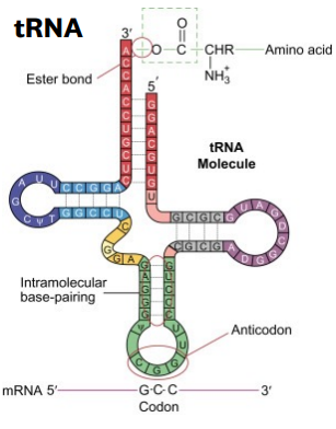
Describe the features and function of tRNA
tRNA are folded RNA molecules, often in a conformation with a hairpin structure. They are transcribed by RNAP II. Their conformation allows them to bind to a ribosome active site at the same time as a certain codon from an mRNA sequence is being “read” by the ribosome. The tRNA strand is translated by the ribosome to an amino acid corresponding to the tRNA and the codon of the mRNA strand, allowing for amino acid chains and then proteins to be formed.
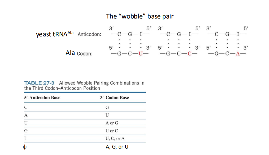
What is another source of redundancy in amino acids that helps maintain the fidelity of the genetic code during protein synthesis
the anticodon on a tRNA binds to mRNA codon, it can recognize >1 codon – the “wobble” base pair. Ensure the fidelity of the genetic code
What is the reaction catalysed by aaRS
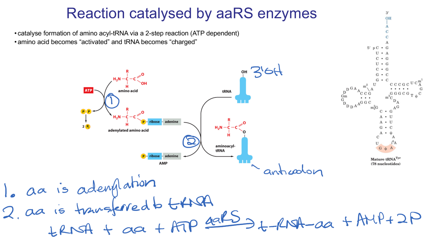
amino-acyl tRNA Synthetases catalyse two reactions. The first is adenylation of an amino acid (the one that corresponds to this particular tRNA). This reaction requires ATP and is called “activating” the amino acid. This amino-acyl group is then transferred onto one of the corresponding tRNA, “charging” the tRNA.
What are isoaccepting tRNAs and what do they help promote?
Isoacceptor tRNAs: tRNAs recognising different codons for the same amino acid. This is how codon redundancy is enforced, since isoacceptor tRNAs will recognise multiple codons for the same amino acid.
Describe the features of the ribosome
Ribonuclear protein complexes protein = scaffold. This brings together the mRNA strand, centring the codon being read at that moment, and brings in a corresponding tRNA.
rRNA = catalyst, catalysis peptide bond formation.
What are the substrates required to initiate translation?
Requires
small ribosomal subunit
capped and polyadenylated mRNA
eIFs (eukaryotic initiation factors)
Met-tRNAi Met (iMet-tRNA)
GTP.
GTP is required as some of the initiation factors are G proteins.
What roles do the eukaryotic translation Initiation Factors perform during the formation of PIC? (6)
Binding to Met-tRNAiMET
Binds to 5’ cap and PABP, RNA unwinding
Binds to A and E sites in 40S subunit
Binds to small subunit
Binds to 43S preinitiation complex
Binds to 60S subunit, displaces other IFs
How is protein fidelity ensured during translation by making sure Met tRNA binds to the correct AUG start codon? (eukaryotes and prokaryotes)
In eukaryotes the correct AUG start codon is flanked by the Kozak sequence which has a higher frequency of certain base pairs. It is not well understood exactly how the kozak sequence helps iMet tRNA bind to the first AUG start codon.
In prokaryotes the relationship is well understood. The Shine Dalgarno sequence is upstream of the first AUG codon and base pairs to a complementary sequence on 16S ribosomal RNA which helps align the iMet tRNA to bind to the correct start codon.
Define Transpeptidation
peptide bond formation
Define Molecular mimicry
when two different molecules can perform similar functions because they have the same shape
Define Peptidyl transfer centre (PTC)
site of peptide bond formation on ribosome
How is translation termination catalysed by eRF complex (and what is eRF)?
eRF is eukaryotic release factor. In Eukaryotes, eRF1 binds to stop codons. Once the stop codon is in the A site, eRF3 (a G protein) binds to eRF1 to help termination, by hydrolysing the ester bond between amino acid and tRNA, releasing the polypeptide chain.
What is the ER import signal for proteins
A hydrophobic Alpha helix on the N terminal of the amino acid chain
How does a protein pass through the ER membrane?
The signal recognition protein (SRP) recognises a hydrophobic Alpha helix at the N terminal of a growing amino acid chain and binds to the amino acid chain and its attached ribosome. The SRP binds to the ribosome A-site, slowing translation. The SRP binds to the ER membrane-bound SRP receptor (SR) via a signal sequence on the SRP and the amino acid chain is fed through the SR into the ER, translation continues and translocation occurs until the full protein is fully translated inside the ER.
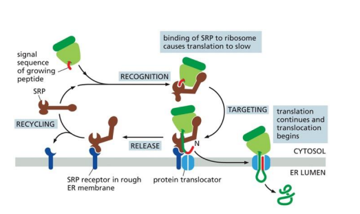
While crossing the ER membrane, how is translation resumed after the SRP binds to the SR?
The SRP protein binds to the ribosomal A-site, inhibiting translation. The SRP and SR are both G proteins. The GTP portion of the SR and SRP bind together, activating GTPase activity that converts GTP to GDP, releasing the SRP and GDP portions of the SR from the ribosome-SR complex. This allows translation to continue and the growing amino acid chain is fed through the SR gate into the ER.
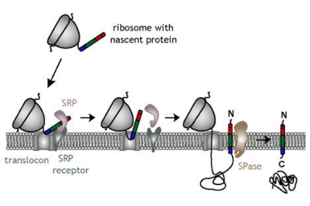
How are transmembrane proteins processed into the ER membrane?
The amino acid chain is fed through the membrane like normal. The transmembrane segement of this chain is stopped when it loops back to go through the membrane again, so the transmembrane sequence stays in the membrane, with the N terminal in the cytosol and the C terminus in the lumen.
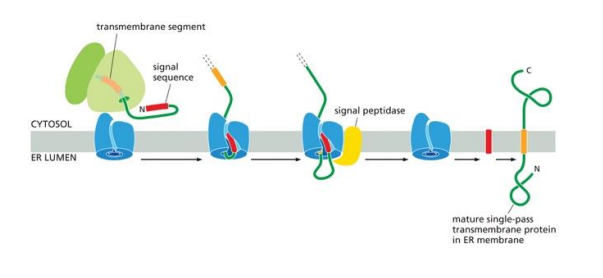
How are proteins always N-glycosylated or otherwise glycosylated?
The Oligosacchayltransferase complex (OST) sits right next to translocons in the ER when means that as proteins are translocated into the ER they are actively glycosylated, so every protein in the ER is subject to glycosylation and other PTM’s.
How is glycosylation used to increase protein diversity?
OST complexes can add up to 10 different sugars. This means there are around 7000 combinations of glycosylations that can be done to a single protein. By randomizing this, thousands more different proteins can be created with different jobs, decreasing the DNA storage capacity requirements for that many different proteins.
What PTM is performed on insulin in humans to make it a mature protein?
Proprotein cleavage is carried out by proprotein convertase enzymes, which have specific recognition sequences of amino acids, which they cleave. Proprotein cleavage is performed on premature insulin peptides. Part C is cleaved after parts A and B have formed disulfide bonds and folded into shape.

What are some reasons to degrade proteins?
Controlling cell signalling/regulation
Recycling amino acids
destruction of foreign proteins
misfolded or damaged proteins
What is the role of ubiquitin and what is the role of the Amino Acids on its N and C termini?
Ubiqitination is the process of attaching a ubiquitin protein to a larger one. Ubiqitin is a protein signaller that tells proteases to break down the protein it is attached to. Usually a chain of many ubiqitin are attached to a protein before it is broken down.
The Glycine on the C-terminus is attached to the substrate and the Lysine on the N-terminus is used to bind to the following Ubiquitin in the chain.
Way to remember. Glycine is just lysine but on the C-terminus, and sequential added lysines push the protein toward being lysed by proteases
What are the steps of ubiqitination
E1 = ubiquitin-activating enzyme: Catalyses attachment of Ub to E1 (Cys residue)
E2 = ubiquitin-conjugating enzyme: Catalyses transfer of Ub from E1 to E2 (Cys residue) Substrate recognition (with E3)
E3 = ubiquitin ligase: Catalyses transfer of Ub from E2 to lysine residue on a substrate (condemned protein) Substrate recognition (with E2)

What determines what kind of signal the ubitination sends to the cell?
The different Lys chains that form peptide bonds in the Ub chain on a protein. Different Lys peptide bond positions between ubiquitin proteins on a ubiqitin chain functions as different cell signals. The one we cover in BIOC221 is homologous Ly48 peptide bonds, which signal proteases to come and lyse the protein that has been ubiquitinised.
Why is ATP required for ubiquitination?
Ubiqitin needs to be activated by binding to E1 which can then promote the binding of this Ub protein to a different protein. This requires ATP for energy and the AMP produced is as an intermediate that binds to Ub before being substituted by E1 itself.
What controls specificity in ubiquitination?
E3 ligases are specific proteins that can recognize a specific protein in solution and bind to add, transferring the Ub bound to it. There is an E3 ligase for every protein.
Why are most genes subject to regulation?
Conserved resources. By limiting the expression of genes not required, bacteria can commit more resources to making ribosomes and maximise growth rate •
Respond to changes in internal and external environments - Adaptability
Ordered development of the cell wall

Break down bacterial transcription/translation and its main difference with eukaryotic transcription.
RNA-polymerase binds to the template strand to synthesise mRNA
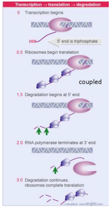
Transcription and translation are coupled in bacteria. The Ribosome will bind to and translate the mRNA as it is being synthesized
Operons
Co-transcribed genes under the control of a single regulated promoter
Why are operons used?
For efficiency. Most operons contain multiple genes with similar functions or in the same pathway, so its makes sense for all of them to be expressed in similar amounts. Sometimes operons contain genes that perform functions for different cell pathways.
ncRNAs
non-coding RNAs, transcribed but not translated. Function alone or as part of RNA-protein complexes • Often facilitate RNA-nucleotide binding
How does Crispr promote immunity?
Crispr is a bacterial ncRNA-protein complex that is used by the immune system to target invading viruses or other genetic elements. CRISPR associated (cas) proteins are expressed from cas genes, Cas proteins form a complex, recognize foreign DNA, cuts and inserts small piece into the CRISPR array

Describe the structure and role of the subunit of DNA dependent RNA polymerases in transcription that allows for initiation
RNA polymerases have a protein subunit called a Sigma factor which binds to promoters upstream of a DNA sequence. This is the site of transcriptional initiation.
Describe the key features of bacterial promoters and how they determine the binding affinity of sigma factors and RNAP
Sigma (σ) factors bind to -35/-10 regions upstream from the transcriptional initiation site (+1). There is variation in sequence, but consensus across -35/-10 sequences. Better match to consensus = stronger promoter = more expression.

How does RNAP bind to the promoter aside from the usual GTFs
Sigma factors on RNA polymerase binds to promoters in DNA if they have good enough match with the transcription site.
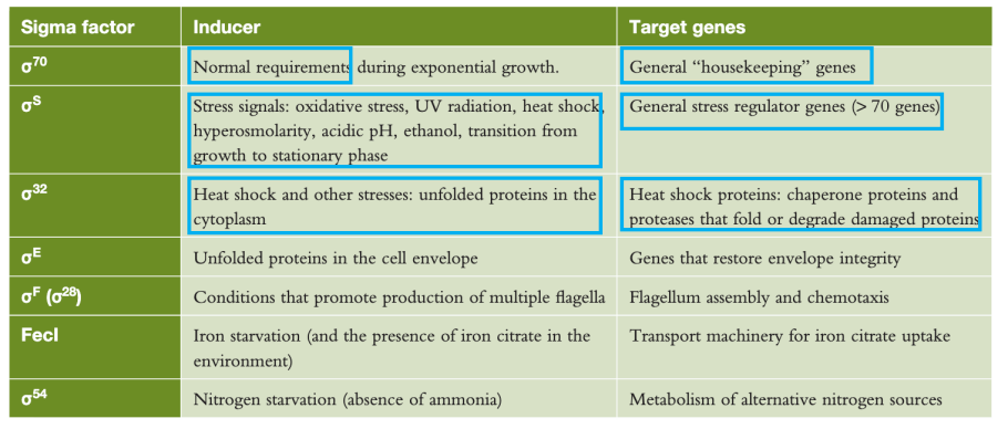
How do sigma factors contribute to gene regulation and response to environmental stimuli?
Sigma70 (σ) is required for the expression of housekeeping genes. Alternative sigma factors, active under different conditions, regulate different genes. Inducing signal and target genes are linked. Different sigma factors recognize different consensus sequences. Ensures that genes are only expressed when needed
Describe the protein subunits that make up DNA dependent RNA polymerases as it binds to DNA to begin initiation
Core enzyme : ⍺⍺ββ’
RpoA: Two subunits from same gene
RpoB/C: Two different catalytic subunits (different genes)
Holoenzyme: Core + sigma factor
Sigma factors: Promoter specificity
RpoZ (sometimes): stability and assembly of the largest subunit, β′, into the core enzyme
Outline the role of DNA dependent RNA polymerases in transcription
1. Non-specific binding of RNAP to DNA-migration to promoter
2. Sigma factor recognize promoter = Closed complex (DNA is still dsDNA)
3. dsDNA is pulled apart = Open complex
4. Transcription initiates at a purine (A/G)
5. Elongation leads to the addition of successive nucleotides
6.Sigma factor is released
Outline with an example how we can inhibit bacterial transcription
Rifampicin is a popular antibiotic for tuberculosis, inhibiting transcription of Mtb. This is by binding to the bacterial RNA polymerase active site and sterically hindering the binding of the RNAP with DNA.
What is the difference between rho-dependent and independent transcriptional termination
Two mechanisms of termination
1. Rho-dependent (RNAP core requires additional proteins to stop transcription)
2. Rho-independent (intrinsic nucleic acid terminator, sometime enhanced by a partner)
Describe rho-dependent transcriptional termination
1. Rho complex binds in open conformation at rut site
2. Rho complex recognizes SBS and recruits to the central channel
3. Rho transitions from inactive open to active closed form
4. Rho uses ATP to pull RNA away from RNAP, leaving behind a completed transcript

What are the differences between RNA and DNA that allow for rho-independent transcriptional termination?
1. RNA base pairing is similar to DNA 1. Except Adenine-Uracil, rather than Adenine-Thymine (2H bonds). G-C is same, with 3H bonds
2. RNA can interact with complementary regions
Interactions lead to stems
Non-interacting sites are loops
Describe rho-independent transcriptional termination
Intrinsic terminator = Termination is driven by RNA interactions forming a hairpin structure.
This is possible as RNA has a G + C–rich region followed by a run of 4 to 10 consecutive A - T base pairs, (A’s on the template strand).
1. Self-complementary G-C rich region is transcribed and forms a hairpin structure (3H bonds)
2. Transcription of run of A = U-A pairing (2H bonds) in RNA-DNA hybrid (2H is weaker than 3H)
3. Hairpin structure leads to RNAP pausing
4. Hairpin is more stable than A-U pairing, pulling RNA away from template strand

Describe negative regulation using lac operon as an example
lacI is the inhibitor of lac operon, Binds operator sequences of lac operon (which surround the promoters and overlap with the +1 transcriptional start site) LacI monomers bind to form dimers that bind to a whole operator sequence and these bind with another dimer to form a tetramer, this wraps up the gene to make sure it can’t be transcribed.
How do transcription factors help regulate gene expression?
Transcription factor exists in an inactive state and can’t bind DNA. If a Ligand binds (often linked to downstream gene), this changes conformation its (allostery). The Transcription factor can now bind DNA and helps RNAP to bind to induce gene expression.
(The image shows positive and negative regulation, negative being similar but the transcription factor blocks RNAP binding, and a ligand binding to the TF releases it to allow gene expression.

In the lac operon example, what triggers the gene to be turned on or off?
Allolactose is the first product made in the metabolic pathway to turn lactose into glucose, and is produced by beta-galactosidase. Allolactose is an inducer of the lac operon, binding to lacI and causing it to detach from the DNA for that gene, allowing gene expression so lactose can be metabolised. Once all the Allolactose has been metabolised, lacI rejoins and represses the lac operon.
How is lactose metabolism started via allolactose synthesis upon ingestion of lactose?
Initial lactose metabolism to allolactose to activate the lac operon is possible due to a weaker constitutive promoter, and leaky transcription where some of the gene is still transcribed when the gene is repressed, which leads to low mRNA production. Despite being repressed, some transcription of the lac gene still occurs which produces trace Beta-galactosidase in cells, thus lactose can be converted to allolactose to kickstart/massively upregulate lactose metabolism
What is the secondary lac operon regulatory system that uses positive transcription factors
glucose inhibits cAMP production, Low glucose = high cyclic AMP (cAMP-starvation signal). High glucose = low cAMP
cAMP binds CRP (cAMP receptor protein, (CRP=Catabolite repression protein))
Active cAMP-CRP complex helps RNAP bind lac promoter
Active cAMP-CRP complex is required to initiate transcription of lac operon
Describe features of gene repression, using trp operon as an example
trpR (inactive) is the inactive form of the repressor of the trp operon, which contains all the genes required for tryptophan synthesis. Tryptophan binds to trpR and activates it, causing it to bind to the inducible promoter and repress gene transcription. This is so the bacteria does not produce tryptophan when it can get it from its environment.
Compare and contrast the regulation of lac and trp operons
Lac operons are regulated via positive transcription factors, cAMP binding to the cAMP-CRP complex, which is required to activate the promoter so RNAP can bind, and it produces its own repressor, which is only made inactive by binding of allolactose, made from lactose.
Trp operon has a constitutive promoter that has a repressor upstream of it, when the trpR repressor is binded to by tryptophan amino acid, it makes it active, so it can bind to the promoter of the trp operon and repress its transcription. It also has transcriptional attenuation mechanism allowing for rho-independent transcriptional termination, it has two trp codingcodons at the start of the trp operon, if [trp] is high in the cell then it will transcribe through very quickly and the unstable 2-3 hairpin will still be formed between those subunits of the RNA, which form a terminator hairpin that peel RNAP off the mRNA. If [Trp] is low in the cell then it will stall at the double trp codon site, leaving enough time for the more stable conformation of the 3-4 basepairing of the RNA. This dsRNA subunit is stable and allows for RNAP to transcribe through it