Integumentary System
1/52
There's no tags or description
Looks like no tags are added yet.
Name | Mastery | Learn | Test | Matching | Spaced | Call with Kai | Chat |
|---|
No analytics yet
Send a link to your students to track their progress
53 Terms
Picture of Skin
The Integumentary System
Largest organ system
Includes: skin, hair, nails, sweat glands, and, sebaceous glands
Function: protective barrier between internal and external enviornment
Functions of the Skin: Protection
Act as a physical barrier against mechanical injury
Prevents excessive water loss and dehydration
Protects against chemical exposure and microbial invasion
Shields underlying tissues from ultraviolet (UV) radiation
Functions of the Skin: Sensation
Contains specialized sensor receptors that detect:
Touch and pressure (mechanoreceptors)
Temperature (thermoreceptors)
Pain (nociceptors)
Functions of the Skin: Thermoregulation
Regulates body temperature through:
Sweat production (evaporative cooling)
Vasodilation and vasconstriction of dermal blood vessels
Function of the Skin: Excretion
Eliminates small amounts of metabolic wastes such as urea and salts through sweat
Functions of the Skin: Vitamin D Synthesis
UV radiation triggers the conversion of precursor molecules in the skin into vitamin D, which is essential for calcium absorption and bone health
Functions of the Skin: Immune Defense
Contains immune cells such as Langerhans cells, which help protect against pathogens
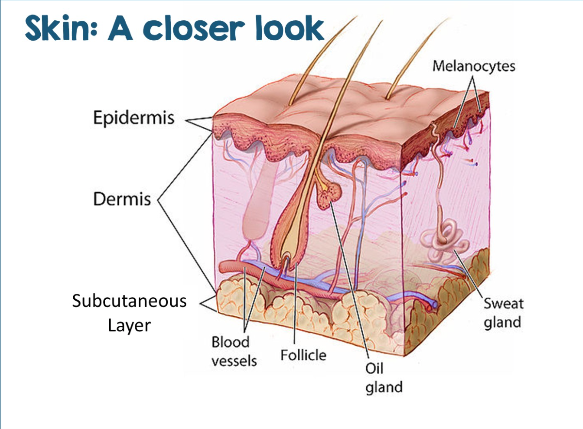
Three Distinct Layers
Epidermis
Dermis
Hypodermis (subcutaneous layer)
Epidermis
Outermost layer of the skin
Consists of keratinized stratified squamous epithelium
Avascular and depends on diffusion from dermis for nurtient
Keratinocytes (Epidermal Cell)
Most abundant; produce keratin for protection
Melanocytes (Epidermal Cell)
produce melanin pigment
Langerhans (dendritic) cells (Epidermal Cell)
Involved in immune response
Merkel Cells (Epidermal Cell)
function in touch sensation
Layers of the Epidermis (Superficial to Deep)
Stratum Corneum
Stratum Lucidum
Stratum Granulosum
Stratum Spinosum
Stratum Basale (germinativum)
Pigmentation and UV Protection
Melanocytes synthesize melanin, which accumulates over the nuclei of epidermal cells, forming a protective “umbrella” that shields DNA from UV damage
Melanocytes
Spider-shaped epithelial cells found in the bottom 2 layers of epidermis (stratum basale & stratum spinosum)
Produces pigment called melanin
Absorbed by nearby epidermal cells
Difference in dark and light skintones
Both have the same number of melanocytes
Difference is the amount of melanin produced
Variations in skin color results from
Differences in melanin production and distribution rather than the number of melanocytes
Excess UV exposure
Potentially leading skin cancers
Basal cell carcinoma (stratum basale)
Squamous cell carcinoma (stratum spinosum)
Melanoma (melanocytes; most dangerous)
Basal cell carcinoma
uncontrolled division of cells in the stratum basale layer
Squamous Cell Carcinoma
uncontrolled division of cells in the stratum spinosum layer
Melanoma
uncontrolled division of melanocytes
Dermis
Lie beneath the epidermis
Provides structural support, nourishment, and sensory input
Composed of dense irregular connective tissue
Rich in collagen and elastic fibers
Components of the Dermis
Blood Vessels
Lymphatic Vessels
Nerve ending and sensory receptors
Hair Follicles
Sweat and sebaceous glands
Dermal Papillae
Uneven junction between epidermis and dermis forms dermal papillae:
increase surface area for diffusion
create fingerprints (epidermal ridges)
Improve grip and tactile sensitivity
Layer of the Dermis
Papillary Layer
Reticular Layer
Papillary Layer (Dermis Layer)
Made of areolar connective tissue
Contains capillaries and sensory receptors
Reticular Layer
Thicker and deeper
Composed of dense collage fiber bundles
Forms lines of cleavage (tension lines)
Clinical Relevance
Incisions made parallel to cleavage lines heal faster and produce less scarring
Vasodilation
During physical activity or heat exposure, vasodilation of the dermal blood vessels increases heat loss
Vasconstriction
During cold exposure = vasoconstriction reduces heat loss
Decubitus Ulcers
Prolonged pressure that restricts blood flow can lead to decubitus ulcers (pressure sores)
Hypodermis
Subcutaneous layer
Lies beneath the dermis
Consists of adipose and areolar connective tissue
Insulates and stores nutrients
Technically not part of skin
Functions of the Hypodermis
Anchors skin to underlying tissues
Acts as an insulating layer
Stores energy as fat
Absorbs shock
Sweat (Sudoriferous) Glands
Skin contains millions of sweat glands that contribute to temperature regulation and waste removal
Eccrine glands (merocrine)
Widely distributed
Produce watery sweat for thermoregulation
Eccrine glands Sweat
Long tubes that open into pores on skin surface
Sweat is 99% water
Salts, vitamins, wastes, and an antimicrobial peptide called dermcidin
Sweat is generally acidic
Apocrine glands (merocrine)
Found in axillary and genital regions
Produce thicker secretions containing proteins and lipids
Become active at puberty
Apocrine glands: Sweat
Contains all sweat components PLUS fatty substances and proteins
Bacertia begin to break down fat & proteins causing body odor
Sebaceous Glands
Secretes sebum, an oily substance that:
Function: Lubricates skin and hair, kills bacteria
Prevents excessive water loss
Inhibits bacterial growth
Sebum production is hormonally regulated and often increases during adolescence
Also based on inheritance
Sebum (oil) Secretion
Usually secreted onto hair → more oil glands on scalp and face
None on palms or soles of feet
Hair
Composed of dead, keratinized cells and varies in thickness and distribution (hard keratin)
Functions of Hair
Protection from UV radiation
Thermal insulation
Sensory detection
Protection of sensitive structures
Hair Growth and Structure
Hair grows from the hair bulb within the follicle (root). Growth cycles include:
Anagen (growth)
Catagen (transition)
Telogen (resting)
Shaft of Hair
Part of the hair that sticks out the skin
Protected by the outermost layer called a cuticle
Conditioner smooths down the rough surface of the cutricle
Hair Bulb
Hair cells divide within the follicle in this region
Cells are filled with keratin and pigments
Arrector Pili
Dead cells are continuously pushed out as new cells are formed
Tiny muscles attached to the hair shaft to make hair “stand on end”
Texture and Color of hair
Texture is based on the shape of hair follicle opening (curly, straight, wavy)
Hair color determined by melanin present at base of hair follicle
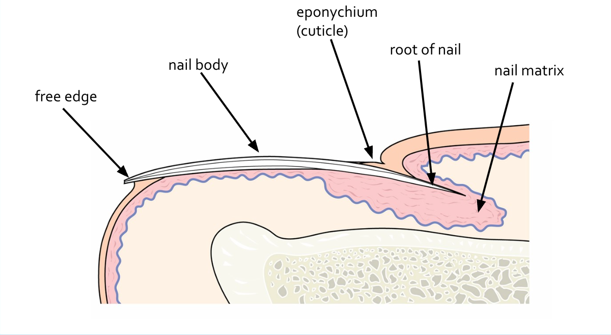
Nails
Nails are modified epidermal structures composed of hair keratin
Structure of a Nail
Protective and useful tools
Example: picking up things, scratching
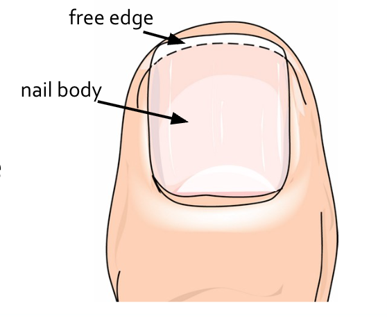
4 Basic Parts:
Free edge & Body (visible)
Root & Nail Bed (not visible)
How are nails formed?
Nail matrix produces heavily keratinized cells, becoming the nail body
Nail body is pink due to the presence of capillaries
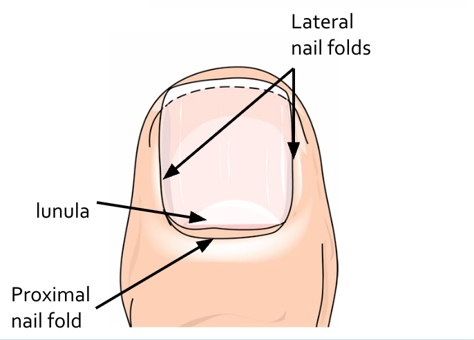
Nail is protected on three sides by nail folds
Lunula - “little moon”; whiter due to thickness of nail
Eponychium
The cuticle
Provides protection seal for the nail matrix
Provides a seal so foreign objects and bacteria can’t enter the nail bed