Opportunistic Intestinal Parasites - Coccidian Parasites
1/26
Earn XP
Description and Tags
Name | Mastery | Learn | Test | Matching | Spaced |
|---|
No study sessions yet.
27 Terms
major cause of endemic and epidemic diarrhea in developing countries
Cryptosporidium parvum
how to kill Cryptosporidium parvum?
resistance to chlorination
use ozonation - use O3 gas to disinfect water, air, food
use desiccation - removal of water
where does Cryptosporidium parvum excyst?
small intestine
enters brush border of epithelial cells
remains extra cytoplasmic
Cryptosporidium parvum causes—
persistent watery diarrhea with cramps
weight loss
fever
nausea/vomiting
Cryptosporidium parvum infectious dose
as few as 10 oocysts - low infectious dose
Cryptosporidium parvum lab diagnosis
modified Kinyoun on fecal concentrate
diarrheal stools tested with EIA Ag detection kits
DFA microscopy morse sensitive
PCR
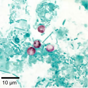
Cryptosporidium parvum oocyst
oocyst contains sporocysts with sporozoites
may have white clearing around sporocyst
honestly idrk how to describe this one
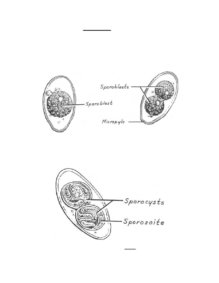
oocyst
infectious, sexual reproductive stage of coccidian parasites
sporocyst
protective sac within an oocyst containing the sporozoites
sporozoites
infective stage of a parasite that proliferates asexually within the sporocyst
Cyclospora cayetanensis is more prevalent at these locations
widespread in tropical or subtropical areas
US cases related to contaminated raspberries and strawberries from Central America
Cyclospora cayetanensis transmission
no direct fecal-oral spread
oocysts that are shed are non-infectious for day-weeks
oocysts release sporocysts in GI tract, invade mucosal cells
incubation period and duration of infection for Cyclospora cayetanensis
incubation for 1 week
duration 10-12 weeks if untreated
Cyclospora cayetanensis causes—
often severe watery diarrhea, anorexia, weight loss, abdominal pain, nausea/vomiting, myalgia, low fever, fatigue
frequent relapses
Cyclospora cayetanensis laboratory diagnosis
collect stool >3 at 2-3 day intervals because of small amounts of oocysts in stool and intermittent shedding
fix in 10% formalin (mod AFB staining)
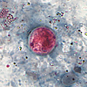
Cyclospora cayetanensis oocyst
oocysts auto-fluoresce blue
8-10 um
Cystoisospora bellii is more prevalent at these locations
worldwide mostly in tropical/subtropical areas
institutional outbreaks in the US
Cytoisospora belli invades —
villous epithelial cells in more commonly immunosuppressed people
Cystoisospora belli causes —
acute, non-bloody diarrhea with cramping and abdominal pain
severe presentation in infants and children and immunosuppressed
eosinophilia is often present (not for other coccidians)
lasts for several weeks
laboratory diagnosis of Cystoisospora belli
stool O&P with 2-3 specimen because of intermittent shedding
wet mount, modified AFB stain, or autofluorescence
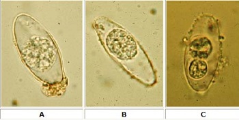
Cystoisospora belli oocysts
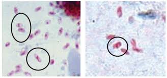
spindle shaped
20-33 um
immature cysts have a single sporoblast
mature cysts have two sporoblasts/cysts
distribution of Microsporidia spp.
worldwide with emergence as opportunist agent on all continents
infectious particle of Microsporidia spp is —
the spore
Microsporidia spp. sizes —
vary by species
Microsporidia spp. causes —
diverse manifestations dependent on species
Enterocytozoon bieneusi - diarrhea
Encephalitozoon intestinalis - diarrhea, dissemination to eye, genitourinary, and respiratory
lab diagnosis of Microsporidia spp.
separate order, so it will not be picked up on stool O&P (special order test)
chromotrope 2R stain
quick-hot gram chromotrope technique
calcofluor white
Microsporidian spp. in chromotrope 2R stain
pink/red spore walls with visible central belt-like stripe