Cardiovascular System: Blood - Lab
 Call Kai
 Learn
Learn Practice Test
 Spaced Repetition
 Match
Match Flashcards
 Knowt Play
1/39

There's no tags or description
Looks like no tags are added yet.
Name | Mastery | Learn | Test | Matching | Spaced | Call with Kai | Chat |
|---|
No analytics yet
Send a link to your students to track their progress
40 Terms
Hematocrit (HCT)
Is the % of erythrocytes, or red blood cells (RBCs), in a whole blood sample
Determined by separating formed elements from plasma and measuring the packed RBCs volume
Ranges of Hematocrit
Adult males: 42-52%
Adult females: 37-47%
Abnormal Elevated Hematocrit
Reasons are
Being at high altitude
Elevated testosterone level
Certain diseases
“Blood Doping”
Blood Doping
Refers to any of several methods used to increase the blood oxygen-carrying capacity.
Hematocrit is one way to measure which of the following characteristics of the blood?
Oxygen-carrying capacity
BLOOD APR:
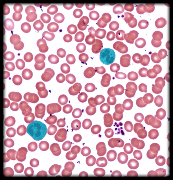
Neutrophil (Blood Peripheral Smear)
Location: Circulating blood
Description:
Leukocyte (white blood cell) with multilobed nucleus (typically 3-5 lobes); lobes connected by nuclear strands)
Fine, specific cytoplasmic granules appear pale pink in blood smears after staining with Wright or Giemsa method
Nonspecific cytoplasmic granules, which do not stain with Wright or Giemsa method, represent lysosomes
Also known as: Polymorphonuclear leukocyte
Neutrophil Function
Function:
Phagocytosis of infectious pathogens, especially bacteria (number in blood increases with bacterial infection)
Release antimicrobial agents
Comments:
Neutrophils represent 60-70% of circulating leukocytes (most numerous granulocyte)
Leukocytes (white blood cells) divided into two groups: granulocytes (neutrophils, eosinophils, and basophils) and agranulocytes (lymphocytes and monocytes)
Phagocytosis (Greek: phago = to eat + kytos = cell + osis = process) is process by which cell engulfs large particles (e.g., microorganisms, cells, and cell fragments) in membrane-bound vesicle

Platelet (Blood Peripheral Smear)
Location: Circulating Blood
Description:
Small, disc-shaped cell fragment (approximately 2 µm in diameter)
Lacks nucleus
Formed by fragmentation of cytoplasm of megakaryocyte (a cell in red bone marrow)
Also known as: Thrombocyte (abnormally low in circulating blood known as thrombocytopenia)
Platelet Function
Function: Contributes to clot formation
Comments: Micrometer (µm), also known as a micron, is 1/1,000 of a millimeter
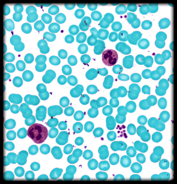
Erythrocyte (Blood Peripheral Smear)
Location:
Blood vessels of body
Heart
Description:
Biconcave disc shape
Mature cells lack nuclei and organelles
Make up 99% of the formed elements of blood
Also known as: Red blood cell (RBC)
Erythrocyte Function
Transports oxygen and carbon dioxide to and from tissues of body

Nucleus of Neutrophil
Location: Neutrophil
Description:
Largest organelle
Typically has 3-5 lobes (multilobed) connected by thin nuclear strands
Nuclear envelope comprised of double membrane (i.e., two lipid bilayers)
Contains cellular DNA and nucleolus
Nucleus Function (Neutrophil, Eosinophil, Basophil)
Functions:
DNA transcription into mRNA
Ribosomal RNA synthesis and ribosome subunit assembly (in nucleolus)
Comments:
Staining of DNA with basic histological dyes like hematoxylin depends on transcriptional activity: DNA undergoing active transcription, called euchromatin, stains lightly because it is less folded and, therefore, less dense; transcriptionally inactive DNA, or heterochromatin, is tightly packed and condensed and, therefore, stains more darkly

Erythrocyte (Neutrophil)
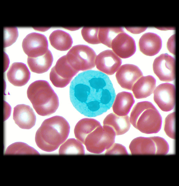
Neutrophil (Neutrophil)
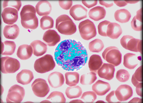
Cytoplasmic Granules in Eosinophil
Location: Cytoplasm
Description:
Large, specific cytoplasmic granules appear deep pink in blood smears after staining with Wright or Giemsa method
Nonspecific cytoplasmic granules, which do not stain with Wright or Giemsa method, represent lysosomes
Cytoplasmic Granules in Eosinophil Function
Function:
Contain agents that act on parasites (e.g., worms)
Contain enzymes that hydrolyze phagocytosed antigen-antibody complexes
Comments:
Leukocytes (white blood cells) divided into two groups: granulocytes (neutrophils, eosinophils, and basophils) and agranulocytes (lymphocytes and monocytes)
Phagocytosis (Greek: phago = to eat + kytos = cell + osis = process) is process by which cell engulfs large particles (e.g., microorganisms, other cells, or cell fragments) and forms a membrane-bound vesicle

Eosinophil
Location:
Circulating blood
Mucous membranes of respiratory, digestive, and lower urinary tracts
Description:
Nucleus typically has two large lobes (bilobed) connected by thin nuclear strand
Large, specific cytoplasmic granules appear deep pink in blood smears after staining with Wright or Giemsa method
Eosinophil Function
Function:
Phagocytosis of antigen-antibody complexes, allergens, and inflammatory chemicals
Releases enzymes that destroy parasitic worms
Comments:
Eosinophils represent 2-4% of circulating leukocytes
Leukocytes (white blood cells) divided into two groups: granulocytes (neutrophils, eosinophils, and basophils) and agranulocytes (lymphocytes and monocytes)
Phagocytosis (Greek: phago = to eat + kytos = cell + osis = process) is process by which cell engulfs large particles (e.g., microorganisms, other cells, or cell fragments) and forms a membrane-bound vesicle
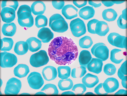
Erythrocyte (Eosinophil)

Nucleus of eosinophil
Location: Eosinophil
Description:
Largest organelle
Nucleus has two lobes (bilobed); connected by thin chromatin filaments
Nuclear envelope comprised of double membrane (i.e., two lipid bilayers)
Contains cellular DNA and nucleolus
Function: Same as other blood cells
Comments: Same as other blood cells
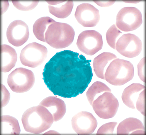
Basophil
Location: Circulating blood
Description:
Nucleus typically large with two lobes (bilobed) frequently described as U- or S-shaped
Large, specific cytoplasmic granules that appear blue-black in blood smears after staining with Wright or Giemsa method
Nonspecific cytoplasmic granules, which do not stain with Wright or Giemsa method, represent lysosomes
Granules frequently obscure nucleus
Basophil Function
Function:
Initiates immune response
Secretes histamine and heparin
Comments:
Basophils represent <1% of circulating leukocytes
Resemble mast cells of connective tissue
Leukocytes (white blood cells) divided into two groups: granulocytes (neutrophils, eosinophils, and basophils) and agranulocytes (lymphocytes and monocytes)

Erythrocytes (Basophil)
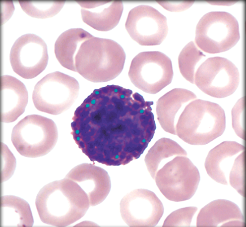
Cytoplasmic granules in Basophil
Location: Cytoplasm
Description:
Large, specific cytoplasmic granules dark appear blue-black in blood smears after staining with Wright or Giemsa method
Nonspecific cytoplasmic granules, which do not stain with Wright or Giemsa method, represent lysosomes
Granules frequently obscure nucleus
Cytoplasmic granules in Basophil Function
Function: Contain histamine, heparin, chemotactic factors for eosinophils and neutrophils, proteases, chondroitin sulfate, and peroxidase
Comments: Leukocytes (white blood cells) divided into two groups: granulocytes (neutrophils, eosinophils, and basophils) and agranulocytes (lymphocytes and monocytes)

Nucleus of Basophil
Location: Basophil
Description:
Largest organelle
Large nucleus with two lobes (bilobed); frequently described as U- or S-shaped
Nuclear envelope comprised of double membrane (i.e., two lipid bilayers)
Contains cellular DNA and nucleolus
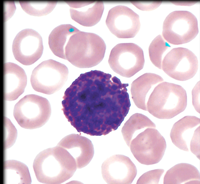
Platelet (Basophil)
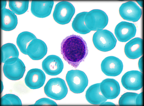
Erythrocyte (Lymphocyte)
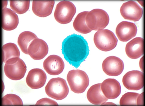
Lymphocyte
Location:
Throughout tissues of body
Circulate in blood and lymphatic vessels between lymphoid tissues and other body tissues
Description:
Three classes: B lymphocyte (B cell), T lymphocyte (T cell), and natural killer (NK) cell
Two main types of T lymphocyte: helper T lymphocyte and cytotoxic T lymphocyte
Lymphocyte Function
B lymphocyte (15-30% of lymphocytes in body) responsible for humoral (Latin: humor = fluid) immune response: activation and differentiation into plasma cells (produce specific antibodies in response to specific antigen as primary immune response) or memory B lymphocytes (secondary immune response)
T lymphocyte (70-85% of lymphocytes in body): responsible for cell-mediated immune response
Natural killer (NK) cell: responds to multiple antigens and can kill wide variety of infected cells and cancer cells
Lymphocyte Comments
Lymphocytes represent 20-25% of circulating leukocytes
Leukocytes (white blood cells) divided into two groups: granulocytes (neutrophils, eosinophils, and basophils) and agranulocytes (lymphocytes and monocytes)
B lymphocytes called "B" because they are derived in chickens from lymphoid organ known as bursa of Fabricius
Antibodies secreted into loose connective tissue enter lymphatic system and then bloodstream
Latin: humor = fluid
Micrometer (µm), also known as a micron, is 1/1,000 of a millimeter

Nucleus of Lymphocyte
Location: Lymphocyte
Description:
Largest organelle
Round or slightly indented; occupies most of cytoplasm
Nuclear envelope comprised of double membrane (i.e., two lipid bilayers)
Contains cellular DNA and nucleolus
Comments: Same as other blood cells
Nucleus of Lymphocyte & Monocyte Function
Function:
DNA replication
DNA transcription into mRNA
Ribosomal RNA synthesis and ribosome subunit assembly (in nucleolus)
Comment:
Staining of DNA with basic histological dyes like hematoxylin depends on transcriptional activity: DNA undergoing active transcription, called euchromatin, stains lightly because it is less folded and, therefore, less dense; transcriptionally inactive DNA, or heterochromatin, is tightly packed and condensed and, therefore, stains more darkly
Micrometer (µm), also known as a micron, is 1/1,000 of a millimeter

Monocyte
Location:
Circulating blood
Connective tissue
Description:
Largest leukocyte (15-20 µm in diameter)
In connective tissue, they transform into macrophages
Monocyte Function
Function: Phagocytosis
Comment:
Monocytes represent 3-8% of circulating leukocytes
Leukocytes (white blood cells) divided into two groups: granulocytes (neutrophils, eosinophils, and basophils) and agranulocytes (lymphocytes and monocytes)
Micrometer (µm) is 1/1,000 of a millimeter
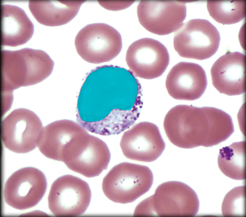
Nucleus of Monocyte
Location: Eccentrically placed in monocyte
Description:
Largest organelle
Large, ovoid or kidney-shaped (horseshoe) nucleus
Nuclear envelope comprised of double membrane (i.e., two lipid bilayers)
Contains cellular DNA and nucleolus

Erythrocyte (Monocyte)

Platelet (Monocyte)