Lab: Cerebral Hemispheres & Cortex
1/78
There's no tags or description
Looks like no tags are added yet.
Name | Mastery | Learn | Test | Matching | Spaced |
|---|
No study sessions yet.
79 Terms
What does the telencephalon become?
cerebral hemispheres
What does the diencephalon become?
thalamus, hypothalamus, epithalamus
What does the mesencephalon become?
mesencephalon
What does the metencephalon become?
pons and cerebellum
What does the myelencephalon become?
medulla
stellate cells
small star-shaped cortical interneurons with numerous dendrites
receive impulses from other areas
pyramidal cells
found in the cerebral cortex, have a triangular cell body and a single, long dendrite among many smaller dendrites
has one apical and 2 basal dendrites and a large axon responsible for sending impulses
Where does white matter lie in relation to gray matter in the brain?
white matter lies internal to grey matter
association fibers
Fibers that connect areas of the cerebral cortex within the SAME hemisphere
commissural fibers
connect one part of cortex to another part of cortex in the OTHER hemisphere
projection fibers
connect the cortex to the thalamus, brainstem, and spinal cord
Corticopedal projection fibers
fibers that come into the cortex from outside of the cortex
Corticofugal projection fibers
fibers that leave from the cortex and go outside of the cortex
molecular layer of cerebral cortex
most superficial (layer 1)
receives dendrites from all internal layers of the cortex and acts as a coordinating center where layers communicate action
external granular layer of cerebral cortex
layer 2
contains stellate cells
receives input from other cortical regions
external pyramidal layer of cerebral cortex
layer 3
contains pyramidal cells
sends output to other cortical regions
What type of fibers are involved in the external granular and pyramidal layers of the cerebral cortex?
association and commissural fibers
internal granular layer of cerebral cortex
layer 4
stellate cells that receive input from the thalamus and other brainstem areas
very thick in sensory areas of the cortex
striate cortex
another name for the primary visual cortex
the internal granular layer of this area is so thick that you can see a line through this layer even if brain is unstained
Internal pyramidal layer of cerebral cortex
layer 5
pyramidal cells that send axons to brainstem and spinal cord
very thick in motor areas of cortex
multiform layer
layer 6
sends axons back to thalamus
works to modulate what information thalamus sends to cortex
modulates what you pay attention to
works as brains equalizer
Brodmann's areas
Histologic regions of the cerebral cortex mapped by Brodmann. Often used to designate functional areas.
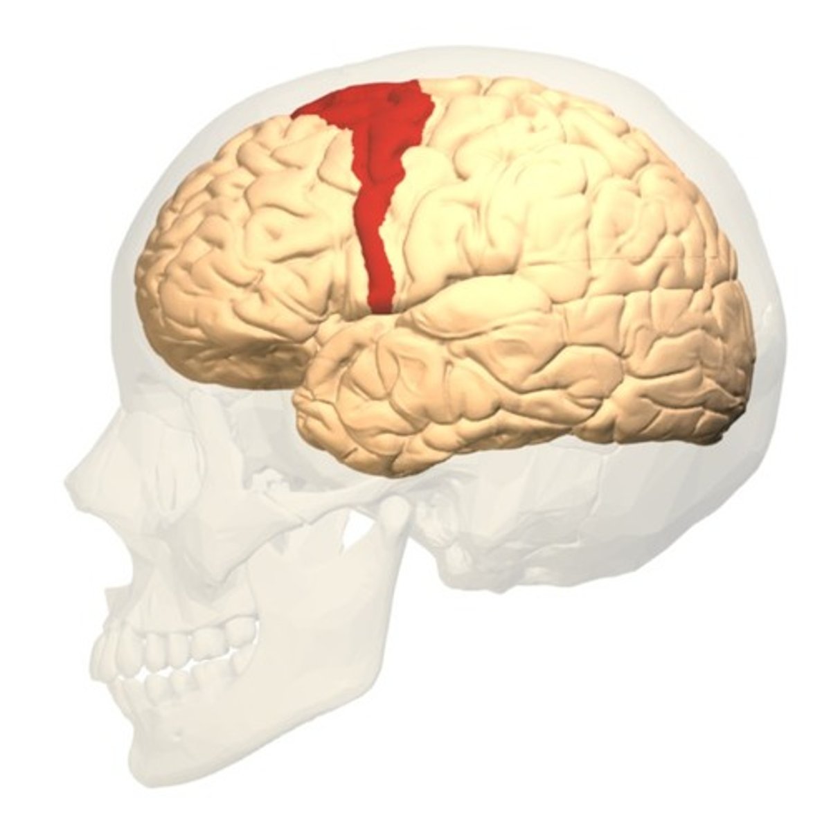
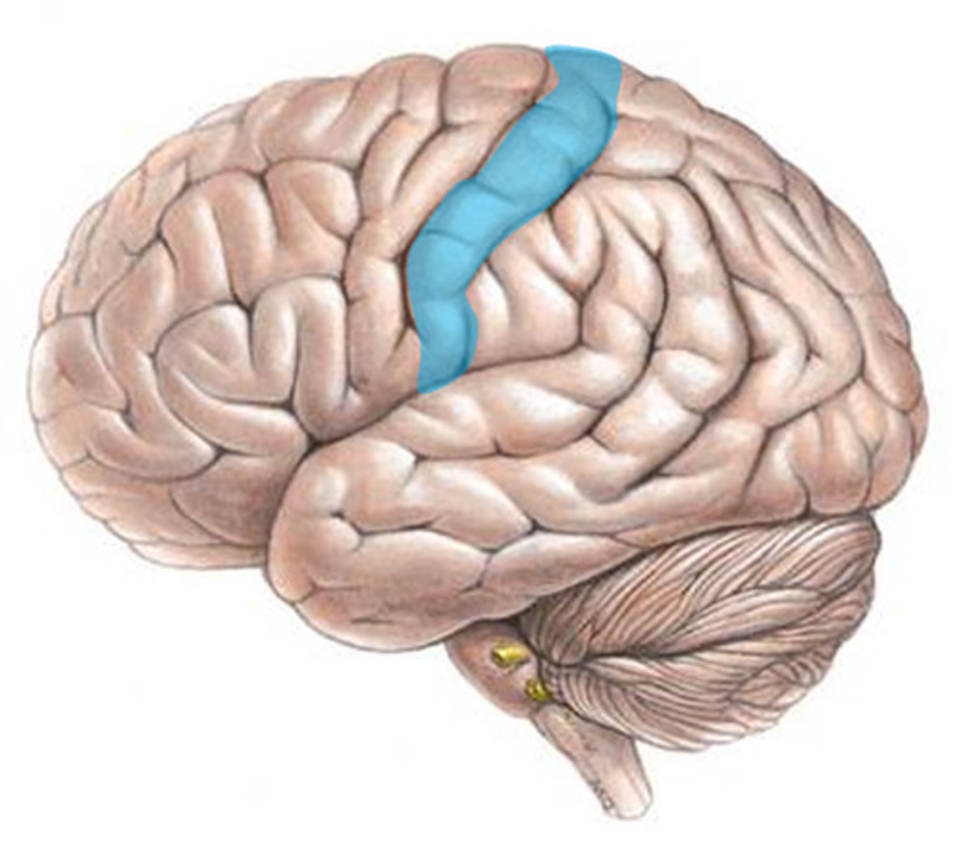
area 4
primary motor cortex (precentral gyrus)
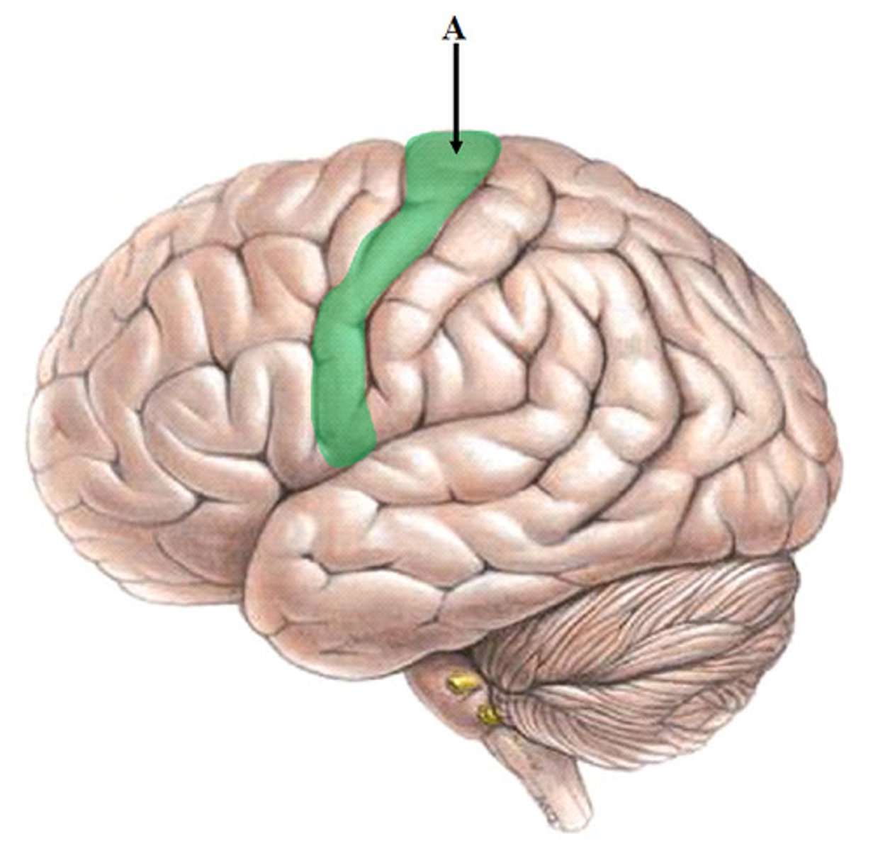
area 6
premotor cortex
Layer 3 of area six synapses with what layer of area four?
Layer 2
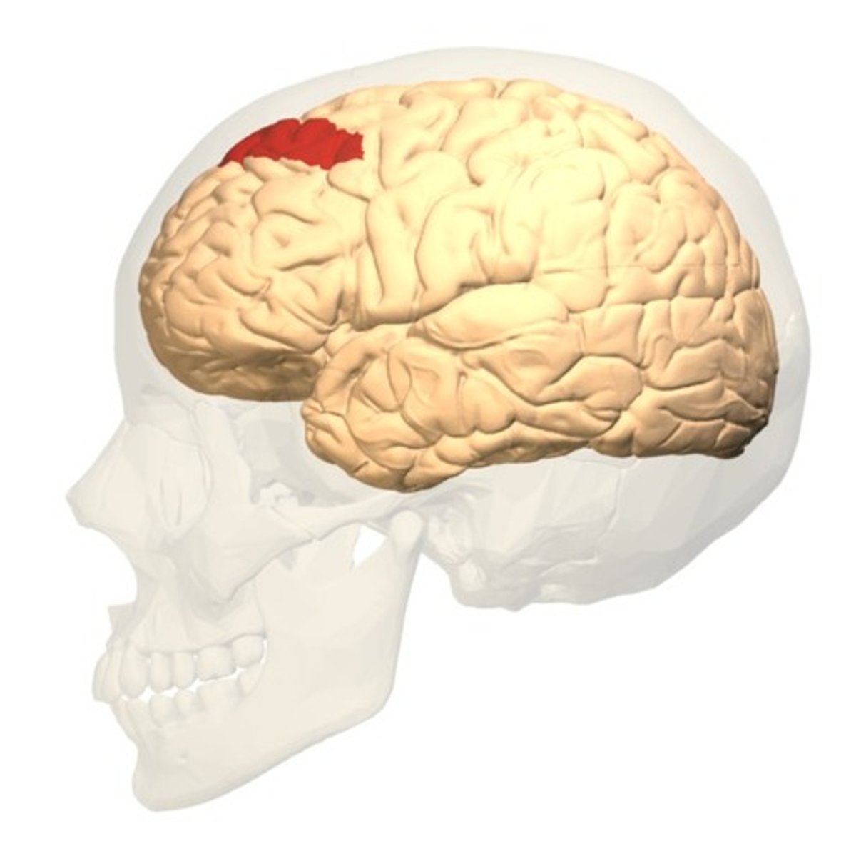
area 8
frontal eye field
responsible for contralateral saccadic eye movements
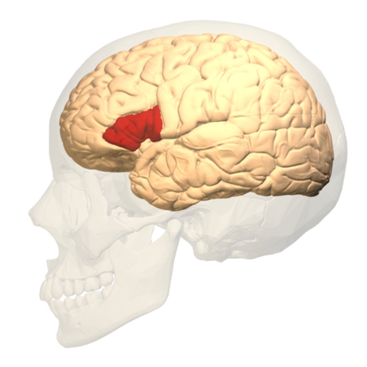
area 44 and 45
Broca's area
responsible for expressive language
Areas 3, 1, 2
primary somatosensory cortex
Which layer of areas 3,2,1 is the thickest?
layer 4
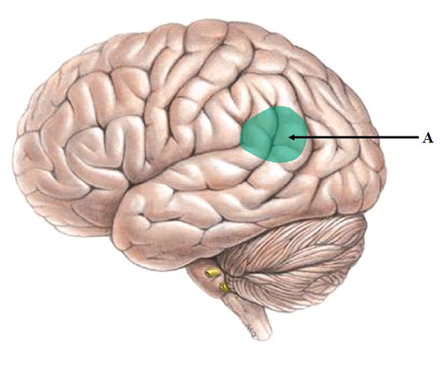
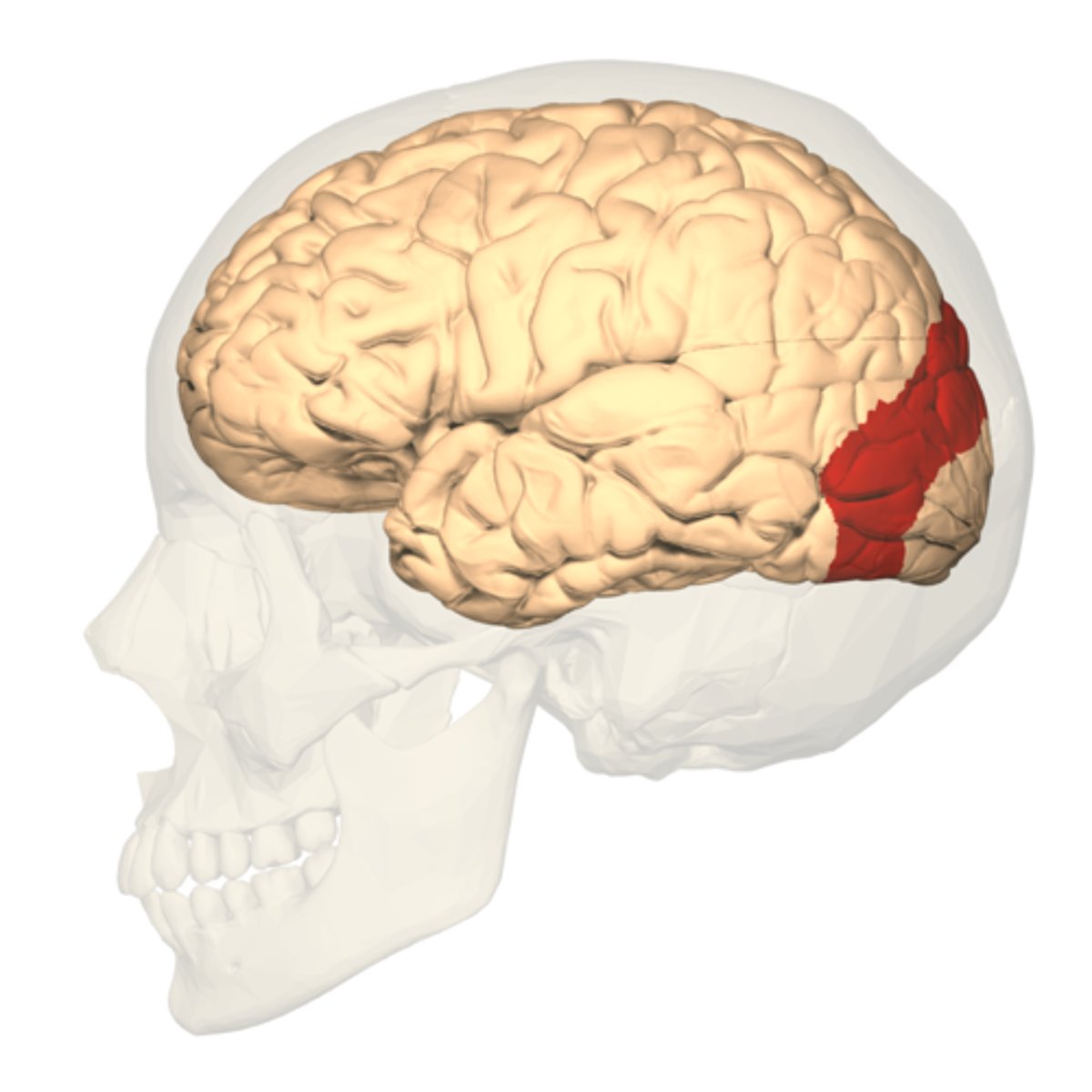
Area 22 and occasionally 39 and 40
Wernicke's area
responsible for receptive language
expressive aphasia
inability to produce language despite being able to understand language
What causes expressive aphasia?
damage to Broca's area (area 44/45 on inferior frontal lobe)
What causes receptive aphasia?
damage to Wernicke's area (posterior 22, some 39 and 40)
receptive aphasia
The inability to understand language despite being able to hear it and produce it
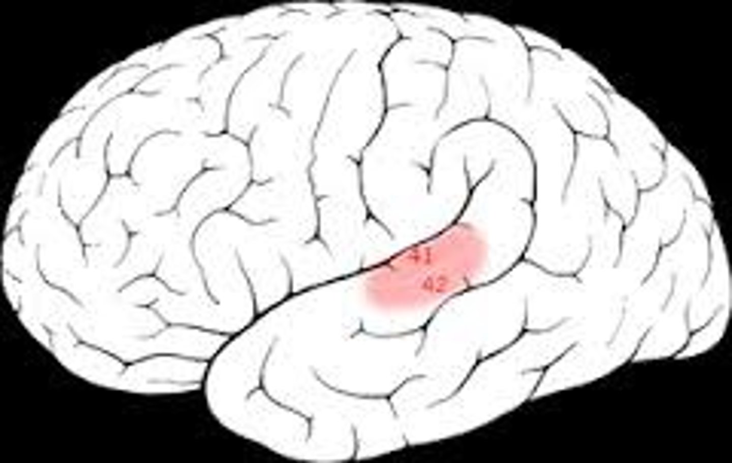
area 41
primary auditory cortex
receives input from MGN and sends to area 42

Which layers are the thickest in area 41?
layer 4 because receiving from the thalamus
layer 2 because projecting to area 42
area 42
auditory association area
processes information here and send to area 22
Which layers are the thickest in area 42?
layer 2
layer 3
area 22
auditory association area
posterior part of Wernicke's
area 17
primary vision cortex
aka V1
processes as very general lines of various orientations and circles of color
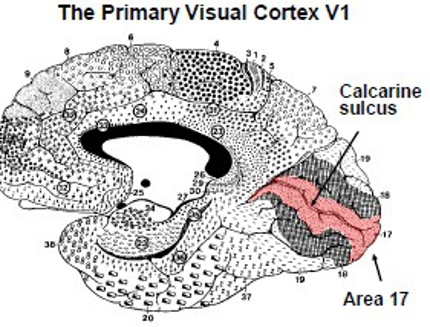
Where does area 17 lies in relationship to the calcarine?
lies half above and half below calcarine processing input from the upper and lower retina respectively
Where does area 17 receive information from?
LGN
area 18
association visual cortex
aka V2
continues to process visual input
Where does area 18 receive information from?
receives input from area 17
Where does area 18 lie?
lies below and above area 17 on the occipital lobe
area 19
association vision cortex
aka V3
furthers visual processing
dorsal stream
visual information pathway from the occipital lobe to the parietal lobe
concerned with WHERE something is, orientation and guiding motor function
ventral stream
visual information pathway from the occipital lobe to the temporal lobe
concerned with WHAT something is
What can occur is the ventral stream is damaged?
agnosias
optic ataxia
can visually identify the orientation of an object, but motor action to the object is inaccurate
due to damage of the dorsal stream
visual neglect
failure to respond to visual stimulation on the side of the visual field that is opposite a brain lesion to the dorsal stream
visual-visual disconnection
caused by lesions of ventral stream
leads to agnosia
agnosias
disruptions in the ability to identify familiar objects
visual-verbal disconnections
due to lesions of the ventral stream
leads to anomias and alexias
anomia
Loss of the ability to name objects or retrieve names of people
alexia
inability to understand written words, difficulty reading
caused by a defect in the occipitotemporal corticofugal bundle (defect in LEFT inferior temporal gyrus)
visual-limbic disconnection
?
achromatopsia
inability to distinguish colors
prosopagnosia
inability to recognize faces
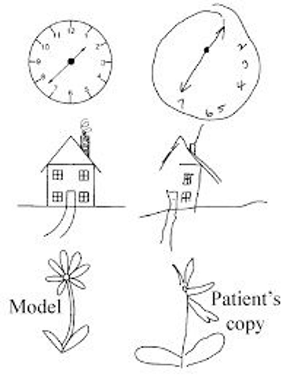
Simultagnosia
an inability to recognize and identify multiple objects in a visual scene; including distinct objects within a spatial layout and distinguishing between "local" objects and "global" objects
ex: seeing a forrest vs a tree
object agnosia
inability to recognize objects
pure alexia
an inability to read
object form topology hypothesis
Object form is represented in ventral temporal cortex continuously in a distributed and overlapping arrangement
locations, faces, written words, objects
T/F: the cortical networks for reading are parallel, simultaneous, and bidirectional
true
What does the rhombencephalon become?
pons, medulla, cerebellum
What does the prosencephalon become?
forebrain (telencephalon and diencephalon)
cerebral hemispheres and thalamus
What are the three subgyri that make up the inferior frontal gyrus?
opercular
trianglular
orbital gyrus
Which gyrus makes a C-shape cap around lateral fissure?
supra marginal gyrus
uncus
on medial surface of temporal lobe (olfactory area)
What lies above the calcarine sulcus?
cuneus
What lies below the calcarine sulcus?
lingual gyrus
What gyri of the temporal lobe are seen from the lateral view?
superior temporal gyrus
middle temporal gyrus
inferior temporal gyrus
What gyri of the temporal lobe are seem from an inferior view?
inferior temporal gyrus
lateral occipitotemporal gyrus
medial occipitotemporal gyrus
parahipocampal gyrus
uncus
What forms the floor of the inferior horn of the lateral ventricle?
hippocampus
What forms the roof of the anterior horn and body of the lateral ventricle?
corpus callosum
What forms the lateral wall of the anterior horn of the lateral ventricle?
head of caudate
What forms the medial wall of the anterior horns and body of the lateral ventricle?
septum pellucidum
Which part of the cerebral hemispheres is responsible for written words
LEFT inferior temporal gyrus
What forms the floor of the anterior horn of the lateral ventricle?
fornix