Immunology Chapter 1
 Call Kai
 Learn
Learn Practice Test
 Spaced Repetition
 Match
Match Flashcards
 Knowt Play
1/99

There's no tags or description
Looks like no tags are added yet.
Name | Mastery | Learn | Test | Matching | Spaced | Call with Kai | Chat |
|---|
No analytics yet
Send a link to your students to track their progress
100 Terms
Innate
Adaptive
Types of immune defenses
Immunity
State of protection
Specific for disease
Vaccines
Kick in adaptive defenses to get to immunity
Skips innate defenses
Antibiotics kill many commensal bacteria —> Pathogenic bacteria take up the space the normal flora previously occupied —> Produce toxins —> Mucosal injury —> RBCs & WBCs leak into the gut between the injured epithelial cells
How can long term antibiotic use lead to further infections (primarily in the gut)
Opportunistic Pathogen
Pathogen that can become harnful when new conditions arise
ie, c.diff, H. pylori
Bacteria
Virus
Parasites
Fungi
Types of pathogens
Protozoans - Eukaryotic
Helminthes - Worms, multicellular
Types of parasites
Innate Defenses
Immediate barriers & response (ie, skin, mucosal surfaces)
Skin
Physical barrier
Sebum
Creates an acidic environment on the skin’s surface
Mucous
Traps pathogens
Mostly in the upper respiratory tract
Cilia
Propels pathogens up and out of the body
Mostly in the upper respiratory tract
Lysozyme
Chemical compound that breaks down bacterial cell walls
Recognition - Cells (neutrophils)/Soluble components (complement) recognize something foreign
Response - Depends on what recognized the pathogen; Neutralize, kill, inflammation, etc
Innate Immune Response Stages
Inflammation
The innate immune response causes [?] at site of infection
Redness (erythema) & Heat (due to vasodilation)
Swelling (edema) (due to increased vascular permeability)
Pain (due to kinins)
S&S of Inflammation
Pathogen cell surface induces cleavage & activation of complement
One complement fragment (C3b) binds to pathogen; Other (C3a) attracts an effector Cell
Complement receptor on the effector cell binds to C3b on the pathogen
Effector cell engulfs the pathogen, kills it, & breaks it down
General: Pathogen-Recognition by effector cell using complement
Cytokines
Hormones of the immune system
Soluble proteins that interact with other cells to trigger the innate immune response
Surface wound introduces pathogen —> Activates effector cells in the area to secrete cytokines
Vasodilation & increased vascular permeability allows fluid, protein, & inflammatory cells to leave the blood and enter the injured tissue
Infected tissue becomes inflamed —> Redness, heat, pain, swelling
Inflammation pathway
Adaptive immune response
The [?] adds to an ongoing innate immune response
Adaptive Immune Response
Specific
Adaptable
Memory
Recognition by T or B cells
Response
Adaptive Immune Response stages
Rapid (within hours)
Fixed
Limited number of specificities
Constant
Characteristics of the recognition mechanisms of innate immunity
Slow (days-weeks)
Variable
Numerous highly selective specificities
Improves during response
Characteristics of recognition mechanism of adaptive immunity
Progenitor cells give rise to a large number of lymphocytes with different specificities DURING DEVELOPMENT
DURING INFECTION, lymphocytes with receptors that recognize the pathogen are activated
Proliferation of pathogen-activated lymphocytes (clonal selection) give effector cells that terminate the infection & produce memory cells to remember the infection
Stages of B/T Cell Response
Effector Cells
Memory Cells
Cell types made through clonal selection when certain lymphocytes proliferate and differentiate
Clonal Selection
Proliferation & differentiation of pathogen-specific lymphocytes —> Stimulated by the antigen
Creates Effector Cells & Memory Cells
Memory Cells
Goal of vaccines
Hematopoietic stem cells
Immune system cells w/ different functions all derive from [?]
Common lymphoid precursor
Common myeloid precursor
Hematopoietic stem cell creates…
Lymphoid lineage
B Cells, T Cells, and Natural Killer cells all come from the [?]
B-Cell
NK/T-Cell Precursor
Common lymphoid precursor creates…
T Cell
NK cell
NK/T Cell precursor creates…
Granulocyte-macrophage progenitor
Megakaryocyte/erthyroid progenitor
Common myeloid precursor creates…
Myeloid & Erythroid
The Common Myeloid Precursor creates the [?] & [?] cell lineages
Macrophage & Dendritic cell precursor
Neutrophil
Eosinophil
Basophil
An unknown precursor
Granulocyte-macrophage progenitor creates…
Mast Cell
The granulocyte-macrophage progenitor creates an unknown precursor which then creates …
Monocyte
Dendritic Cell
Macrophage & dendritic cell precursor creates…
Macrophage
Dendritic Cell (maybe)
Monocytes create…
Megakaryocyte
Erthyroblast
Megakaryocyte/erythroid progenitor creates…
Platelets
Megakaryocytes create…
Erythrocyte (RBC)
Erythroblasts create…
Neutrophils
Most abundant WBC
1st Responders
Short-lived - Last ~3 days
Kill Pathogens
Produce cytokines to start an inflammatory response
2 Major functons of the Neutrophil
Eosinophils
Fight parasites
(particularly helminthes
Mast Cells & Basophils
Primarily responsible for allergies
Dendritic Cells
Wander around looking for pathogens
Good at starting adaptive immune response
Phagocytosis = primary function
Monocytes & Macrophage
Killing pathogens through phagocytosis
Produce cytokines
Wander around blood stream (monocytes) & tissues (macrophages) looking for pathogens to kill
Macrophage
Monocyte that has entered the tissue
Natural Killer Cell
Type of lymphocyte
Larger than T & B Cells
Natural ability to kill tumor cells & fight viruses
Fighting viral infections
Tumor surveillance
Primary functions of natural killer cells
T & B Cells
Adaptive cells
Go through formal selection process (Clonal selection)
Bone marrow
Large reserves of neutrophils are stored in the [?] & are released when needed
Macrophages
As neutrophils die in the tissue after killing bacteria, they are then engulfed and degraded by [?]
Dead neutrophils
Pus is composed primarily of [?]
Binding of bacteria to phagocytic receptors on macrophages
What induces the engulfment & degradation of bacteria
Phagosome
Membrane-bound sac that forms around foreign pathogens during phagocytosis
Phagolysosome
Cytoplasmic body formed by the fusion of a phagosome and lysosome during phagocytosis in order to destroy engulfed pathogens
Synthesis & release of cytokines & initiation of inflammatory response
The binding of components of bacteria to signaling receptors on macrophages induces what?
Antibody
Structures on the surface of B Cells
Bind to pathogens & cause them to be inactivated or destroyed
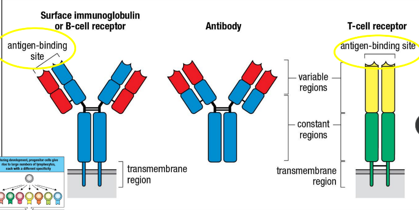
Antigen-binding site
Variable regions
Constant regions
Transmembrane region
Top to bottom: Structures on the surface of B (antibodies) and T cells (T-Cell Receptor)
Antigen
Part of pathogen
Triggers immune response when binging to antibody
Effector cells - Plasma cells
Memory cells
Clonal Selection: B Cell + Antigen =
Effector cells - Cytotoxic T Lymphocytes (CTL) & helper cells
Memory cells
Clonal Selection: T Cell + Antigen =
Cytotoxic T Lymphocyte
Produced after T cells discover an antigen
Killer cells
Plasma Cells
Produced when B cells encounter antigens
Make antibodies
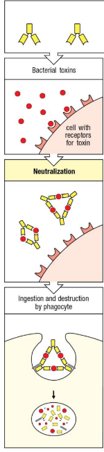
Toxin attaches to cell w/ receptors for toxin
Neutralization - Antibodies surround toxins
Antibodies w/ toxins gather near surface of phagocyte, leading to ingestion & destruction of toxins
Antibody response to bacterial toxins
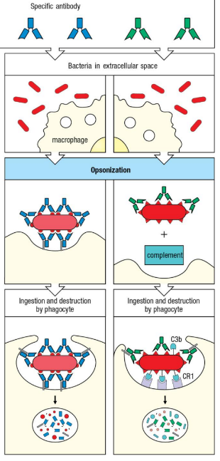
Macrophage or dendritic cell encounter bacteria
Opsonization
Antibodies surround the bacteria & bring it closer to macrophage surface
Antibodies gather around bacteria and complement binds to bacteria
Ingestion & Destruction of bacteria
Phagocyte surrounds bacteria w/ antibodies
Complement binds to receptors on phagocyte, anchoring the bacteria for phagocytosis
Antibody response to full bacteria in extracellular space
Thymus
Primary lymphoid organ
Where T cells develop & mature
Bone Marrow
Primary lymphoid organ
Where B Cells develop & mature
Spleen
Peyer’s patches (in small intestine)
Adenoids
Tonsils
Lymph nodes
Where clonal selection happens
Secondary lymphoid organs
Clonal selection
Secondary lymphoid organs are determined as where [?] happens
Spleen
Where lymphocytes in the blood are circulated
Left Subclavian vein
Where lymph drains into to return to the heart
Lymphatic Capillaries
Structure where lymphocytes enter the tissue space
Afferent lymphatic vessel
Where lymphocytes exit the lymph node
Efferent lymphatic vessel
Where lymphocytes enter the lymph node
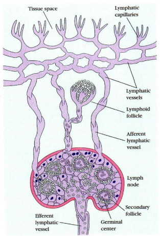
Lymphatic Capillaries
Lymphatic vessels
Lymphoid follicle
Afferent lymphatic vessel
Secondary follicle
Germinal center
Efferent lymphatic vessel
Lymph node structures
is not an antigen
The lymphocyte leaves the lymph node when there [is/is not] an antigen
Valves
There are [?] along the afferent and efferent lymphatic vessels to prevent backflow
Secondary lymphoid tissues
Adaptive immunity is initiated in [?]

Dendritic cells carrying pathogens
The adaptive immune response is activated by [?] being drained into the lymph node
Paracortex
In the lymph node, the T-cell area is called the [?]
Cortex
In the lymph node, the B-cell area is called the [?]
Germinal Center
Active center of B cell activity in the lymph node
Medullary Sinus
In the lymph node, Plasma cells mostly reside in the [?]

Lymphoid follicle/cortex (B-cell area)
Medullary sinus
Paracortex/T-cell area
Germinal center
Marginal Sinus
Afferent & Efferent lymphatic vessels
Artery & Vein
Structures of the Lymph node
Tissue
Lymph nodes drain [?] of pathogens
Blood
Spleen drains [?] of pathogens
Get rid of old RBCs
Start immune response
Functions of the Spleen
White pulp
[?] of the spleen initiates immune responses
Red pulp
[?] of the spleen is where RBCs are destroyed
B-Cell corona - young
Germinal center - mature
Area where B-cells are in the white pulp of the spleen
Periarteriolar lymphoid sheath
Area where T-cells are in the white pulp of the spleen

Perifollicular zone
Marginal zone
B-Cell Corona
Germinal Center
Central arteriole
Periarteriolar lymphoid sheath
Structure of the white pulp of spleen
M cell
Dendritic cells
In the gut, antigens get into lymphoid tissue through the [?] in the epithelium surrounding the gut lumen, activating [?]
Efferent lymphatic vessels to a larger lymph nodes
Gut-associated lymphoid tissues drain through the [?]
Opsonization
Coating of antibodies on pathogen facilitate phagocytosis by attaching to receptors on phagocytes
Neutralization
Antibodies bind tightly to a site on a pathogen to inhibit its growth, replication, or interaction with human cells
Tonsils
Adenoids
Appendix
Peyer’s patches
Gut-Associated Lymphoid Tissues