Muscles Test
 Call Kai
 Learn
Learn Practice Test
 Spaced Repetition
 Match
Match Flashcards
 Knowt Play
1/110

Earn XP
Description and Tags
11/20/25
Name | Mastery | Learn | Test | Matching | Spaced | Call with Kai | Chat |
|---|
No analytics yet
Send a link to your students to track their progress
111 Terms
What are the seven ways we name skeletal muscles?
Direction
Location (near a bone or body region)
Relative Size
Number of Origins (Heads)
Shape
Location of Origin and/or Insertion
Action of Muscle (describes what muscle does)
Relative Size Examples (3)
Maximus = Largest (Gluteus Maximus)
Longus = Longest (Fibularis Longus)
Brevis = Shortest (Extensor Pollicis Brevis)
Direction Examples (3)
Rectus = Parallel (Rectus Abdominus)
Transverse = Perpendicular (Transverse Abdominus)
Oblique = @ 45 degree Angle (External Obliques)
Location Examples (2)
Frontalis is on frontal bone
Tibialis Anterior is next to Tibia
Number of Origins (Heads) Examples (2)
Biceps = 2 Origins
Triceps = 3 Origins
Shape Examples (2)
Deltoid = Triangular
Trapezius = Trapezoid
Location of Origin and Insertion Example
Sternocleidomastoid
> Origin: Sternum
> Insertion: Mastoid Process
Action of Muscle Example
Flexor Digitorum: Flexes Fingers
Prime Mover (Agonist)
Major responsibility for producing a specific movement
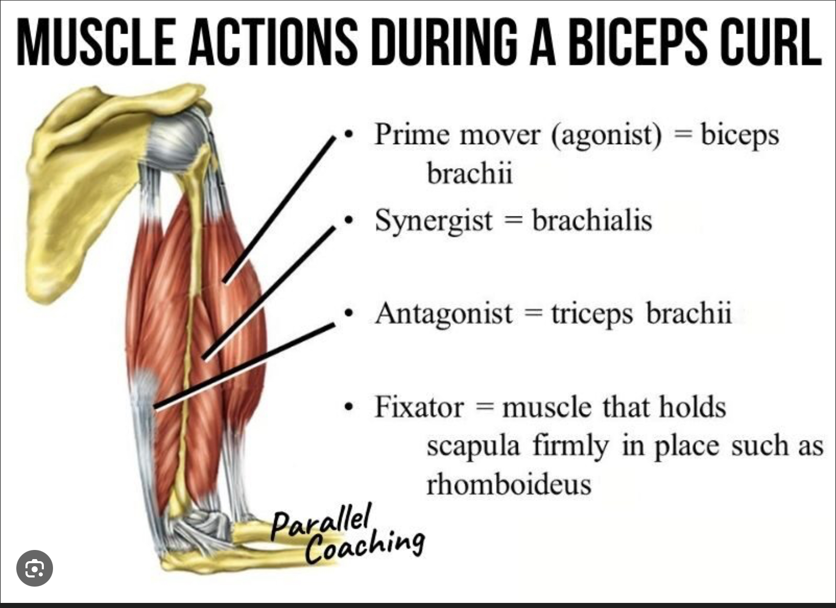
Antagonist
Muscle that oppose or reverse a particular movement
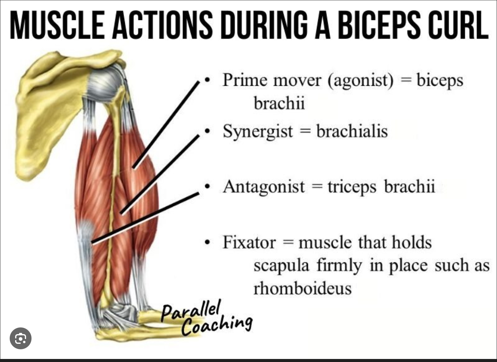
How are antagonists contracted?
They’re contracted slightly to provide some resistance to slow or stop movement
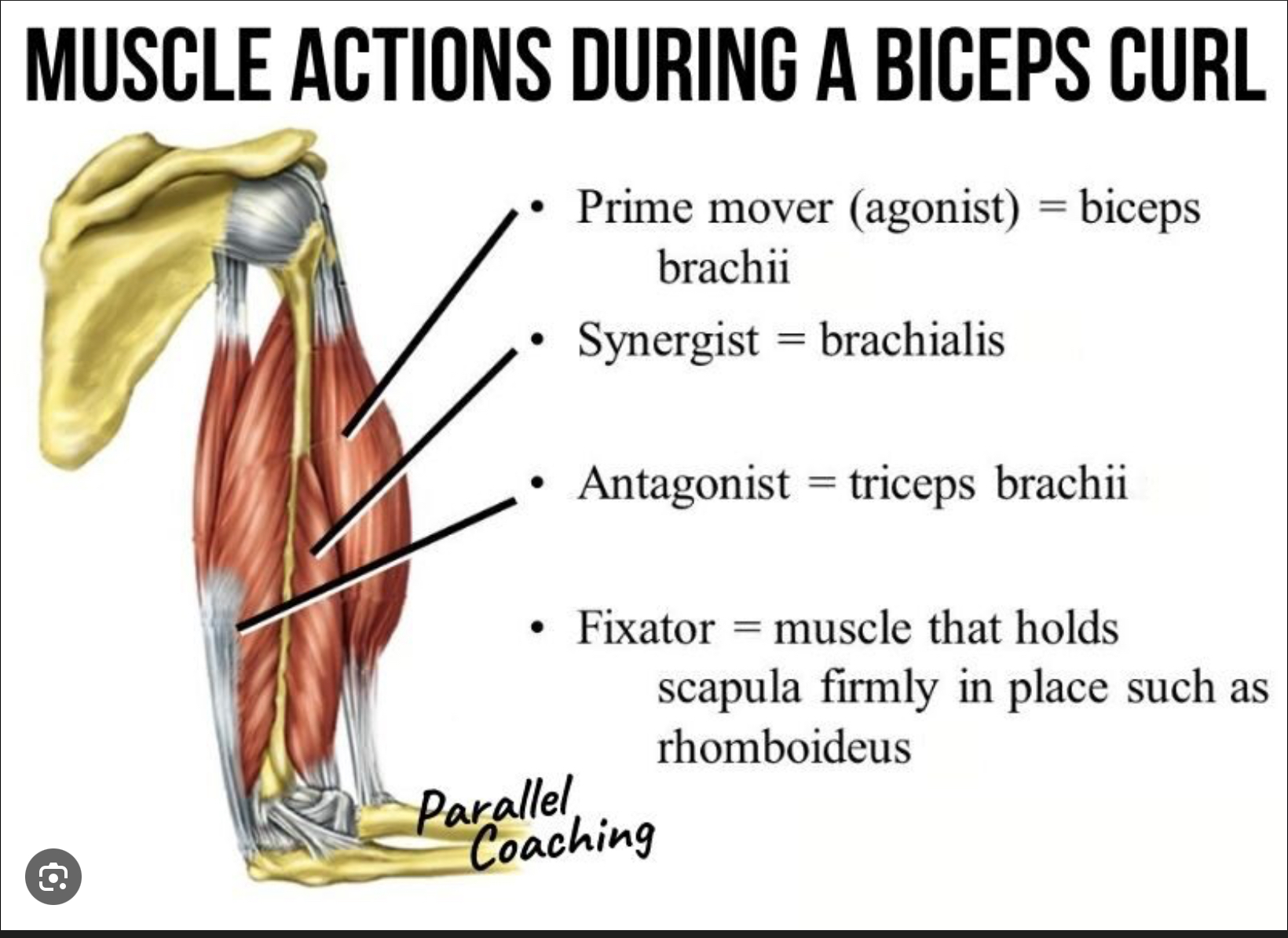
Synergists
Help prime mover by adding little extra force to movement and reduce unnecessary movement that occurs as a prime mover contracts
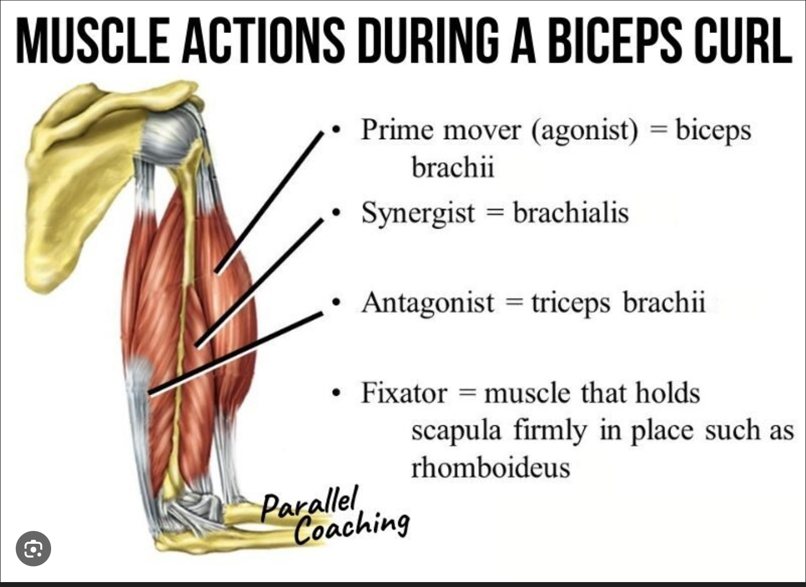
Fixators
Immobilize the bone
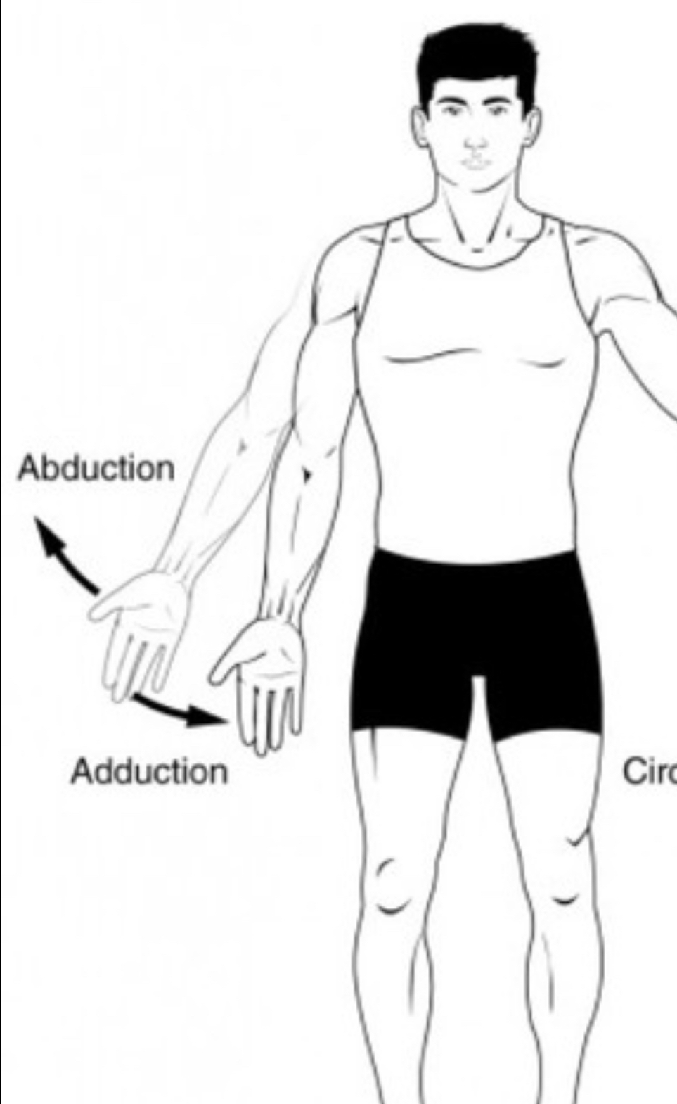
Adduction
Towards the midline or center
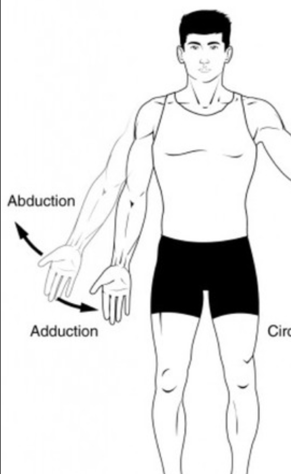
Abduction
Away from midline or center; spreading out
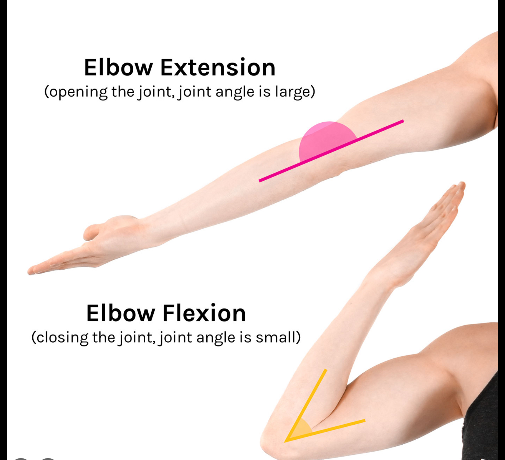
Extension
Straightens the joint
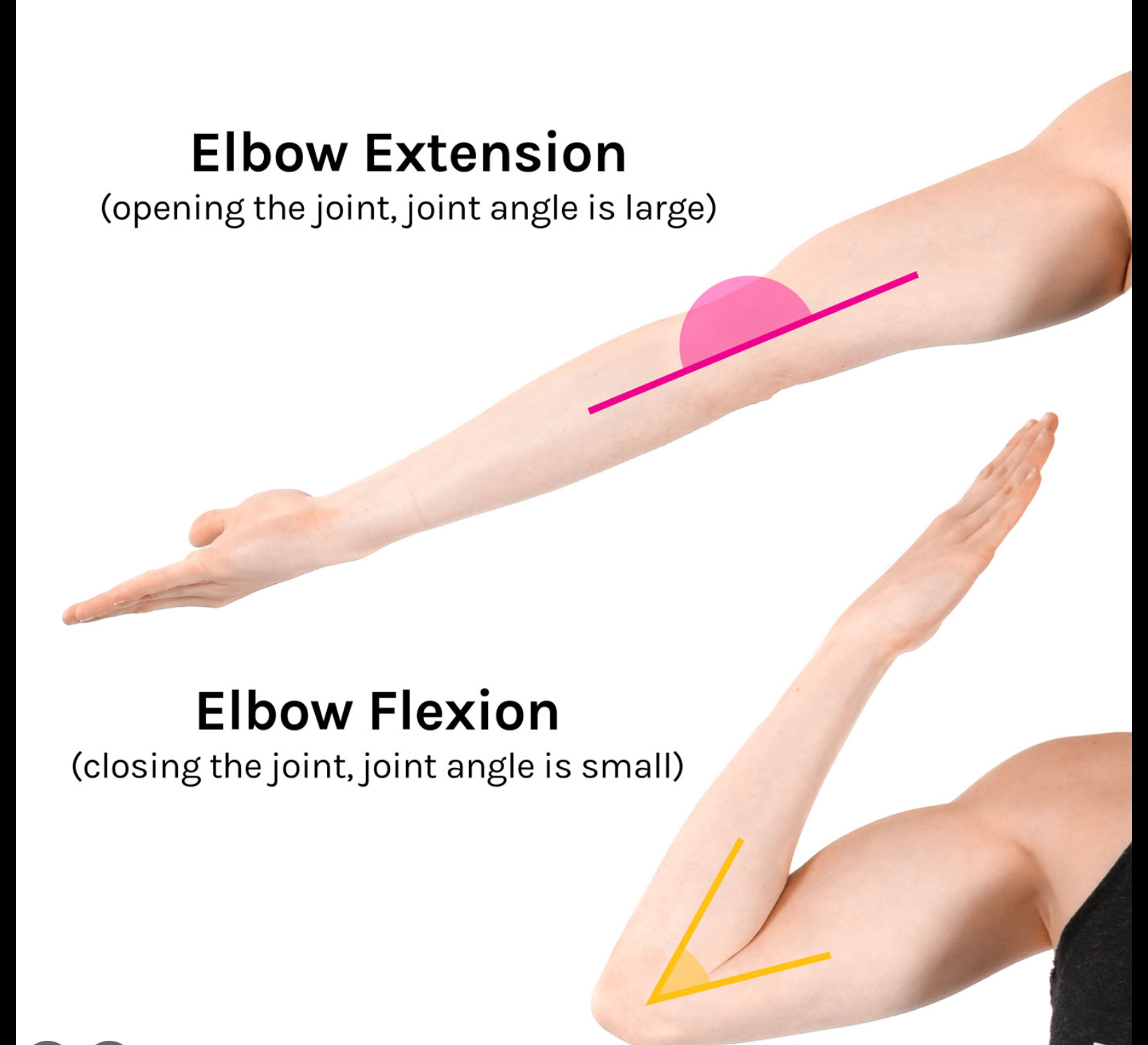
Flexion
Bends the joint
Circumduction
Circle or full rotation
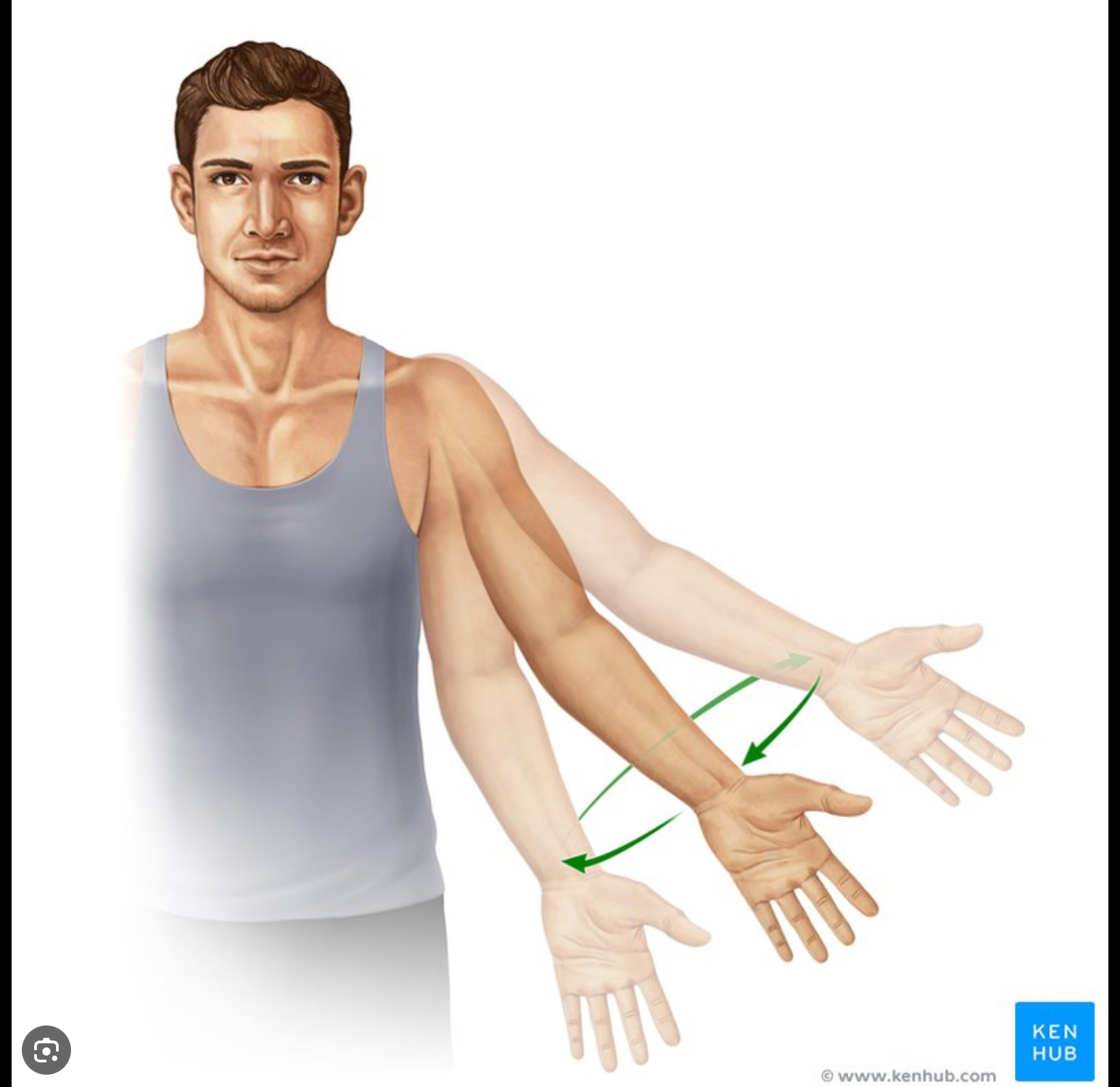
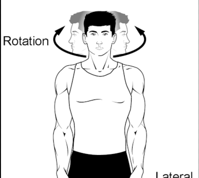
Rotation
Head “yes” or “no” movements
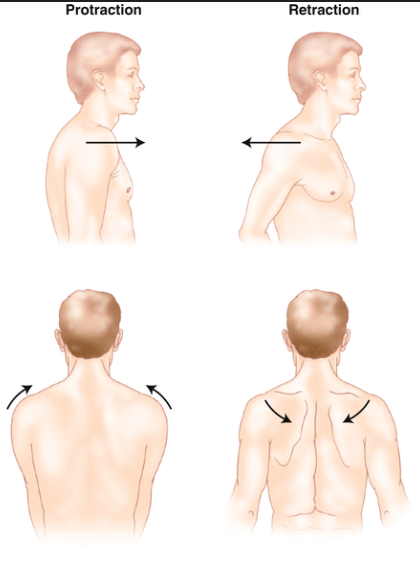
Retraction
Pull head (chin) back
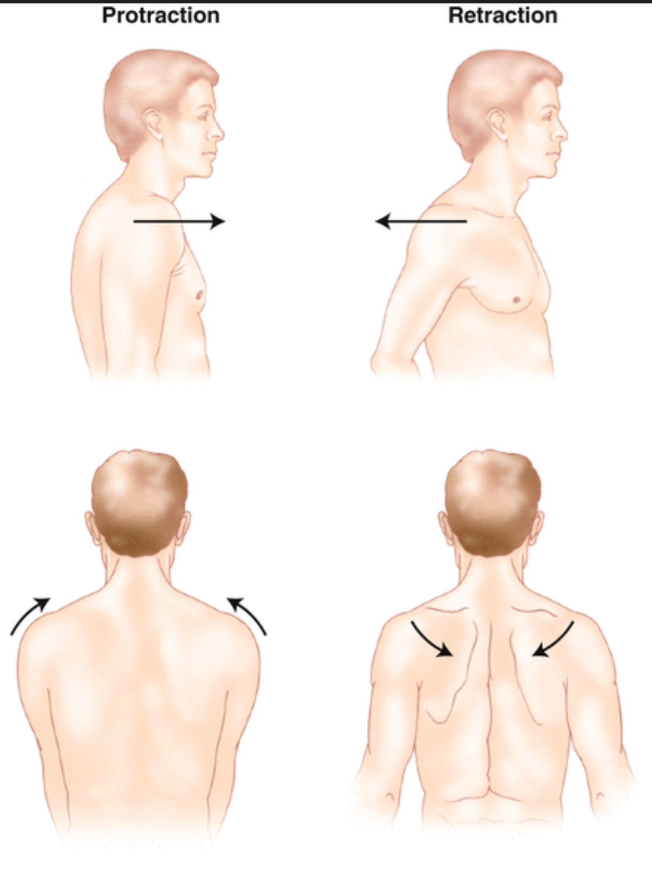
Protraction
Push head (chin) forward
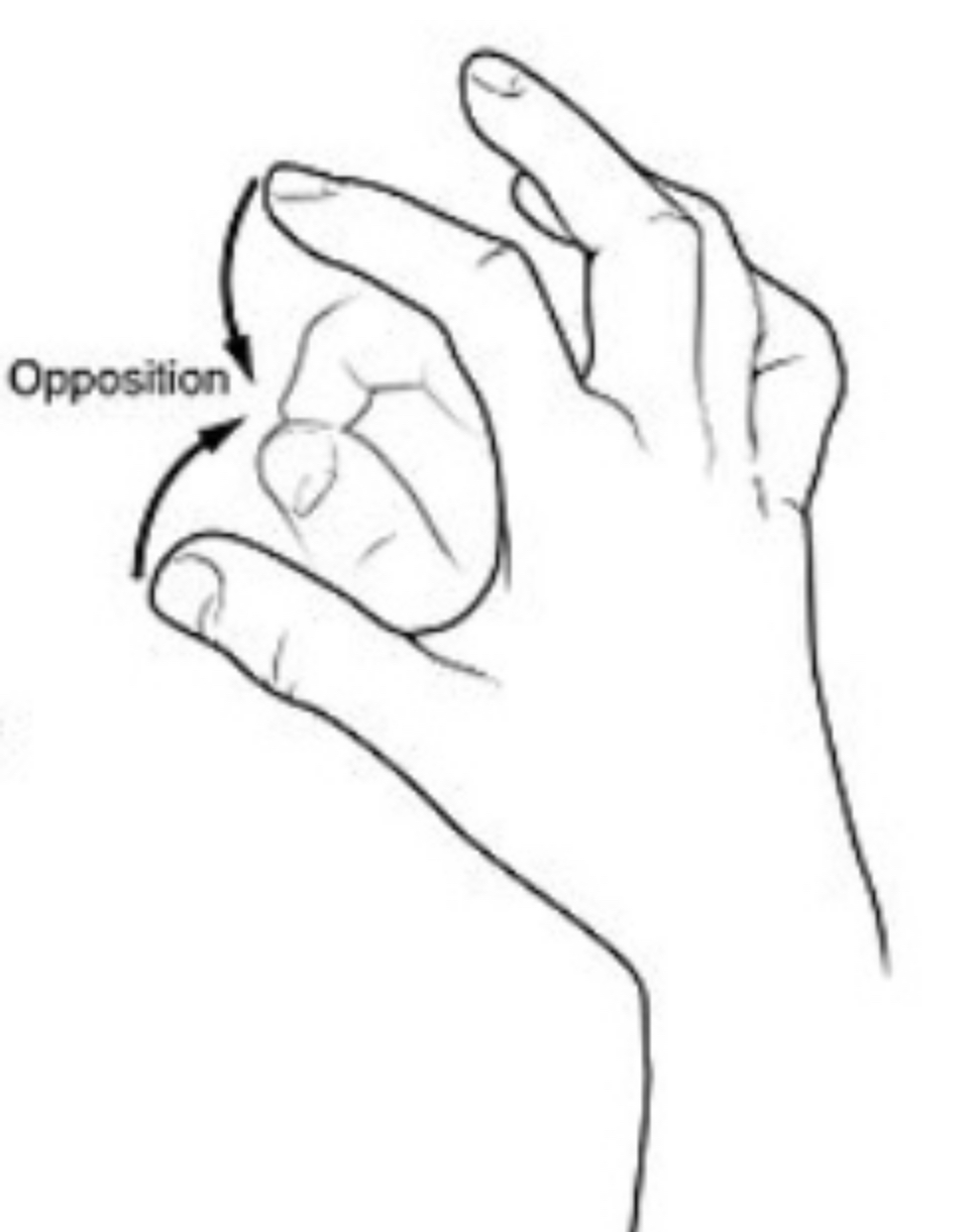
Opposition
Thumb to finger
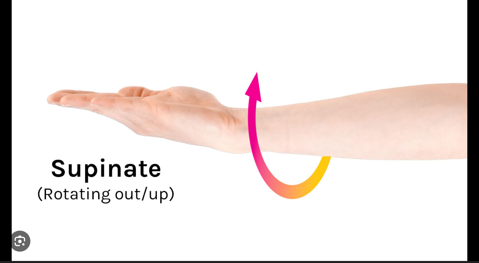
Supination
External rotation or flip hand over (palm up)
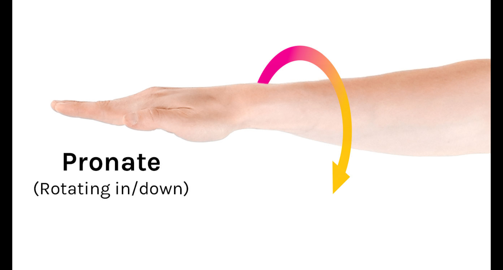
Pronation
Internal rotation or flip hand back (palm down)
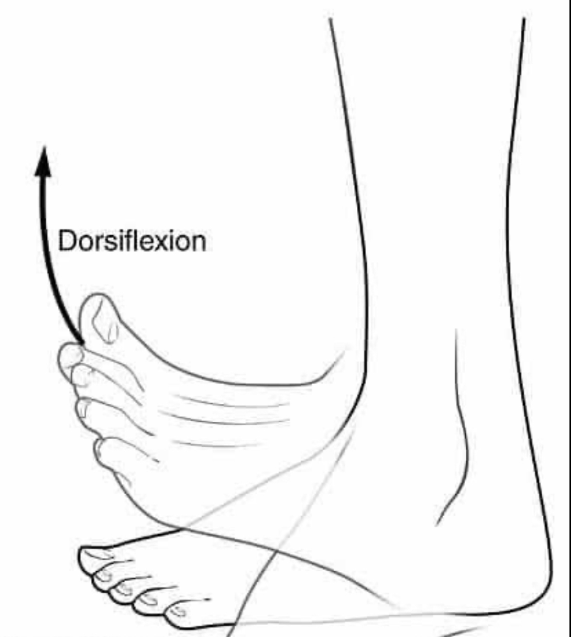
Dorsiflexion
Bending foot up
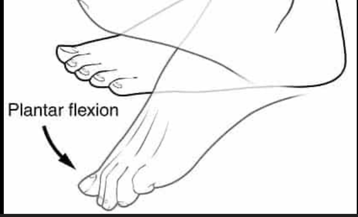
Plantar Flexion
Bending foot down, point toes
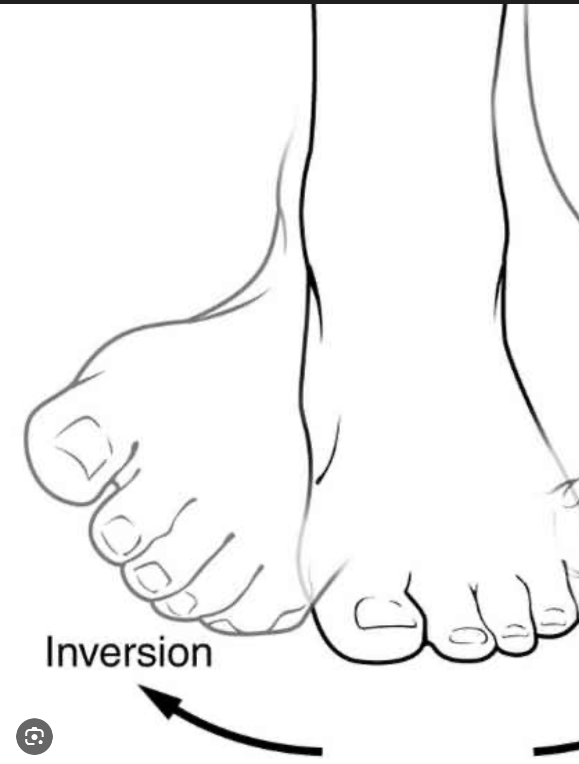
Inversion
Foot move in towards body
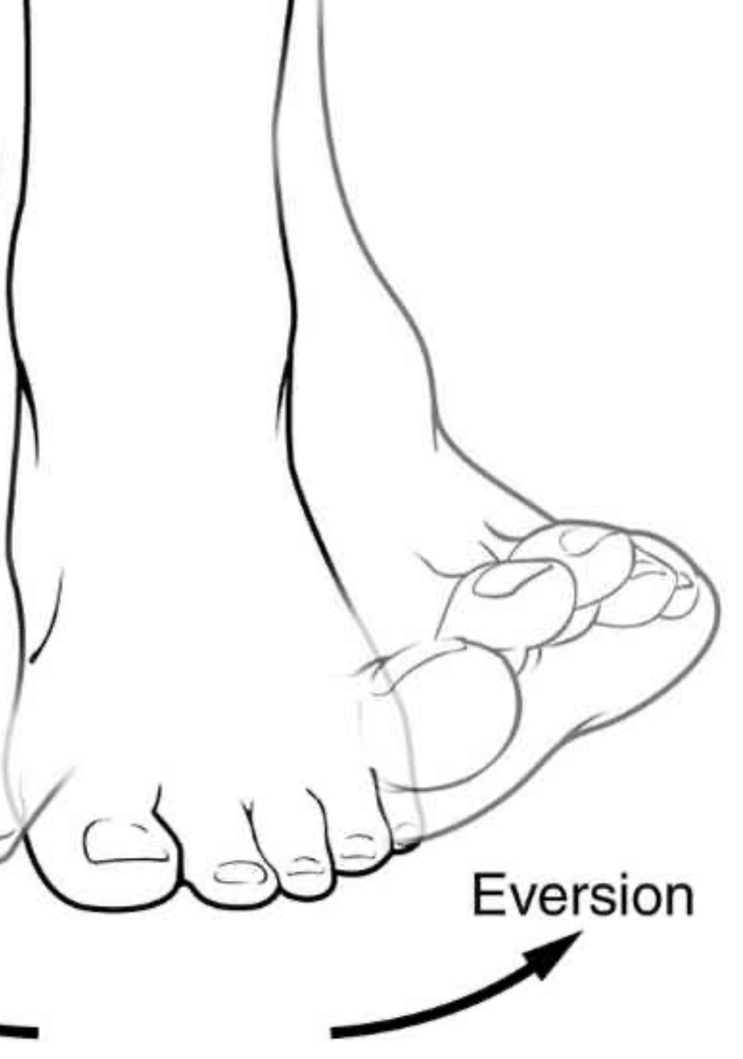
Eversion
Foot turned out from body
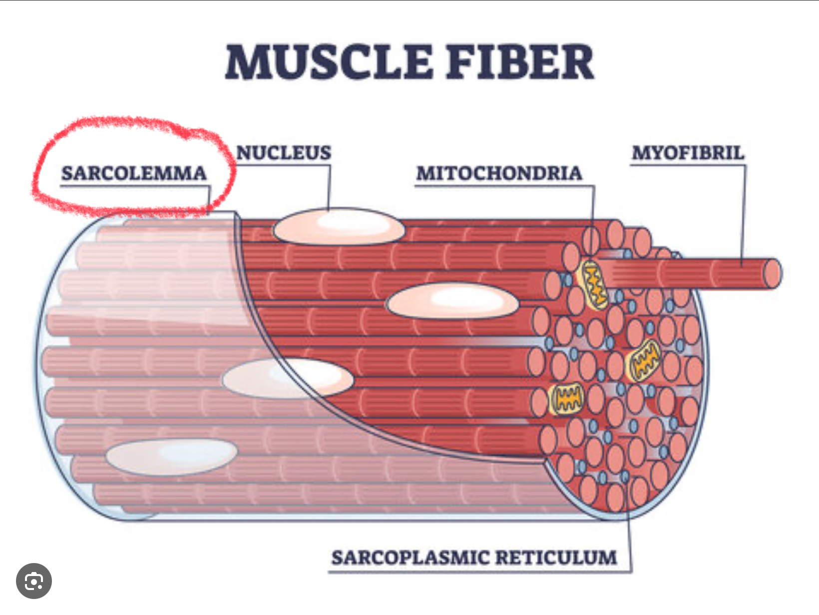
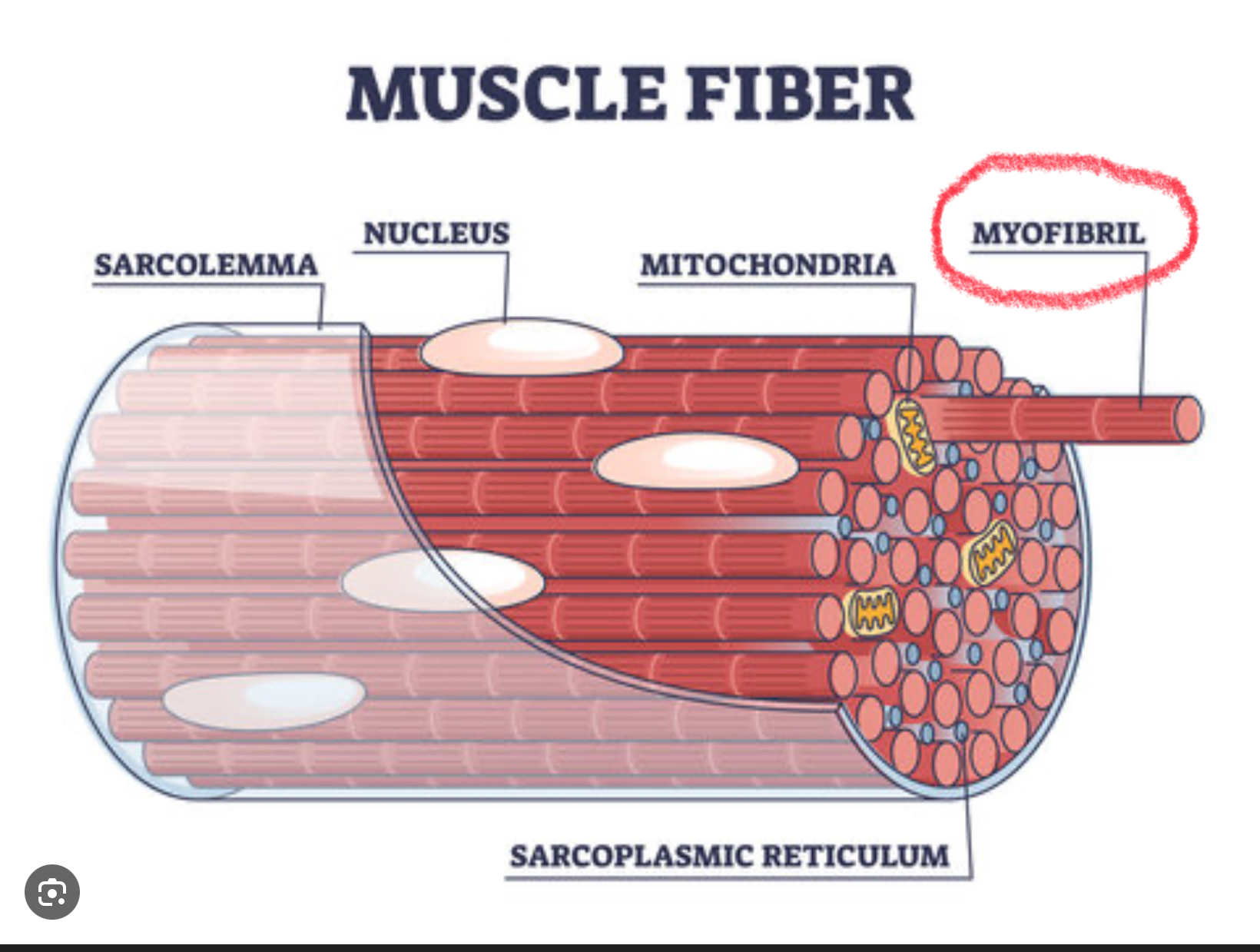
Sarcolema
Cell membrane of a muscle fiber
Myofibrils
Contractile fiber in striated muscle cells
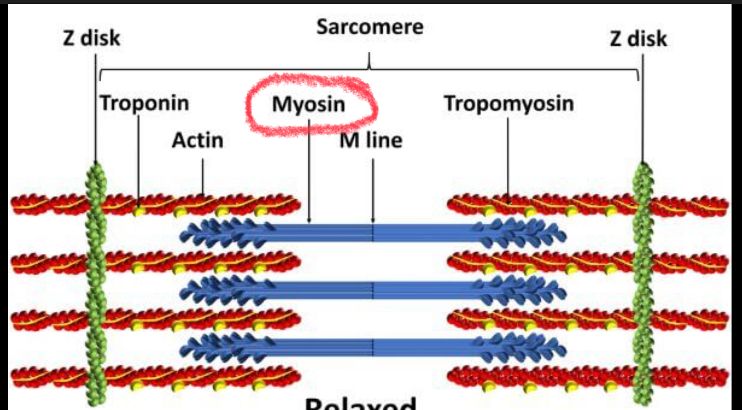
Myosin
Protein in a muscle fiber that forms the thick filaments and pulls on actin to help muscle fibers contract
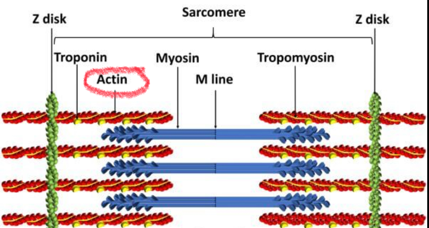
Actin
Protein in a muscle fiber that forms the thin filaments that slide between thick filaments of the protein myosin, contracting muscle fibers
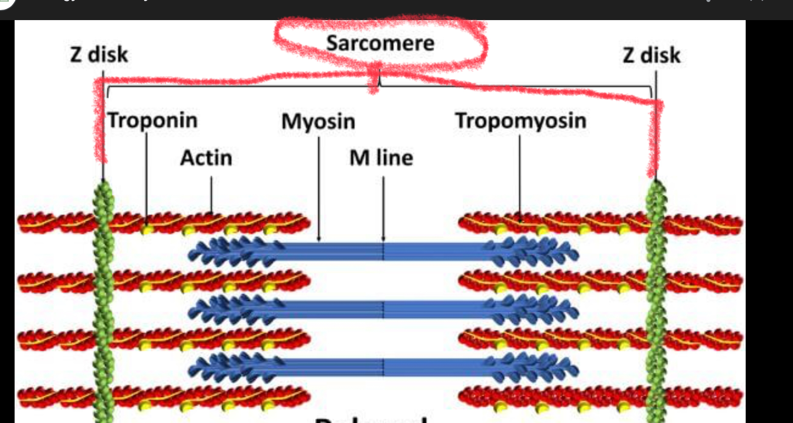
Sarcomere
Structural unit of a myofibril and the functional unit of muscle contraction
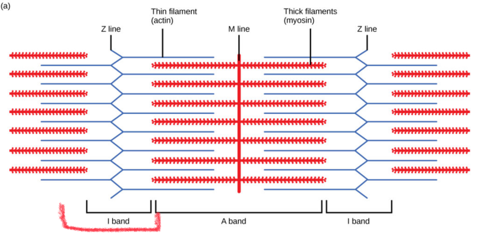
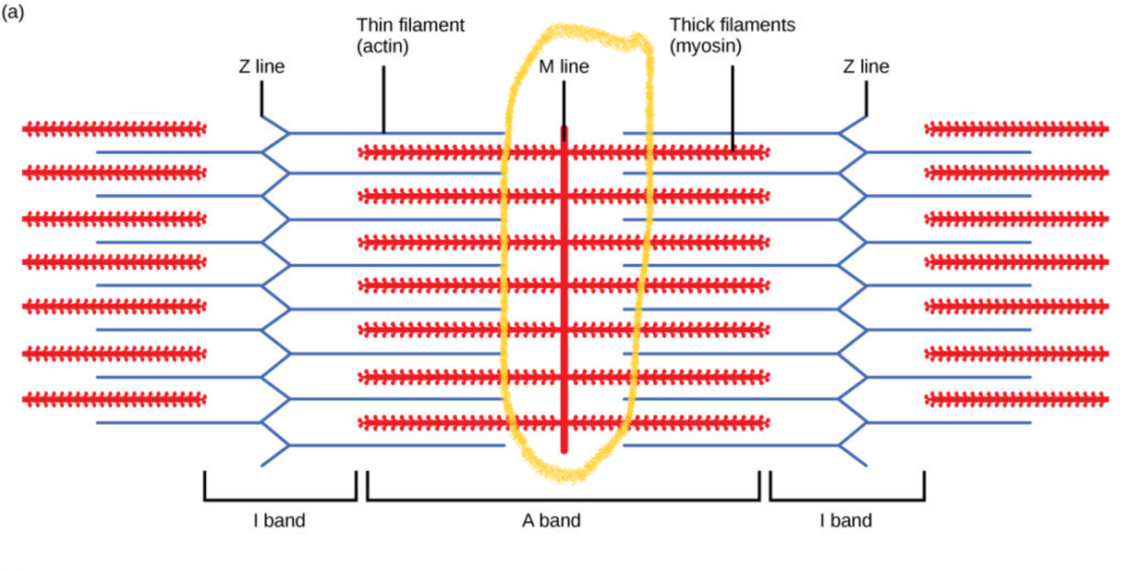
I Bands
The light, thin bands on the striation pattern of skeletal muscle fibers composed of thin filaments (actin) directly attached to Z Lines
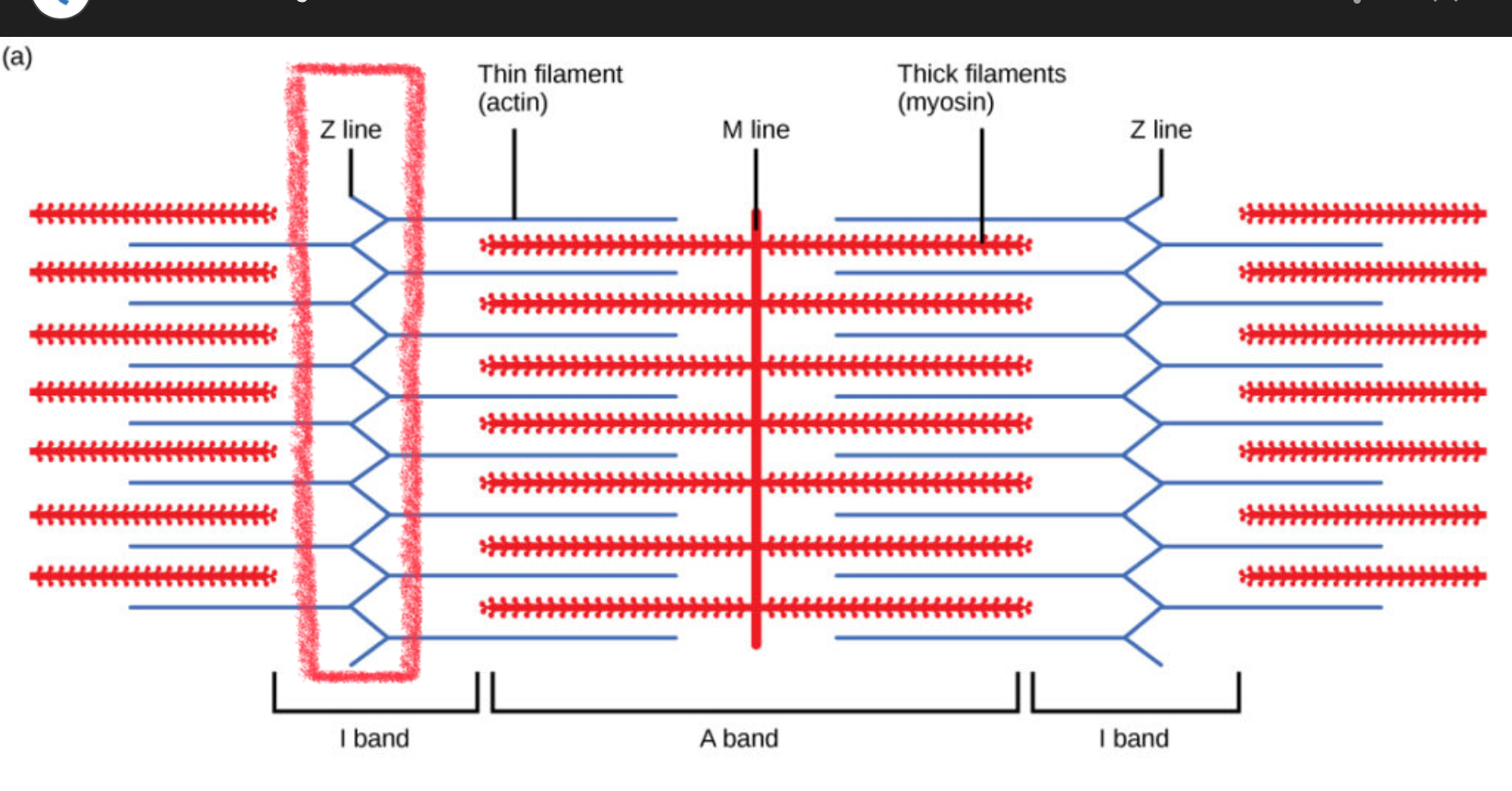
Z Line
Forms a line (boundary) for the sarcomere
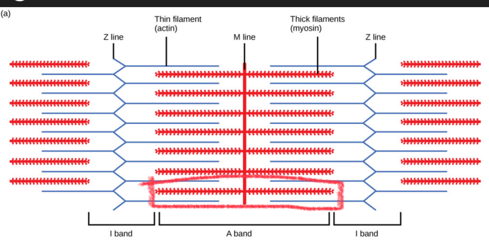
A Bands
The dark stripes in a muscle fiber where thick filaments (myosin) are found. They stay the same length during muscle contraction
H Zone
Center of the A band where only thick filaments (myosin) are present. It gets smaller when the muscle contracts.
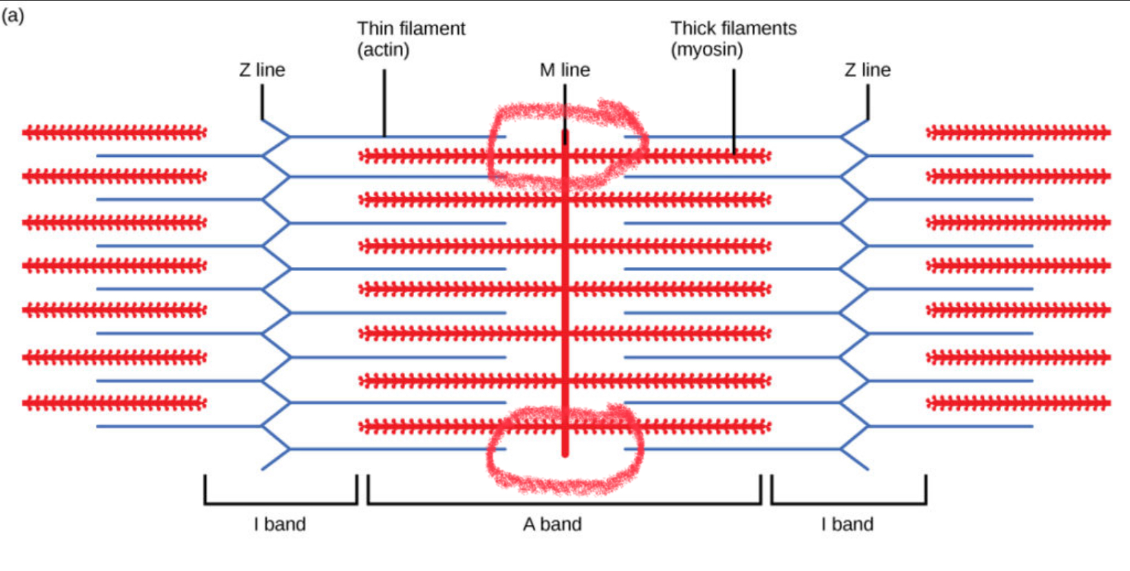
M Line
The middle of the sarcomere within the A band. It holds the thick filaments (myosin) together and helps keep them aligned during contraction.
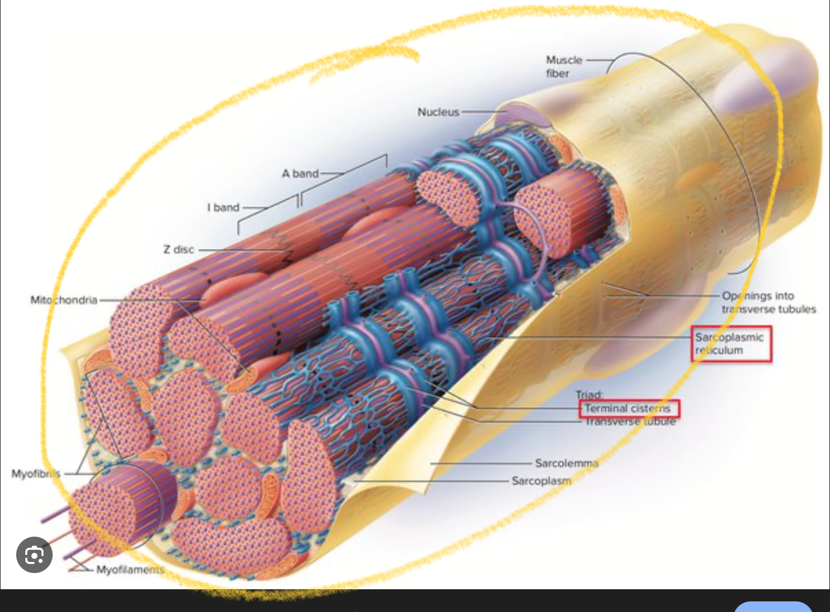
Sarcoplasmic Reticulum
The sarcoplasmic reticulum is a specialized network of tubes in a muscle fiber that stores and releases calcium ions (Ca²⁺), which are essential for muscle contraction
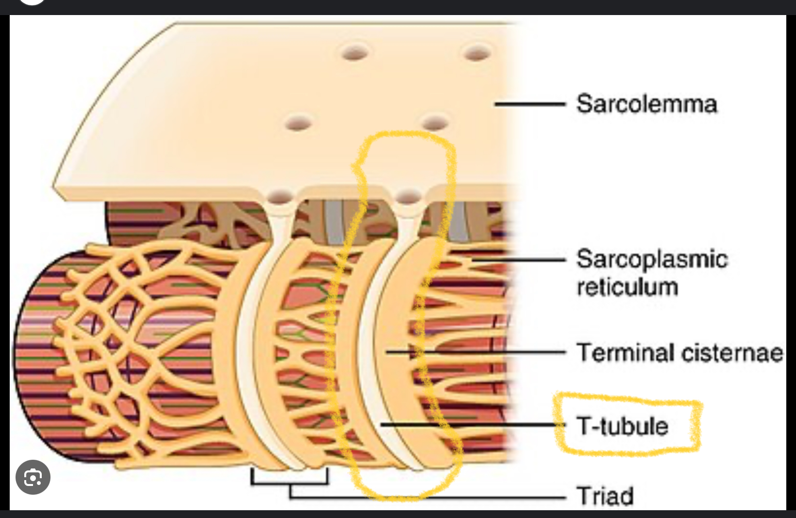
Transverse Tubules (T-Tubules)
Tiny tubes in a muscle fiber that carry electrical signals (action potentials) deep into the muscle so all parts of the fiber can contract at the same time
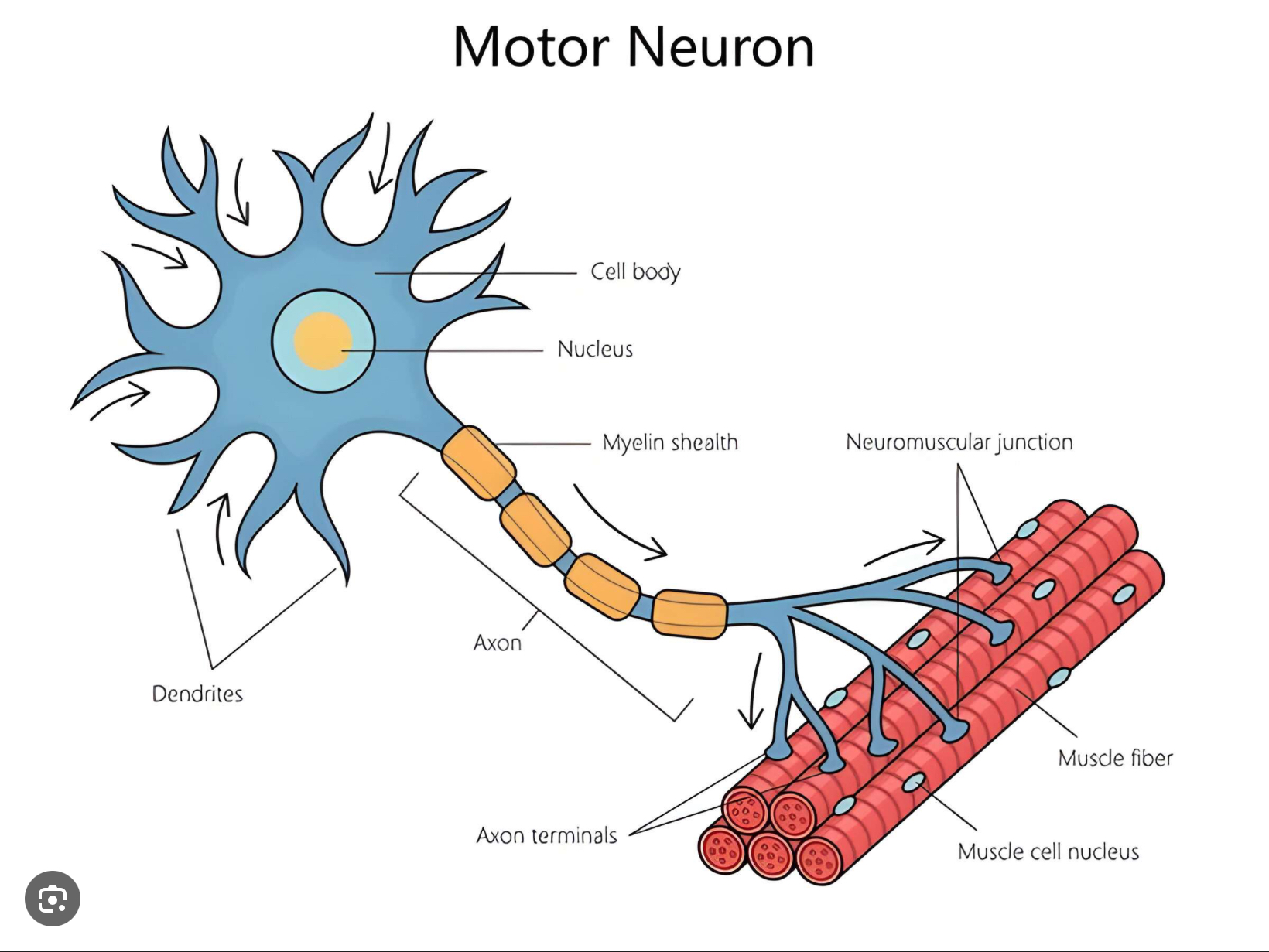
What are the 5 parts of the Neuromuscular Junction?
Motor Neurons
Synapse
Neurotransmiter
Neuromuscular Junction
Synaptic Cleft
Motor Neuron
Neurons that control effectors (such as muscles)
Synapse
The functional connection between a neuron and another cell
Neurotransmitter
The chemical neurons released to communicated with the cells they control
Neuromuscular Junction
The synapse between a motor neuron and the muscle fiber that it controls
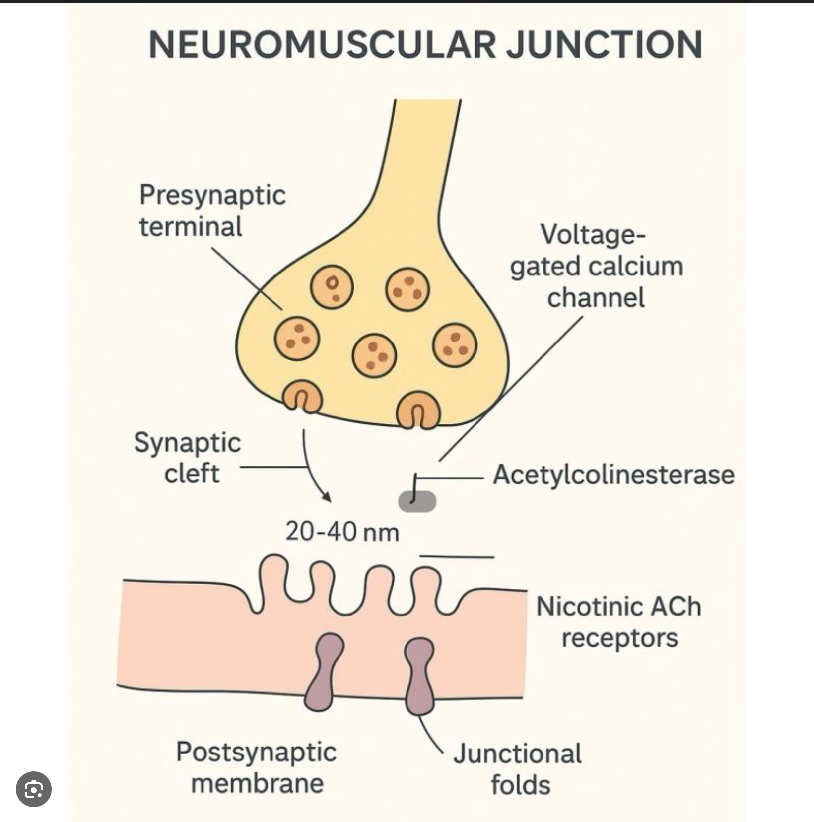
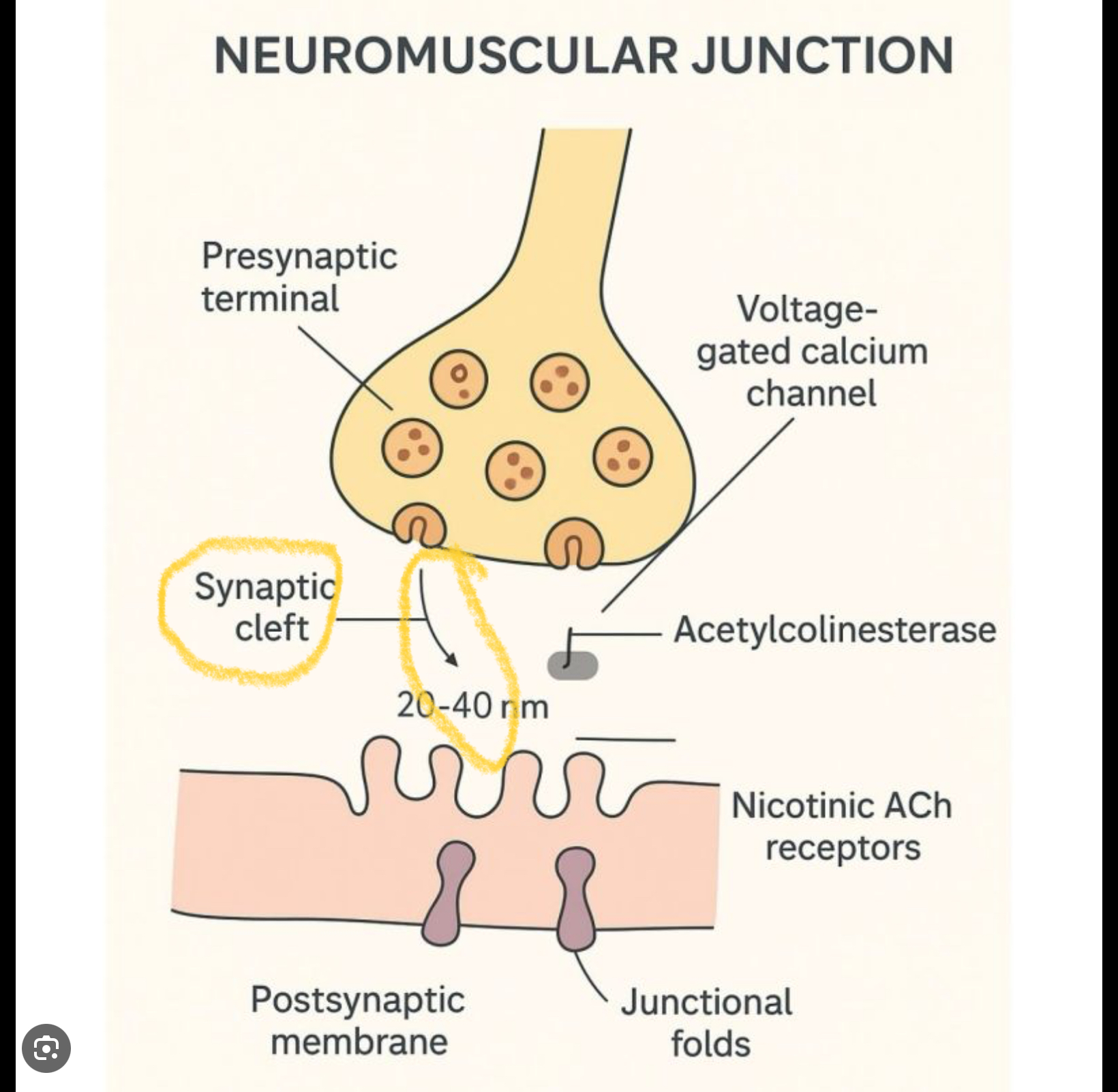
Synaptic Cleft
A small gap that separates the membrane of the neuron and the membrane of the muscle fiber
What are the two types of myofilaments?
Thick
Thin
Describe the thick filaments (type of myofilament)
Protein myosin (the most abundant)
Describe the thin filaments (type of myofilament)
Made of actin, plus regulatory proteins troponin and tropomyosin
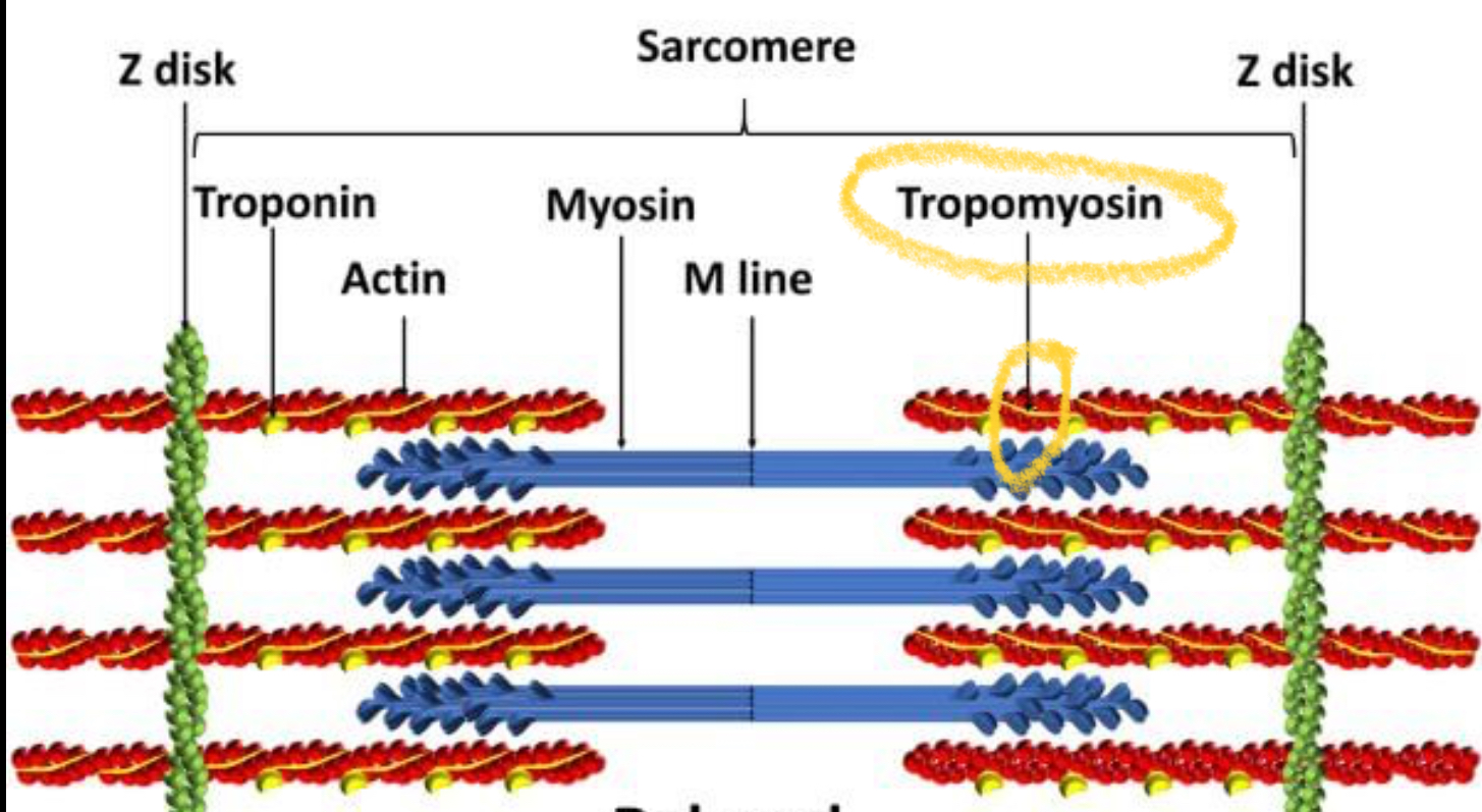
Tropomyosin
A protein on the thin filament (actin) that blocks myosin from binding to actin when the muscle is relaxed
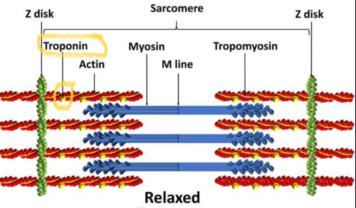
Troponin
Binds to tropomyosin on actin and calcium
What do troponin and tropomyosin control?
Interactions between myosin and actin
Sarcomere Units (2)
Contractile units of muscle
Structural units of myofibrils
What do sarcomeres form?
Patterns in myofibrils = Muscle striations
Zone of overlap in sarcomeres
The area where thick filaments (myosin) and thin filaments (actin) lie on top of each other
What causes muscle contraction?
Interactions of thick and thin filaments
What determines the interactions that cause muscle contractions?
Structures of protein muscles
Sliding Filament Theory
Muscles contract because thin filaments (actin) slide past thick filaments (myosin)
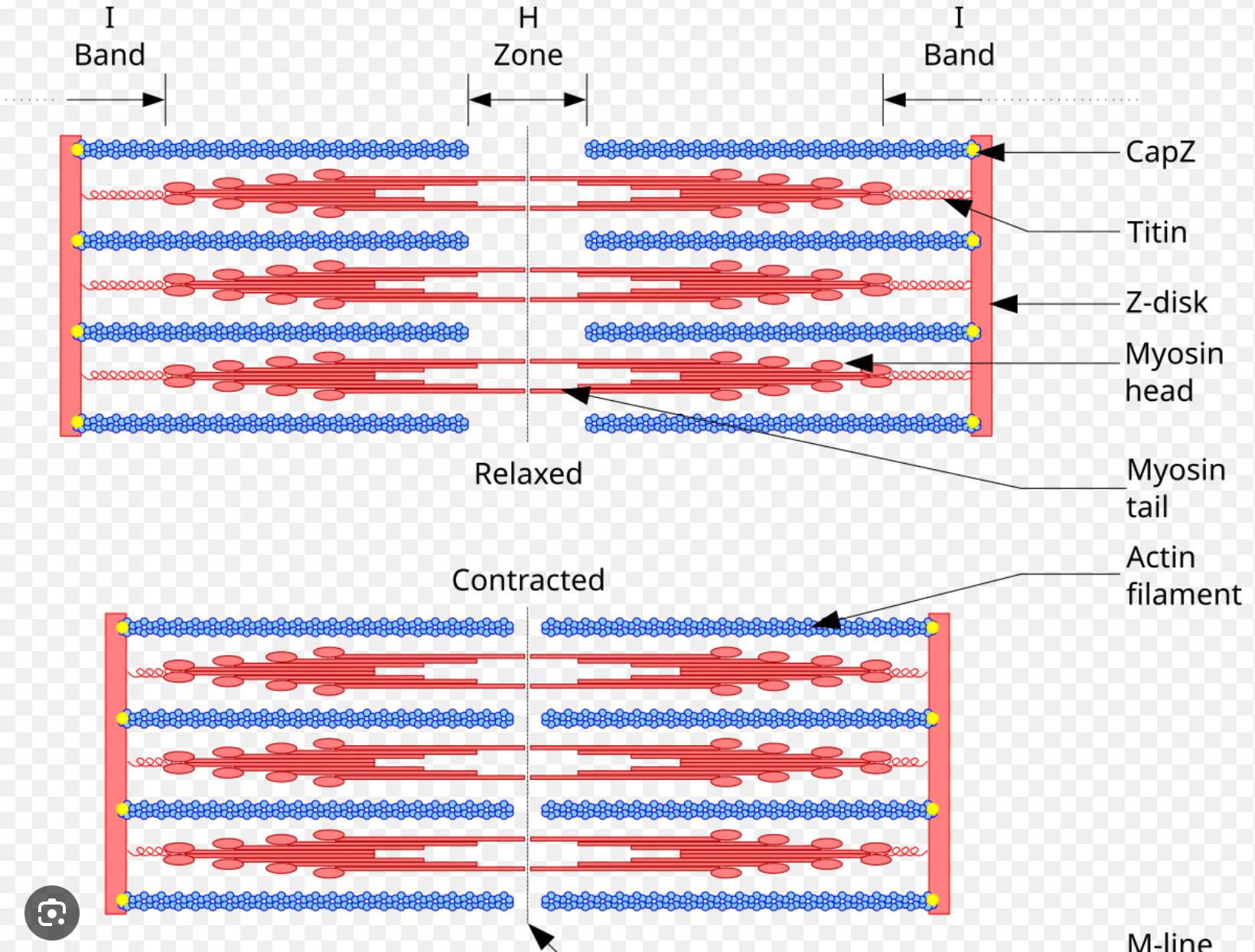
What happens to the zones and lines of the sarcomere in the Sliding Filament Theory?
A band: same
I band: smaller
H band: smaller
Zone of overlap: bigger
Z lines: moves towards m line
M line: stays put
What are the steps to induce a skeletal muscle to contract?
A motor neuron sends an electrical signal to the muscle.
The signal reaches the neuromuscular junction (where nerve meets muscle).
The nerve releases acetylcholine (ACh), a chemical messenger.
ACh makes the muscle fiber create its own electrical impulse.
This causes the muscle cell to release calcium.
Calcium triggers the sliding filament mechanism → the muscle contracts.
Step 1 of the Contraction Cycle
Cross-bridge formation
Myosin head attaches to actin (when calcium has moved tropomyosin out of the way).
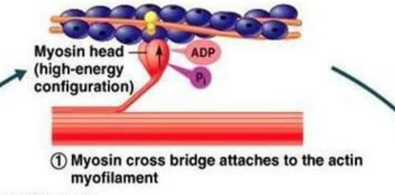
Step 2 of the Contraction Cycle
Power stroke
The myosin head pulls the actin filament inward → this uses energy from ATP.
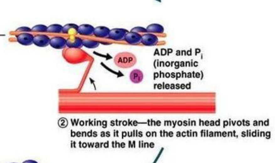
Step 3 of the Contraction Cycle
Cross-bridge detachment
A new ATP binds to myosin → myosin lets go of actin.
Step 4 of the Contraction Cycle
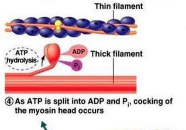
Myosin reactivation (re-cocking)
ATP is split (ATP → ADP + P), giving energy to reset the myosin head to its “ready” position.
Step 5 of the Contraction Cycle
Repeat as long as calcium + ATP are present
Myosin keeps grabbing, pulling, releasing, and resetting → muscle stays contracted
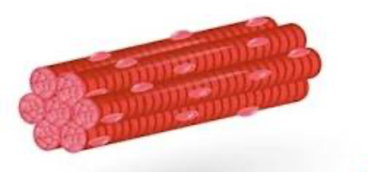
Skeletal Muscle Description
Long
Thin
Striated
Multi-Nucleated
What proteins cause the striations of the skeletal muscle
Myosin
Actin
Troponin
Tropomyosin
Is Skeletal Muscle voluntary or involuntary?
voluntary or involuntary
Skeletal Muscle Location
Attached to bones by tendons for movement
Skeletal Muscle Contraction Type
Sustained and controlled
Skeletal Muscle stimulation of contraction
Released calcium binds troponin, which causes tropomyosine to shift to expose actin site that allows myosine head to attach. forms cross-bridge (with ATP available) to perform a power stroke
What is the smallest functional unit of contraction?
Sarcomere
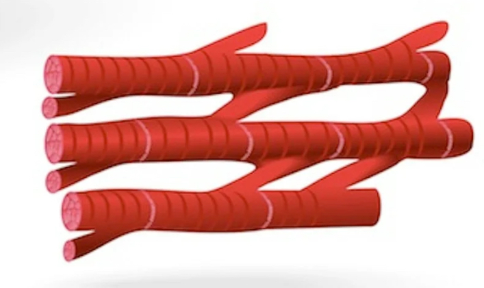
Cardiac muscle description
striated
1 nuclei
branched
intercalated disks
located only in heart
Cardiac muscle voluntary or involuntary?
Involuntary
Cardiac muscle contraction type
Rythmic Contractions = All or Nothing
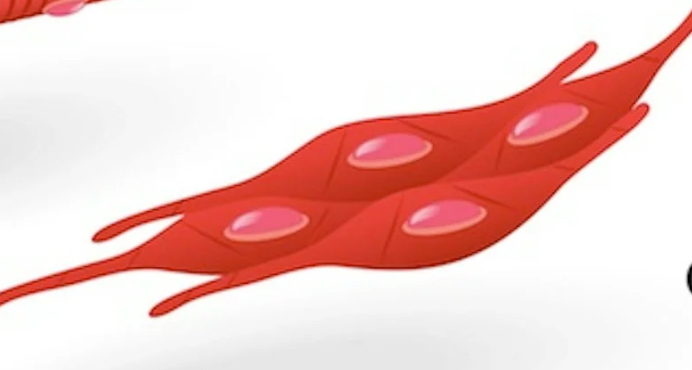
Smooth Muscle description
lacks striations
1 nucleus
thick/thin filaments arranged diagnolly
intermediate filaments
What are the 2 types of smooth muscle?
Visceral and Multi-Unit
Where is visceral smooth muscle found?
Hollow Organs
Visceral Smooth Muscle contraction type
Slow & Sustained
muscle fibers work together AS A GROUP
Where is multi-unit smooth muscle found?
Iris of eyes
Blood vessels
Multi-Unit smooth muscle contraction type?
Rapid
muscle fibers work INDEPENDANTLY
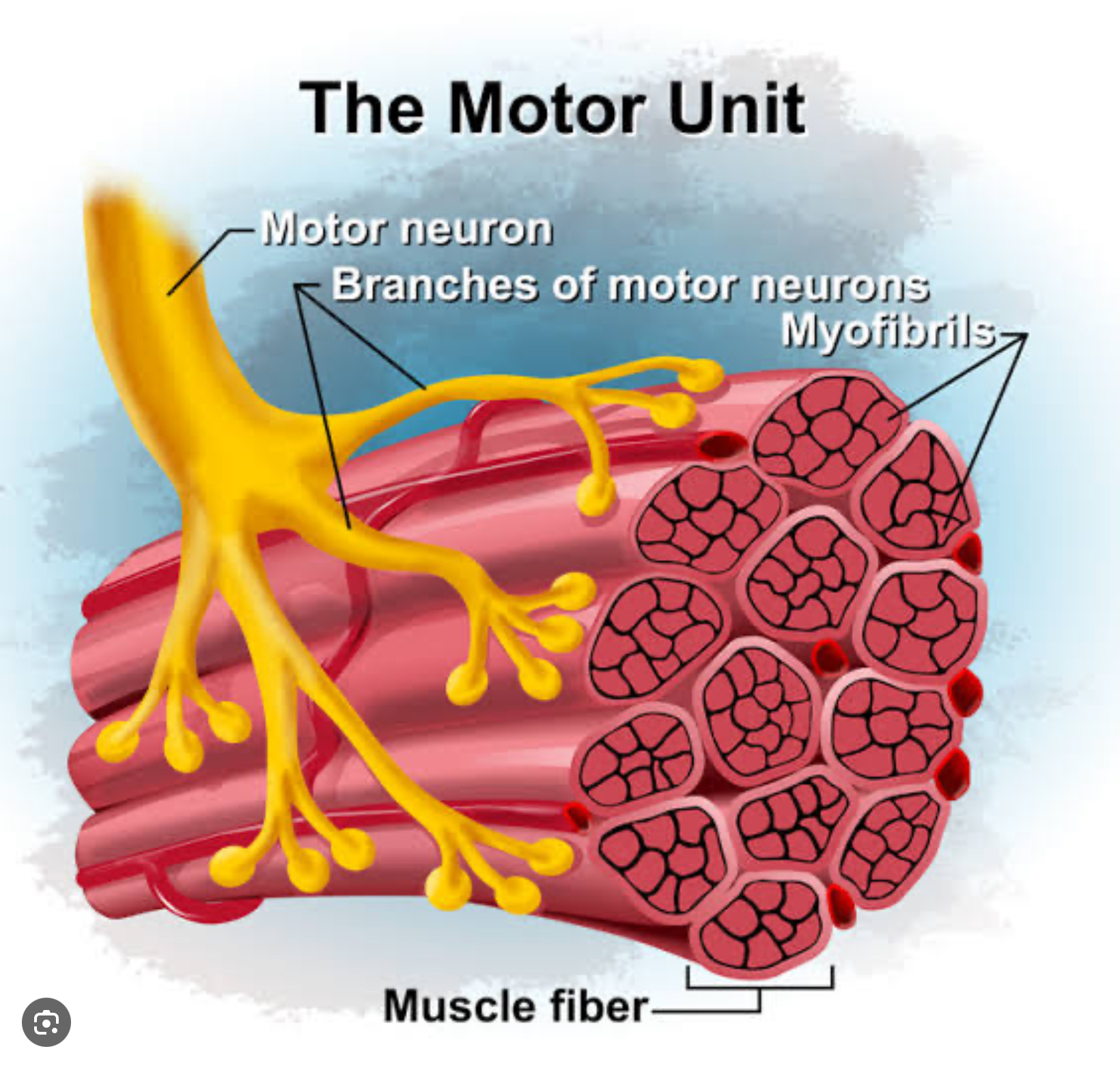
Motor Unit Function
Motor neuron tellls muscle fibers to innervate (move)
Smooth muscle location example
Digestive system
smooth muscle example function
Peristalsis: Esophagus moves food down
smooth muscle contraction type
slow & sustained
What stimulates smooth muscle contractions
A lack of troponin, so calcium binds to the protein calmodulin to activate contracts
Smooth Muscle contraction neurotransmitters (2)
Norepinepherine
Acetycholine
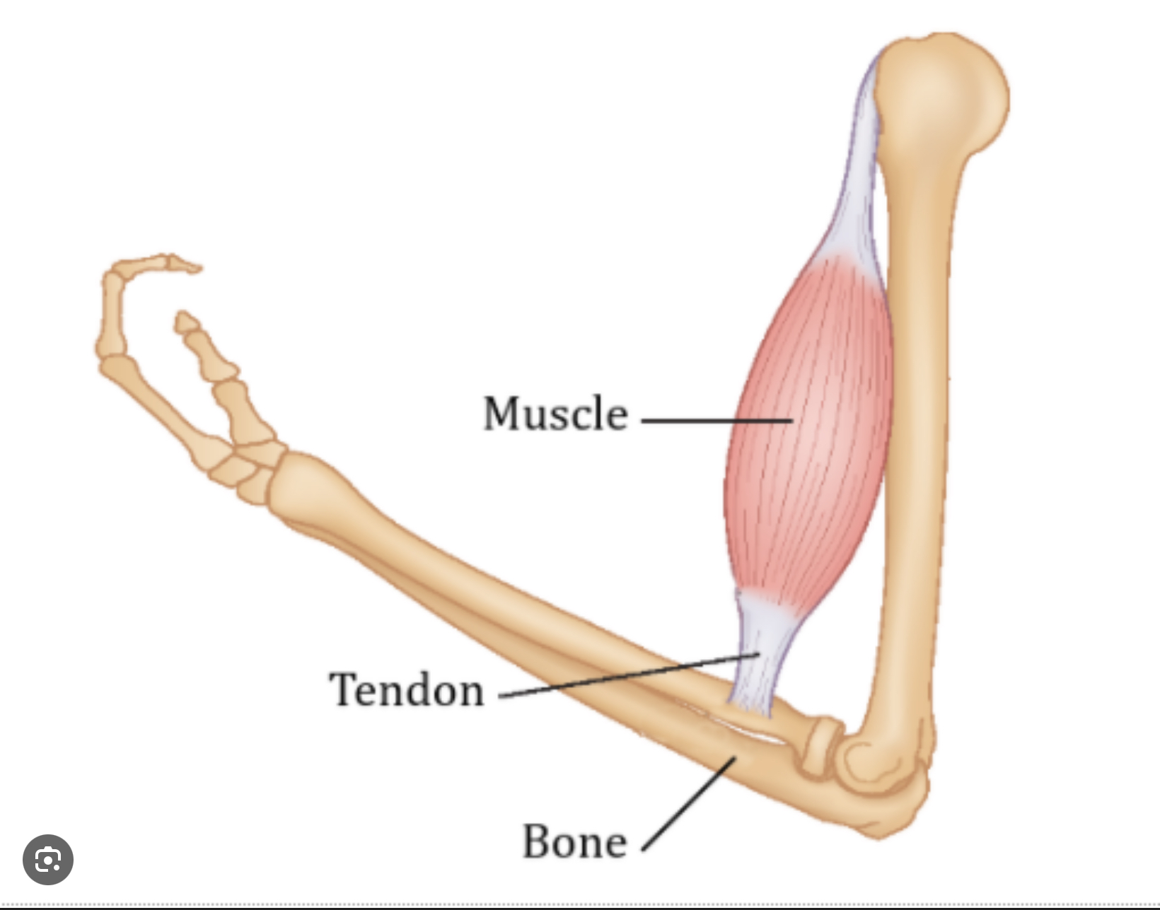
How do bones connect to muscle
By tendons
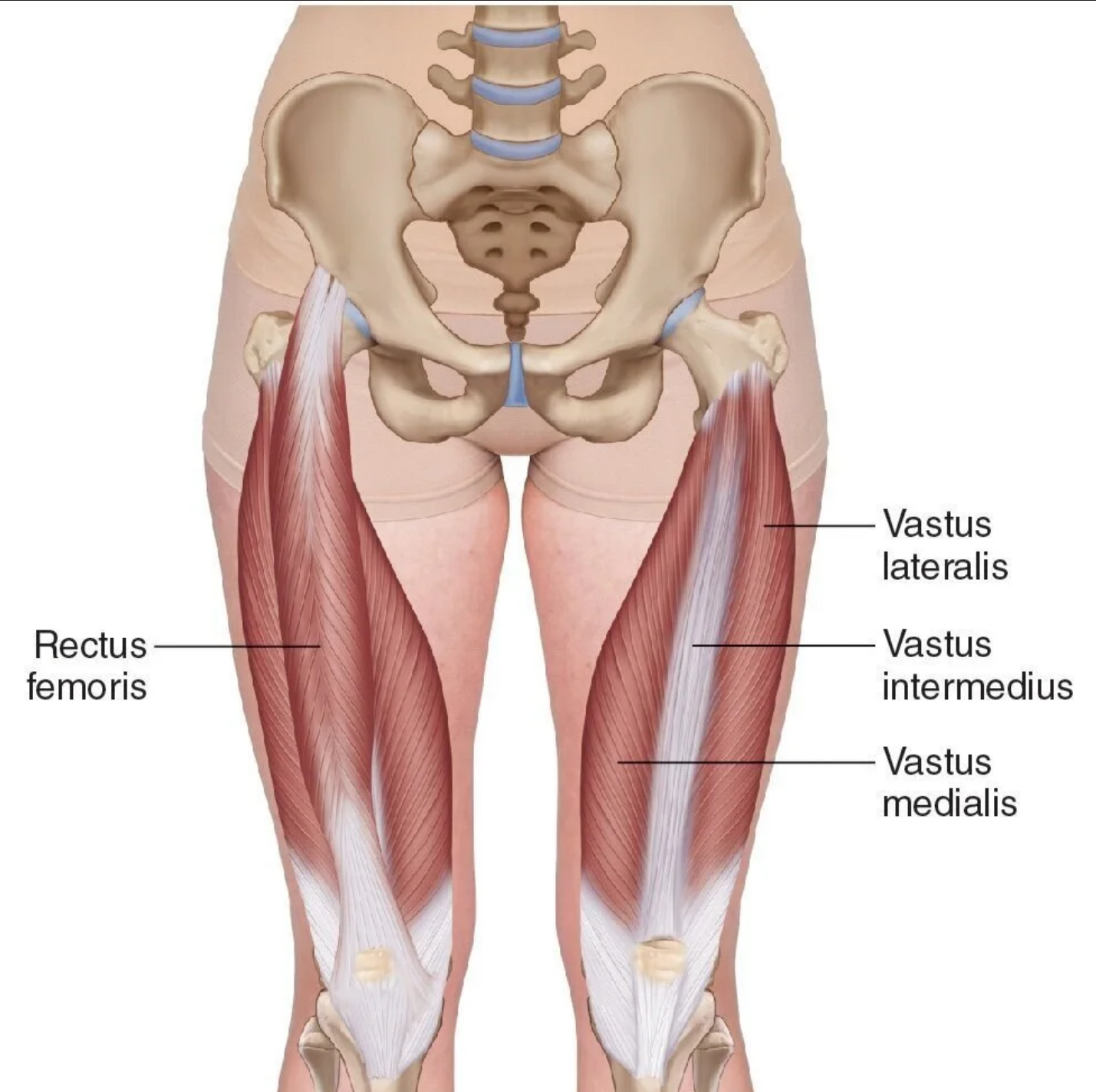
Quadriceps (Front upper leg) muscles (4)
Rectus Femoris
Vastus Lateralis
Vastus Medialis
Vastus Intermedius
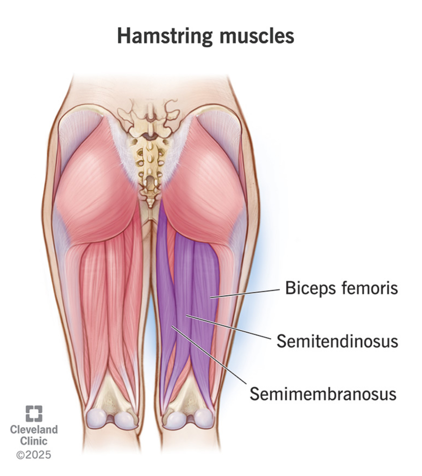
Hamstring (back upper leg) muscles (3)
Biceps Femoris
Semitendinosus
Semimembraneous
Antagonist example
Triceps brachii opposing the biceps during arm bending
Agonist example
Biceps Brachii during arm bending
What muscles are involuntary?
Smooth & Cardiac
What is the most abundant muscle protein?
Myosin
When does the main force of a contraction occur?
During the sliding of myosin along actin, which causes the muscle fibers to shorten
What IS a motor unit?
A group made up of a motor neuron and all the muscle fibers it controls
What are the I bands made of?
Actin filaments
What does the H zone contain?
Only myosin filaments
During contraction of sarcomere, what do calcium ions bond with?
Troponin
What is the MAIN neurotransmitter for muscle contraction?
ACh