BIO 201 Lecture Exam 2
 Call Kai
 Learn
Learn Practice Test
 Spaced Repetition
 Match
Match Flashcards
 Knowt Play
1/40

Earn XP
Description and Tags
Lymphatic, Immune, & Respiratory Systems
Name | Mastery | Learn | Test | Matching | Spaced | Call with Kai | Chat |
|---|
No analytics yet
Send a link to your students to track their progress
41 Terms
Immunity
set of diverse processes that protect the body from both cellular injury and disease-causing cells and molecules (pathogens)
Pathogen
anything that causes disease in cells
Lymphatic & Immune Systems…
…Work together
Cellular (Living) Pathogens
Parasites (ex. tapeworm)
Protozoa (ex. malaria)
Fungi (ex. Athlete’s foot)
Prokaryote (Bacteria; ex. leprosy)
Acellular (Non-Living) Pathogens
VIirus (ex. HIV)
Prion (ex. CJD)
Immune System
Function
Works to defend the body against internal and external threats
Characteristics
Contains no organs or tissues
Consists of cells and proteins located in the blood and tissues of other systems
Including the lymphatic system
Includes formed elements known as leukocytes, or white blood cells (WBCs), and immune proteins located in the plasma
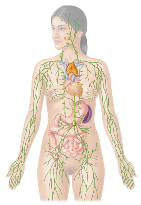
Lymphatic System
Functions
Group of organs and tissues that work with the immune system
Participate in several functions
1. Regulate the volume of interstitial fluid (body tissue fluid)
2. Absorption of dietary fats
3. Immune Functions
Two Main Components:
Lymphatic Vessels
System of blind-ended tubes
Lymphatic Tissue and Organs
Clusters of lymphoid follicles
Ex. Mucosa-associated lymphatic tissue (MALT)
Tonsils, Peyer’s patches, and appendix
Lymph nodes, spleen, and thymus
Regulating the volume of interstitial fluid
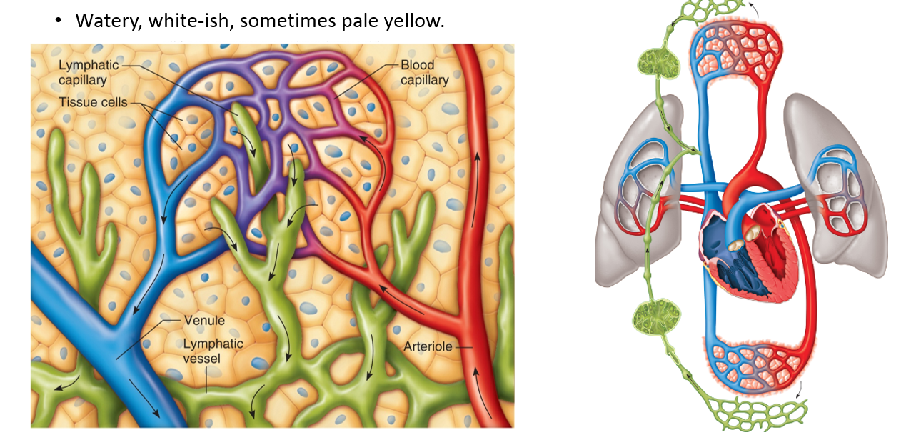
One of the main functions of the lymphatic system
Net filtration pressure (NFP) in blood capillaries favors filtration over reabsorption
Overall: more water exits capillaries and enters body tissues
Results in an overall loss of water from blood plasma to interstitial fluid
About 2-4 liters per day
Lymphatic vessels pick up fluid and return to circulation
Pick up excess fluid in extracellular space, transport it through the body, and deliver it back to the cardiovascular system
Factors of Fluid Loss
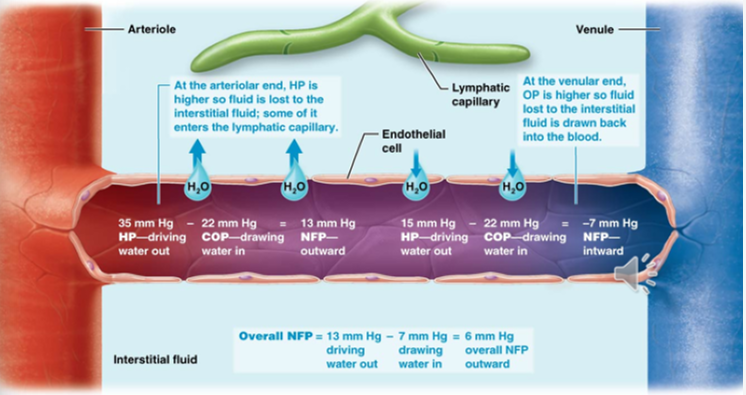
Colloid osmotic pressure pulls water into blood vessels
Blood is rich in protein (especially albumin)
Hydrostatic pressure is great in the arterioles —> pushes water out
Pressure is reduced in the venule, so there is some inward flow
Net Effect is Fluid Loss
Lymph
Fluid that enters the lymphatic vessels from the extracellular space
Similar in composition to interstitial fluid
Watery, white-ish, sometimes pale yellow
Sometimes contains cell debris, pathogens, or cancer cells
Absorption of Dietary Fats
One of the main functions of the lymphatic system
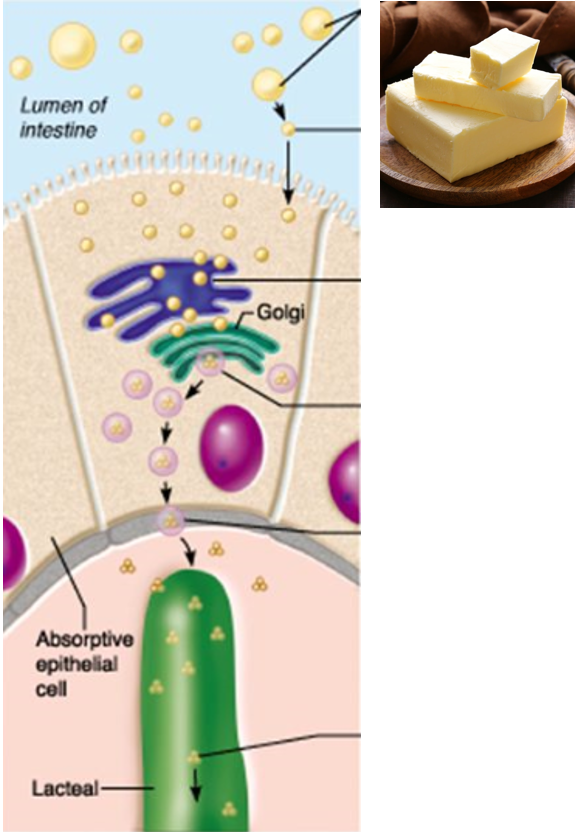
Break down products of fats in the diet that are too large to pass between the endothelial cells of blood capillaries
Dietary fats instead enter small lymphatic vessels in the small intestine
Travel through lymphatic vessels and are delivered to the blood with lymph
Immune Functions of the Lymphatic System
The lymphatic system plays an important role in protecting the body from infection.
Lymphoid organs filter pathogens from lymph and blood
House several types of leukocytes
Plays a role in their maturation
The lymphatic system helps to create a memory of specific pathogens that the body has encountered before
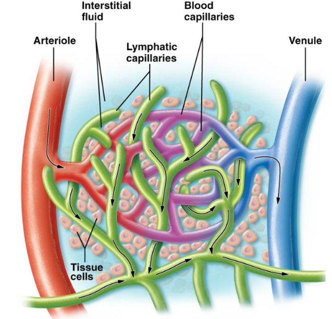
Lymphatic Capillaries
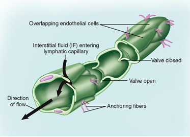
Lymphatic vessels begin in tissues with tiny capillaries
Form a web-like network that surrounds blood capillary beds
Differ in structure & function from blood capillaries
Blind-Ended
Lymphatic vasculature is a one-way system that only moves lymph away from other tissues
Let things leak into them but not out
Valve-like cells of lymphatic capillaries
Allow cells such as macrophages and other immune cells to enter lymph
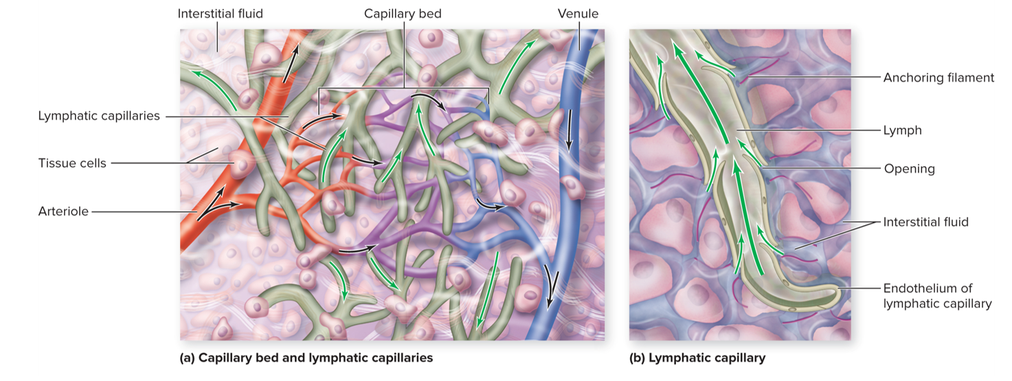
Cells of Lymphatic Capillary Walls
These cells are able to open and close (valve-like)
Increase in Interstitial Fluid Pressure
Forces lymphatic endothelial cells apart
Allows large volumes of fluid to enter, but not exit, lymphatic capillaries
Decrease in Interstitial Fluid Pressure
Endothelial cells flap shut
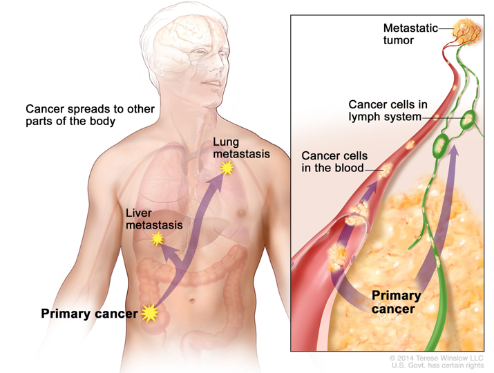
Metastasis Clinical Connection
Wandering cancerous cells establish secondary tumors
Develop in other locations in the body
Cancerous cells break free from the primary tumor
Transported in the lymph
End up in the Brain, Bones, Liver, and Lungs!
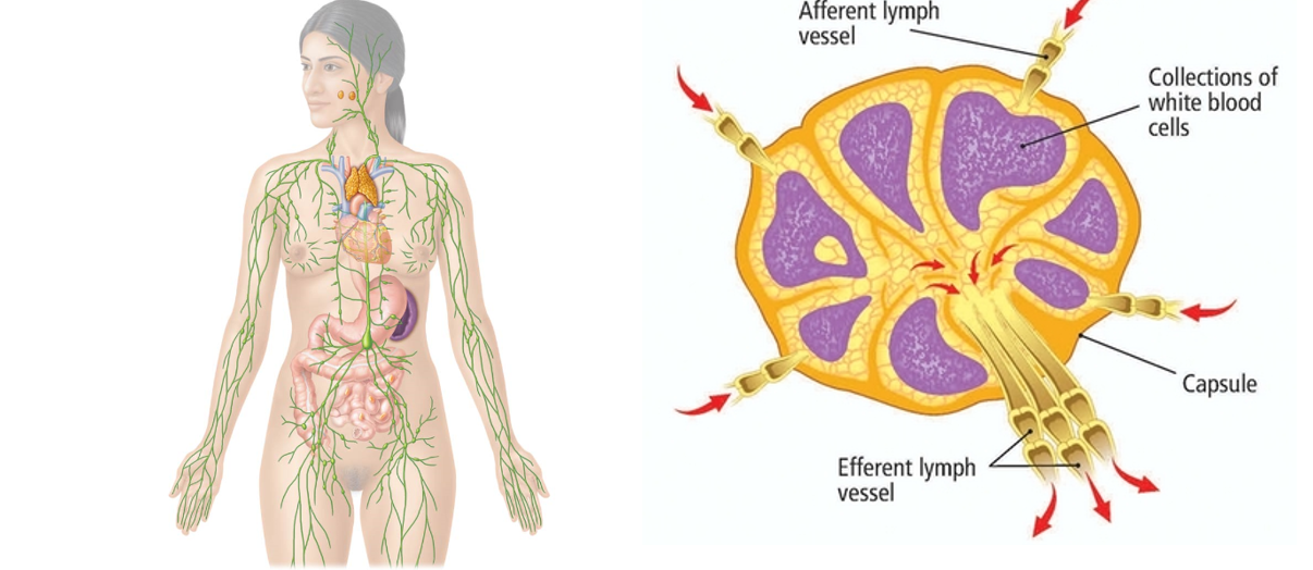
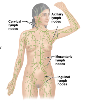
Lymph Nodes
Small, vaguely bean-shaped clusters of lymphatic tissue located along lymphatic vessels throughout the body
Limit the spread of pathogens through the body by acting as filters.
Trap pathogens and prevent them from traveling elsewhere
Specific clusters include:
Axillary Lymph nodes (in axillae, under armpit)
Cervical Lymph nodes (in neck)
Inguinal Lymph nodes (in the groin)
Mesenteric Lymph nodes (in abdominal cavity)
Normally, they are small and barely distinguishable
Become swollen when inflamed
Lymphatic Vessels
Fed by lymphatic capillaries
Located adjacent to arteries and veins
Have valves to prevent pooling and backflow of lymph
Lymphatic system lacks a pump; moves lymph using:
Skeletal muscles and respiratory pumps
Pulsatile movement of blood in nearby arteries
Rhythmic contraction of smooth muscle in larger lymph vessel walls
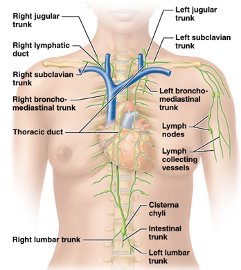
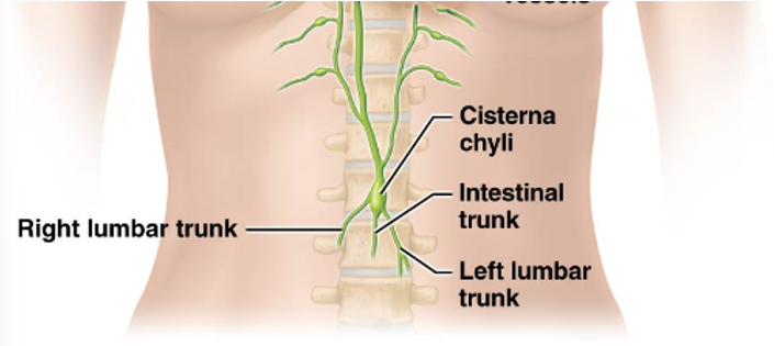
Lymphatic Trunks
Interstitial fluid drained into these
Fed by lymphatic vessels
Located in many different locations of the body
Jugular Trunks
Subclavian Trunks
Bronchomediastinal Trunks
Intestinal Trunks
Lumbar Trunks
Jugular Trunks
Left and right lymphatic trunks that run along the jugular veins
Drain lymph from the head and neck
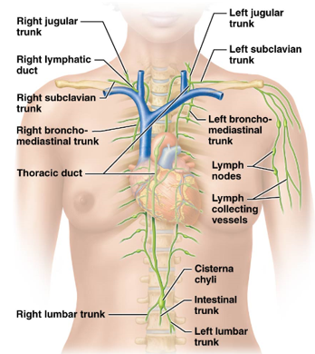
Subclavian Trunks
Left and right lymphatic trunks that run along the subclavian arteries & veins
Drain lymph from the upper limbs, breasts, and superficial thoracic wall
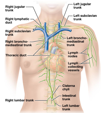
Bronchomediastinal Trunks
Left and right lymphatic trunks that run along the area of the lungs and mediastinum
Drain lymph from deep thoracic structures
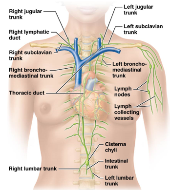
Intestinal Trunk
Lymphatic trunk located around the intestines
Drain lymph from most abdominal structures
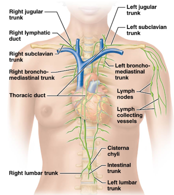
Lumbar Trunks
Left and right lymphatic trunks located in the lumbar region
Drain lymph from the lower limbs, abdominopelvic wall, and pelvic organs
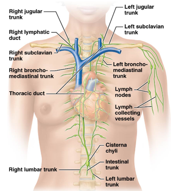
Cisterna Chyli
large swollen vessel in which the intestinal trunk and lumbar trunks drain into
Drains into the thoracic lymphatic duct
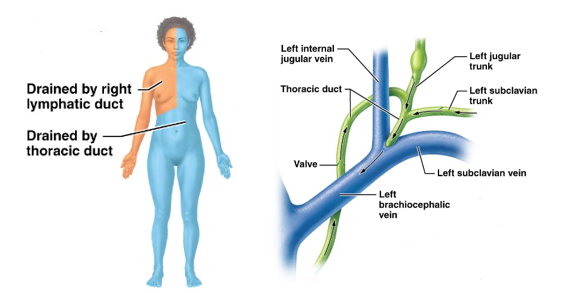
Thoracic Duct
drains lymph from the cisterna chyli and trunks from the left side of the body
Drains all of the lower body and left side of upper body
Largest lymphatic duct
Runs along anterior vertebral column from diaphragm to junction of the left subclavian and left jugular veins
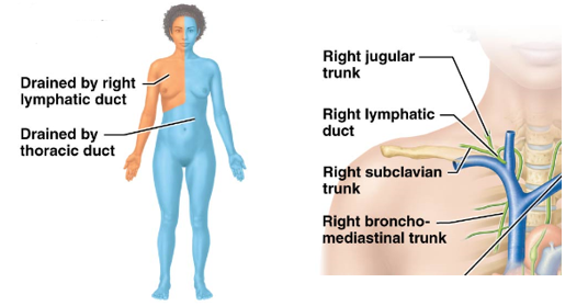
Right Lymphatic Duct
remaining lymphatic trunks drain into this tiny duct located near the right clavicle
Right Jugular Trunk, Right Subclavian Duct, Right Bronchomediatinal Trunk
Drains into junction of right internal jugular and right subclavian veins
Drains upper right side of body
Lymphedema
Accumulation of interstitial fluid due to interference with lymphatic drainage
Causes swelling and pain in the affected area
Usually the legs due to gravity
May interfere with wound healing and contribute to infection
Mostly caused by blockage of lymph vessels
Ex. trauma or infection of lymph vessels or tumors
Controlled with compression garments, exercise, and massage
Extreme cases known as elephantiasis
Caused by infection of parasitic worms
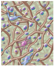
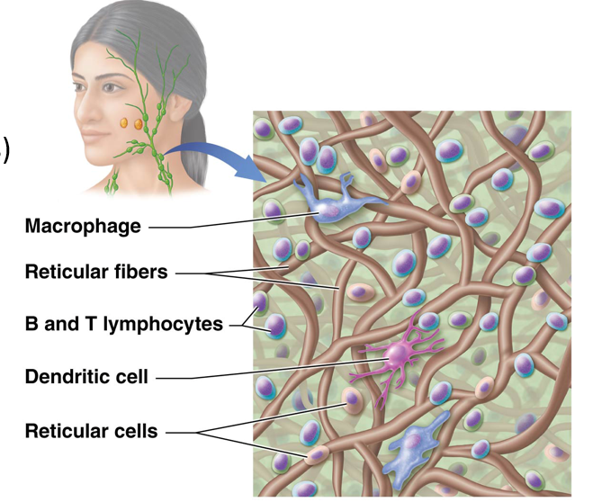
Reticular Tissue (Lymphoid Tissue)
Loose connective tissue that is the predominant tissue type of the lymphatic system
Contains specialized cells and thin reticular fibers
Interweave to form “nets” that trap disease-causing pathogens
Found in lymphoid organs and also as independent clusters
Cells of Most Lymphoid Organs & Tissues
Leukocytes:
Macrophages (mature monocytes)
B and T lymphocytes
Dendritic Cells
Reticular Cells
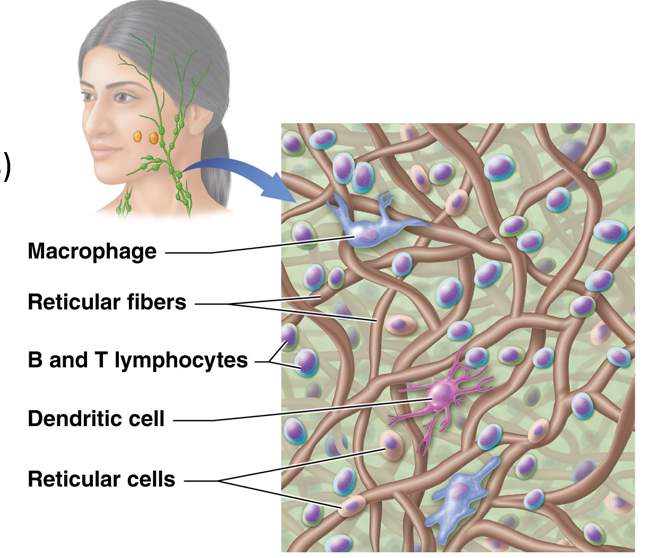
Dendritic Cells
Immune cells with spiny processes resembling dendrites of neurons.
Originate in the bone marrow or connective tissue
Responsible for moving fluid around
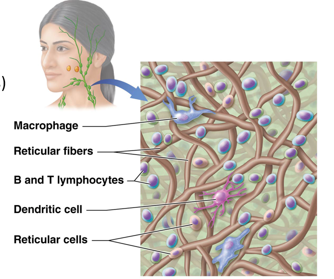
Reticular Cells
Abundant cells in organs such as the spleen and lymph nodes
Modified fibroblasts (fiber-forming cells)
Produce reticular fibers
Help anchor lymph tissue in place
Lymphoid Nodules
Clusters of lymphoid cells with some extracellular matrix
Not entirely surrounded by a connective tissue capsule
Scattered around the body
Found in every body organ
Help defend against infection
In some areas, group together to form larger structures
Example: MALT
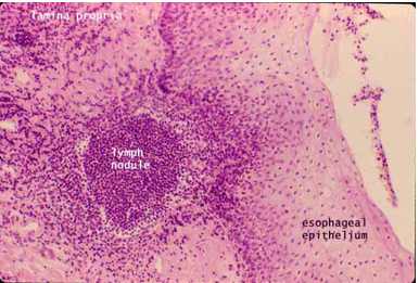
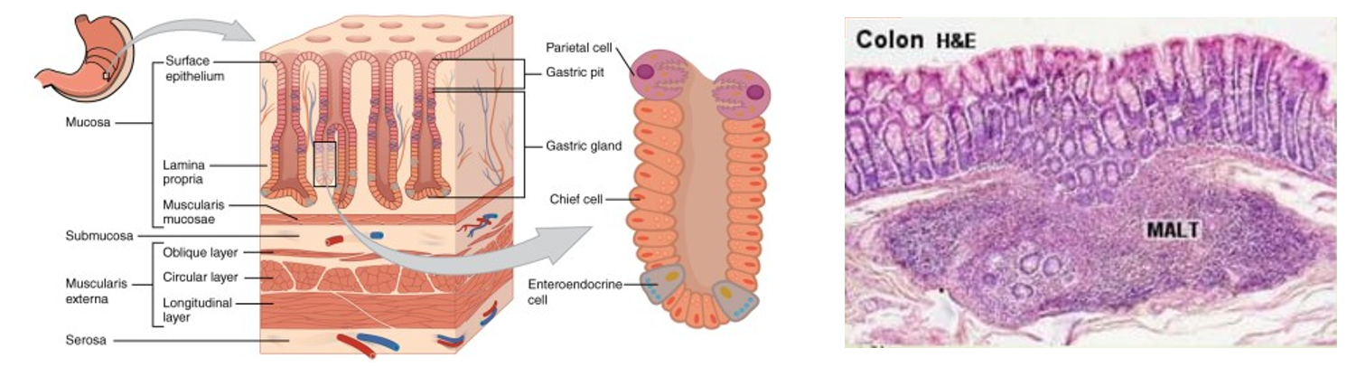
Mucosa-Associated Lymphatic Tissue (MALT)
Located in areas that are regularly exposed to large amounts of pathogens
Gastrointestinal Tracts
Respiratory Tracts
Genitals
Urinary Tracts
Oral & Nasal Cavities
Found in the lamina propria of the mucosa
Help defend against foreign substances
Chief cells can help
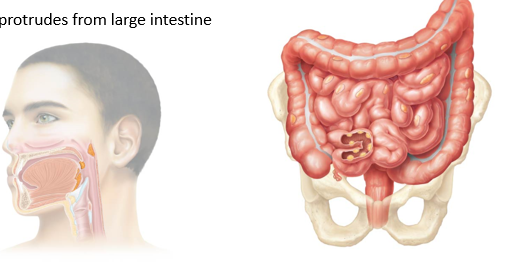
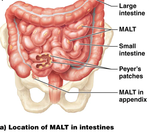
Specialized MALT Found in Three Locations of the GI Tract
Tonsils - located around oral and nasal cavities
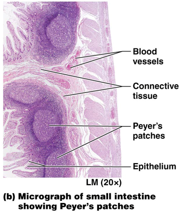
Peyer’s Patches (aggregated lymphoid nodules) - located in the last portion of the small intestine (aka ileum)
Appendix - protrudes from large intestines
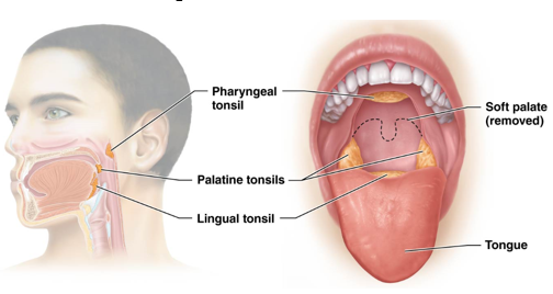
Three Main Tonsils
Pharyngeal Tonsil (adenoid) - located in posterior nasal cavity (nasopharynx)
Palatine Tonsils - in posterolateral oral cavity
Lingual Tonsil - at the base of the tongue
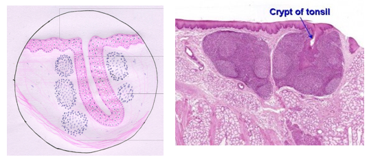
Tonsillar Crypts
Epithelium lining tonsils indents deeply in several locations
Trap bacteria and debris
Tonsil location puts them into contact with a large number of potential pathogens
Commonly become inflamed when they trap too much
Tonsilitis
Peyer’s Patches
defend against any bacteria that have escaped from the large intestine
Appendix
Part of the cecum defends the body from bacteria in the large intestine, specifically those that could be pathogenic
In plant-eating animals
Houses bacteria that can be useful for digesting tough plant fibers
Lymph Node Components
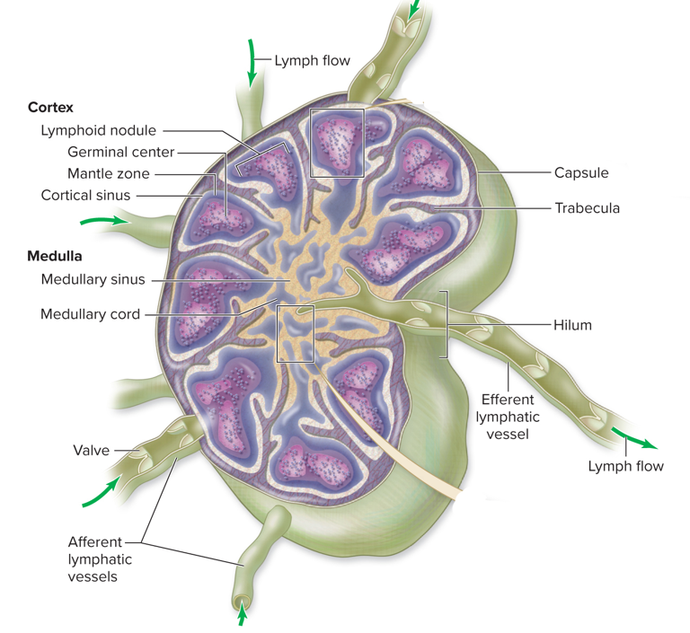
Afferent Lymphatic Vessels
Bring lymph into node
Efferent Vessels
Drains lymph node
Located at the hilum (involved portion of node)
Dense connective tissue capsule encloses node
Trabeculae of capsule subdivide node into compartments
Outer cortex, inner medulla
Cortex of Lymph Nodes
Contains lymphoid nodules
Reticular fibers support a germinal center
Houses proliferating B lymphocytes and some macrophages
Surrounding mantle zone contains T lymphocytes, macrophages, and dendritic cells
Cortical sinuses are tiny open channels limited by macrophages ppo33333333333333333333333333333333333333333333333333333333333333333333333333333333333333333333333333333333333333333333333333333333333333333333333333333333333333333333333333333333333333333333333333333333333333333333333333333333333333333333333333333333333333333333333333333333333333333333333333333333333333333333333333333333333333333333333333333333333333333333333333333333333333333333333333333333333333333333333333333333333333333333333333333333333333333333333333333333333333333333333333333333333333333333333333333333333333333333333333333333333333333333333333333333333333333333333333333333333333333333333333333333333333333333333333333333333333333333333333333333333333333333333333333333333333333333333333333333333333333333333333333333333333333333333333333333333333333333333333333333333333333333333333333333333333333333333333333333333333333333333333333333333