summer studying lecutre 13 a/b/c/d
 Call Kai
 Learn
Learn Practice Test
 Spaced Repetition
 Match
Match Flashcards
 Knowt Play
1/127

There's no tags or description
Looks like no tags are added yet.
Name | Mastery | Learn | Test | Matching | Spaced | Call with Kai | Chat |
|---|
No analytics yet
Send a link to your students to track their progress
128 Terms
What are the general characteristics of the cardiovascular system?
Consists of heart and blood vessels
What is the heart?
Hollow, cone-shaped muscular pump; generates force to transport respiratory gases, nutrients, and wastes through body
What are blood vessels?
Transport blood throughout body
consists of arteries, veins, capillaries
What are arteries?
Transport blood away from ventricles of heart
What are veins?
Receive blood from venules and carry it back to atria of heart
What are capillaries?
Transport blood between arteries and veins, perform nutrient, gas, and waste exchange
What is the pericardium (pericardial sac)?
Covering over heart and proximal ends of large blood vessels
consists of fibrous, parietal, visceral pericardium
consists of pericardial cavity
What is fibrous pericardium?
Tough outer layer that surrounds double-layered serous membrane
What is the parietal pericardium?
Deep to fibrous pericardium; outer layer of serous membrane
What is the visceral pericardium?
Inner layer of serous membrane; attached to surface of heart
aka epicardium
reduces friction
What is the pericardial cavity?
Space between visceral and parietal layers of serous pericardium
What are the 3 layers of the heart?
epicardium
myocardium
endocardium
What is the myocardium?
Middle layer of heart; thickest layer
composed of cardiac muscle tissue
What is the endocardium?
Innermost layer of heart; thin
forms inner lining of all heart chambers
What are the 9 steps to the path of blood flow to the heart?
right atrium receives blood from superior and inferior venae cavae and coronary sinus
blood flowers from right atrium through open tricuspid (right AV) valve into right ventricle
Pulmonary valve is pushed open when right ventricle contracts, allowing blood to enter pulmonary trunk
blood travels with right and left pulmonary arteries to alveolar capillaries in lungs, where CO2 is unloaded and O2 is loaded
blood travels within right and left pulmonary veins from lungs to left atrium
Blood in left atrium flows through open mitral (left AV) valve into left ventricle
aortic valve is pushed open when left ventricle contracts, allowing blood to enter aorta
blood flows from aorta to systemic capillaries where O2 is unloaded and CO2 is loaded
blood returns to right atrium from body tissues and myocardium (via venae cavae and coronary sinus)
What is the pulmonary circuit?
Portion of circulatory system that carries deoxygenated blood from heart to the lungs for oxygenation and returns oxygen-rich blood back to heart
What is the blood flow pathway of the pulmonary circuit?
starts in Right Atrium and goes through tricuspid valve, to the right ventricle, and to the pulmonary valve
Blood flows into pulmonary trunk, where it flows to both the left and right pulmonary arteries
The left blood supply will flow to alveolar capillaries in left lung; the right blood supply will flow to alveolar capillaries in right lung (now oxygenated)
Both blood flows will respectively go to left/right pulmonary veins
both veins will flow into left atrium
from the left atrium, blood flows to the mitral valve, into the left ventricle, to the aortic valve
Finally arrives at the aorta
What do the right and left coronary arteries do?
Supply heart with oxygenated blood
What is the systemic circuit?
Part of circulatory system that carries oxygen-rich blood from left ventricle to aorta, to all tissues, then returns oxygen-depleted blood to the right atrium via venae cavae
What is the blood flow pathway of the systemic circuit?
begins in left ventricle, where blood travels to the aortic valve, then into the aorta
From the aorta, systemic capillaries in the upper body tissues, myocardium, and lower body tissues receive blood
deoxygenated blood then flows back into the superior vena cava, coronary sinus, and inferior vena cava
all of the cava/sinuses then flow back into the right atrium, where it is then carried through the tricuspid valve, right ventricle, into the pulmonary valve
blood finally arrives at the pulmonary trunk
Where does the systemic capillaries of the upper body tissues flow?
Superior vena cava
Where does the systemic capillaries of the myocardium flow into?
Coronary sinus
Where does the systemic capillaries of the lower body tissues flow into?
inferior vena cava
What are the actions of the heart?
systole
diastole
both contribute to the cardiac cycle
What is systole?
contraction of a heart chamber
What is diastole?
relaxation of a heart chamber
What is the cardiac cycle?
atria contract (atrial systole) while the ventricles relax (ventricular diastole)
Then ventricles contract (ventricular systole) while atria relax (atrial diastole)
Then both chambers relax briefly
What is a heart transplant?
Replacement of most of a failing heart w/ a donor heart
What is an LVAD?
Left Ventricular Assist Device
mechanical half-heart, used in some cases temporarily until donor heart is available
What is an implantable replacement heart?
Titanium and plastic artificial heart, used in people that cannot have a heart transplant, and do not have long to live
What is stem cell technology?
Cardiac muscle tissue can be cultured from altered somatic cells; may allow stem cell heart patches in the future
need to match up intercalated discs for action potentials to travel
What is the cardiac conduction system?
Specialized group of cardiac muscle cells, which initiate and distribute cardiac action potentials through myocardium
What are the major components of the cardiac conduction system?
Sinoatrial Node
Internodal Atrial Muscle (atrial syncytium)
Junctional Fibers
Atrioventricular Node
Atrioventricular Bundle (of His)
Left and Right Bundle Branches
Purkinje Fibers
Ventricular syncytium
What is the Sinoatrial Node?
Pacemaker, initiates rhythmic contractions of the heart
What is the internodal atrial muscle?
Conducts impulses from SA node to atria
What are junctional fibers?
Conduct impulses from SA node to atria
What is the Atrioventricular Node?
Conducts impulses to AV bundles; delays impulse, so atria finish contracting before ventricles contract
What is the Atrioventricular Bundle (of His)?
Conduct impulses rapidly between SA node and bundle branches
What are the left and right Bundle Branches?
Split off from AV Bundle, conduct impulse to Purkinje fibers on both sides of heart
What are the Purkinje Fibers?
Large fibers that conduct impulses to ventricular myocardium; conduct impulses to apex first
whorled pattern of muscle in ventricles contract w/ twisting motion
What is an electrocardiogram (ECG, EKG)?
A recording of electrical charges that occur in myocardium during cardiac cycle
used to assess ability of heart to conduct impulses
includes P wave, QRS complex, and T wave
What is the P wave?
Atrial depolarization; occurs just prior to atrial contraction
What is the QRS complex?
3 waves→ ventricular depolarization that occurs just prior to ventricular contraction
record of atrial repolarization is hidden in large QRS complex as ventricular depolarization is much larger
What is the T wave?
Ventricular repolarization that occurs just prior to ventricular relaxation
What is the ECG Pattern?
initial impulse from SA node
then contraction in right atria into septum to left atria→ by end of P wave, electrical impulse has moved across into AV node
Signal then moves from AV node to AV bundle into bundle branches→ signal moves back to myocardium of ventricles
QRS complex then finishes when impulse has made its way through all muscle
T wave then starts and brings ventricular repolarization
What does a prolonged QRS complex indicate?
Indicates disruption of ventricle→ AV bundle damage
What does an absent P wave indicate?
SA node issue
What does an absent T wave indicate?
Issue with repolarization in ventricles
issue w/ myocardium in ventricle
What are the properties of pressure and volume changes during cardiac cycle?
Pressure in heart chambers rises and falls
pressure changes open and close the valves
What happens to pressure/volume in ventricular diastole?
ventricular pressure is lower than atrial pressure
atria and ventricles are relaxed
AV valves open, and semilunar valves are closed
about 70% of blood flows passively from atria into ventricles
What happens to pressure/volume during atrial systole and ventricular diastole?
Atria contract and ventricles are relaxed
AV valves open and semilunar valves close
Atrial systole pushes remaining 30% of blood into ventricles, causing ventricular pressure to increase
What happens to pressure/volume during ventricular systole and atrial diastole?
when ventricular pressure rises above atrial pressure, AV valves close
chordae tendineae prevents cusps of valves from bulging too far backward into atria
atria relax
blood flows into atria from venae cavae and pulmonary veins
ventricular pressure continues to increase, and opens semilunar valves
blood flows into pulmonary trunk and aorta
What regulates the cardiac cycle?
Cardiac center in medulla oblongata performs neural regulation of heart
SA node (pacemaker) normally controls heart rate→ heart rate changes occur due to factors that influence SA node
sympathetic/parasympathetic fibers modify heart rate in response to physical exercise, body temp, fight-or-flight, concentration of K+ and Ca2+
How do the parasympathetic impulses influence the heart rate?
Reach heart via vagus nerves
lower SA node rate of 100 beats/min to 60/80 beats/min
decrease heartrate (due to influence on SA and AV nodes)
How do the sympathetic impulses influence the heart?
Reach heart on accelerator nerves
increases heartrate due to influence on SA and AV nodes, atrial and ventricular myocardium
What are the baroreceptor reflexes?
Involve cardiac control center medulla oblongata
balance inhibitory/excitatory effects of parasympathetic and sympathetic fibers
contains cardio-inhibitor reflex and cardio-accelerator reflex
What are the characteristics of the baroreceptor reflex?
A rapid negative feedback loop where elevated blood pressure causes heartrate and blood pressure to decrease
a decrease in blood pressure leads to an increased heart rate→ returning blood pressure to normal levels
What are the steps to the baroreceptor reflex?
stretch receptors located in the wall of the aortic arch and carotid sinus will sense stretch caused by inc blood pressure
sensation will trigger baroreceptors to fire action potentials at higher than normal rate
Inc activity is sent via vagus and glossopharyngeal nerves to nucleus of tractus solitarius (NTS) in brainstem
NTS activates the parasympathetic system and inhibits the sympathetic system→ allows same direction to maximize blood pressure reduction
PSNS releases ACh which impacts pacemaker cells of SA node
Inhibition of SNS causes vasodilation, decreased HR and stroke volume
If blood pressure is decreased, NTS will inhibit PSNS and activate SNS
SNS will release norepinephrine, which accelerates HR and BP via SA node
What happens to blood pressure when standing up from sitting?
Decrease in blood pressure is sensed by baroreceptors as decrease in tension
baroreceptors fire at a lower than normal rate and info is transmitted to NTS
NTS then inhibits PSNS and activates SNS
SNS will then release Norepinephrine which acts on SA node to inc heart rate and blood pressure, and vasoconstriction
What parts of the SA node are impacted by norepinephrine and acetylcholine?
cardiomyocytes, smooth muscle cells of blood vessels
What are arrhythmias?
Altered heart rhythms
consists of 4 types
Tachycardia
fibrillation
bradycardia
flutter
What is fibrillation?
Uncoordinated, chaotic contraction of small areas of myocardium
atrial fibrillation not life-threatening; ventricular fibrillation is often fatal
What is tachycardia?
Abnormally fast heartbeat
>100 bpm at rest
What is bradycardia?
abnormally slow heartbeat
>60 bpm at rest
What is a flutter?
Rapid, regular contraction of a heart chamber
250-350 bpm
What is premature beat?
beat that occurs before expected in normal cardiac cycle; often originates from ectopic regions of heart (other than SA node)
What is the ectopic pacemaker?
Damage to SA node may lead to AV node taking over→ acts as secondary pacemaker
40-60bpm
What is an artificial pacemaker?
Device used to treat disorders of cardiac conduction system; implantable and battery-powered
What does a ventricular fibrillation look like?
WRS complex is absent
P wave are present
ventricles not contracting

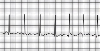
What does tachycardia look like?
QRS complexes present
R to R is much smaller than normal pace
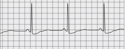
What does bradycardia look like?
QRS complexes present
R to R is much bigger than normal pace
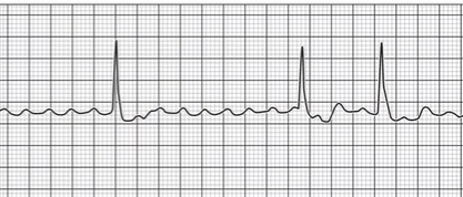
What does an atrial flutter look like?
Lots of P waves are present (continuous stimulation)
QRS firing prematurely or long period of nonfiring
What are venules?
receive blood from capillaries and conduct it back to veins
What are arterioles?
Receive blood from arteries, carry it back to capillaries
What is angiogenesis?
formation of new blood vessels
mainly controlled Vascular Endothelial Growth Factor (VEGF)
Regulated in the body since excess, deficient, or inappropriate blood vessel formation can cause common diseases
how is angiogenesis promoted?
Body secretes VEGF in response to blocked coronary artery
if not sufficient, VEGF may be able to be delivered in time-release capsules or gene therapy→ delivers genes encoding VEGF to area that lacks oxygen
How is angiogenesis prevented?
Tumor secrete VEGF to nourish themselves→ antiangiogenesis drugs are used to treat cancer and age-related macular degeneration
What percent of blood is in systemic veins?
60-70% at any time
What percent of blood is in the lungs?
10-12% at any time
What percent of blood is in the heart?
8-11% at any time
What percent of blood is in systemic arteries?
10-12% at any time (arteries)
What percent of blood is in capillaries?
4-5% at any time
What is atherosclerosis?
Deposits of cholesterol plaque form in inner lining of walls of arteries and arterioles
What is an aneurysm?
A bulge in the wall of an artery, formed when blood pressure dilates a weakened area of the vessel; can burst wall of artery
What is phlebitis?
Inflammation of a vein, very common
What are varicose veins?
Abnormal and irregular dilations in superficial veins; most common in legs
What is blood pressure?
The fource the blood exerts against the innner walls of the blood vessels
circulates the blood
usually refers to pressure of systemic arteries
exists throughotu tthe vascular system
blood moves from higher to lower presure throughout the system
What is arterial blood pressure?
Rises when ventricles contract; fals when ventricles relax
What is arterial systolic pressure (SP)?
The maximum pressuer reached during ventricular contraction
What is arterial diastolic pressure (DP)?
The minimum pressure remaining before next ventricular contraction
What is the blood pressure format?
SP/DP
What is Pulse Pressure (PP)?
Difference between systolic and diastolic blood pressures
SP-DP
What is Mean Arterial Pressure (MAP)?
Avereage pressure in arterial system; representst average force driving blood to the tissues
(2DP+SP)/3
What is stroke volume (SV)?
Volume of blood that enters the arteries w/ each ventricular contraction (70mL/beat)
1.85xPP (pulse pressure)
What is cardiac output (CO)?
volume of blood discharged from a ventricle each minute; blood pressuer varies w/ cardiac output
CO= SV*HR
What does an increase in SV (stroke volume) imply?
HR (heart rate) is increased, which increases CO (cardiac output)→ inc blood pressure (BP)
What is peripheral resistance (PR)?
Force of friction between blood and wals of blood vessels
measured as Total Peripheral Resistance (TPR)=MAP/CO
blood pressure must overcome PR in order to flow
factors that change PR also change BP
What does vasoconstriction do to arterioles?
Increases peripheral resistance, which increases BP
How does blood go against PR (peripheral resistance)?
When blood is pumped out of ventircles, arteries swell
rapid elastic recoil sends blood through arteries, against PR in arterioles and capillaries
How does viscosity impact the cardiovascular system?
Difficulty w/ which molecules of fluid flow past each other
greater the viscosity, greater the resistance Etonian blood flow
blood cells and plasma proteins inc the viscosity of blood
any facter that alters conc of blood cells or plasma proteins also alters blood viscosity