ekg/ecg pictures
1/71
There's no tags or description
Looks like no tags are added yet.
Name | Mastery | Learn | Test | Matching | Spaced | Call with Kai | Chat |
|---|
No analytics yet
Send a link to your students to track their progress
72 Terms
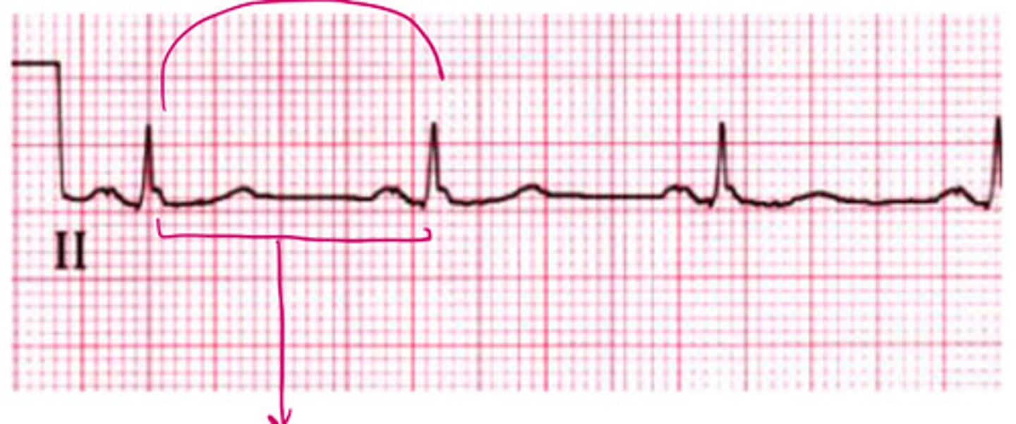
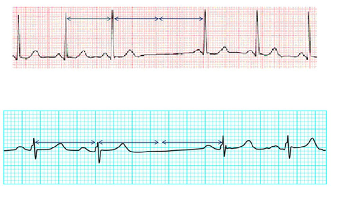
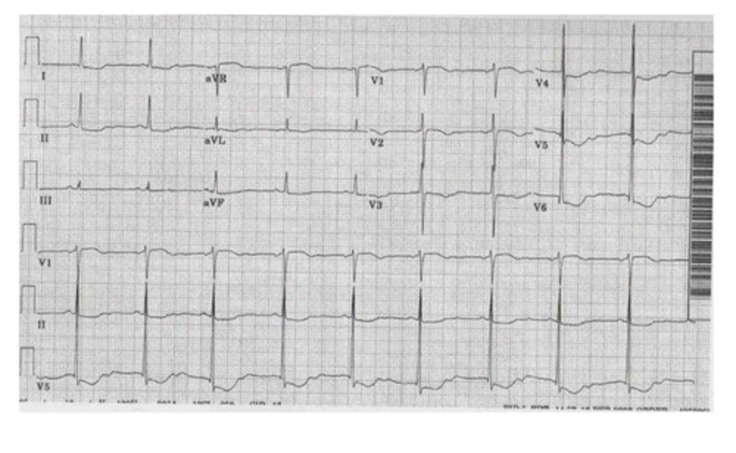
~68
what is the HR of this
stepwise approach to interpreting ECG
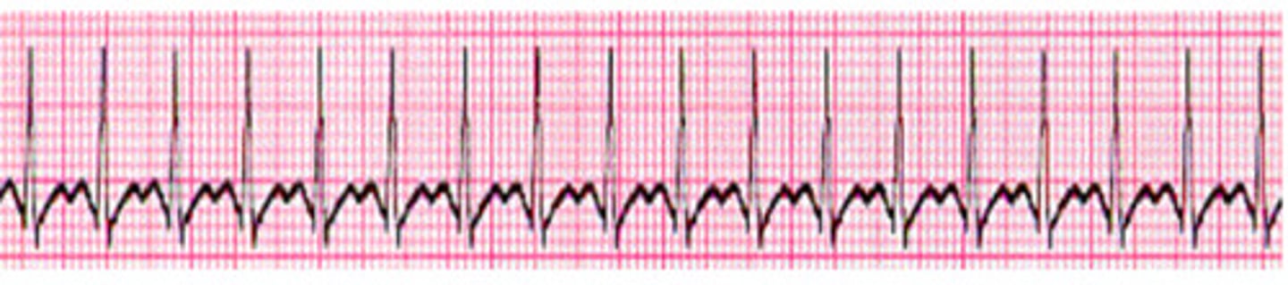
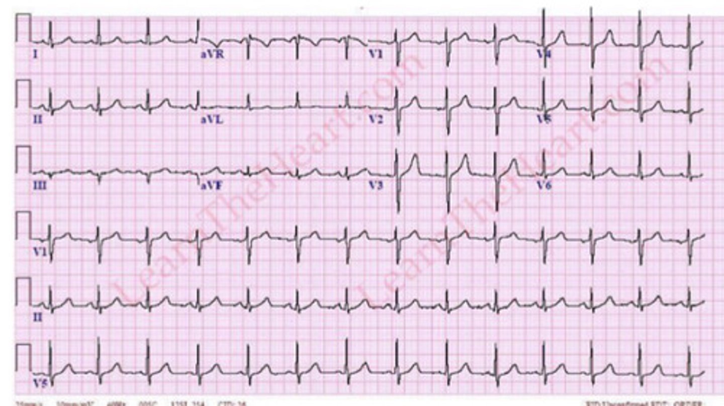
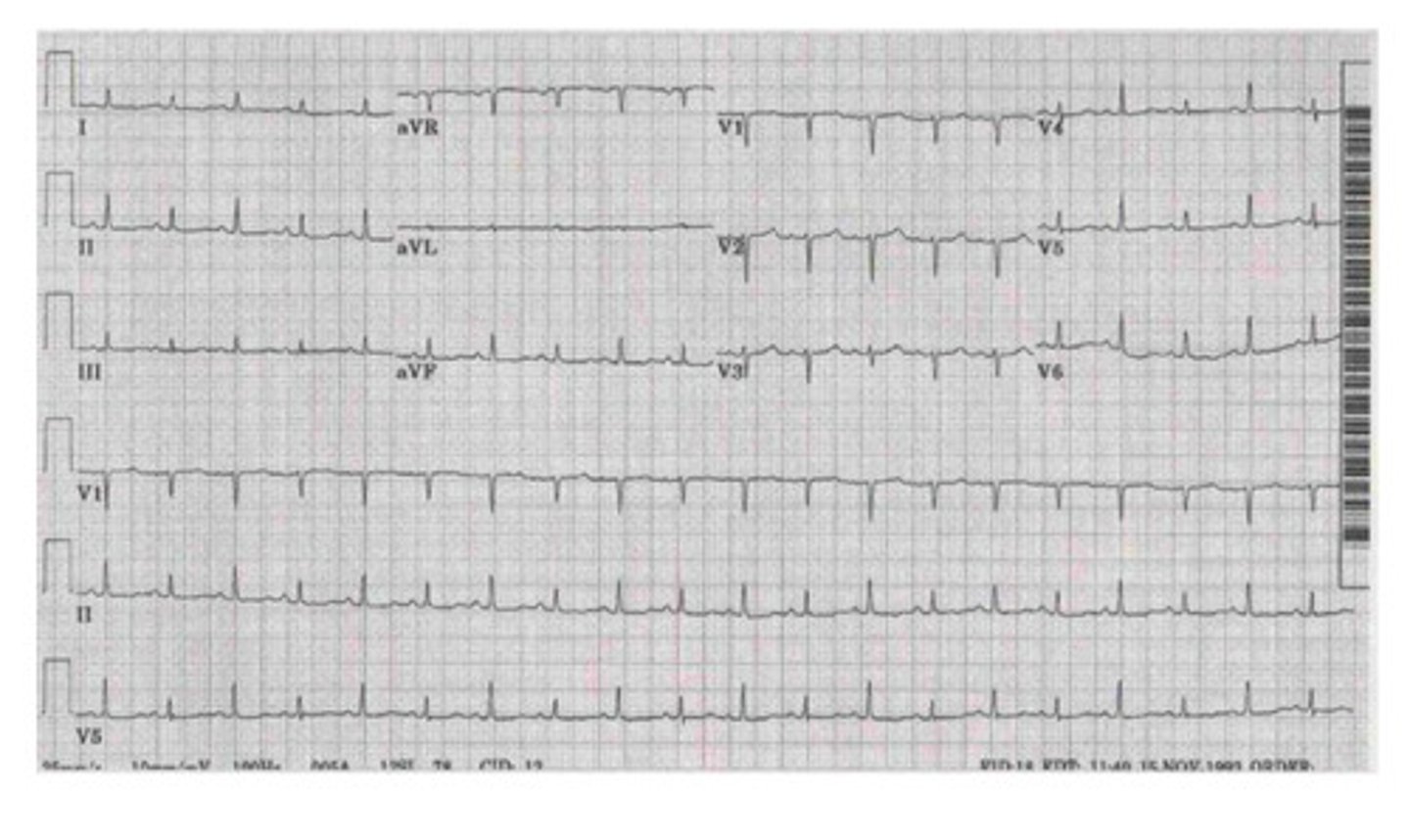
~187.5
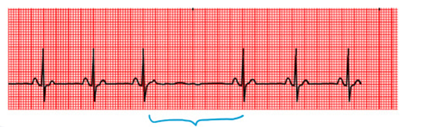
what is the HR of this
0.8 (normal)
what is the QRS interval of this
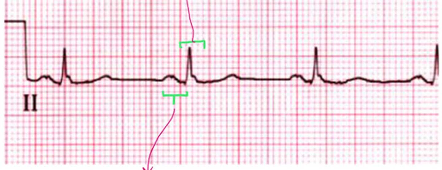
0.16 (normal)
what is the PR interval of this

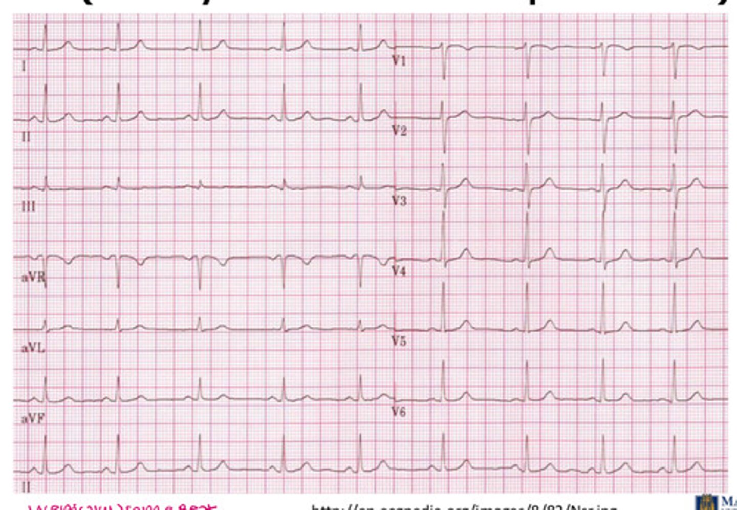
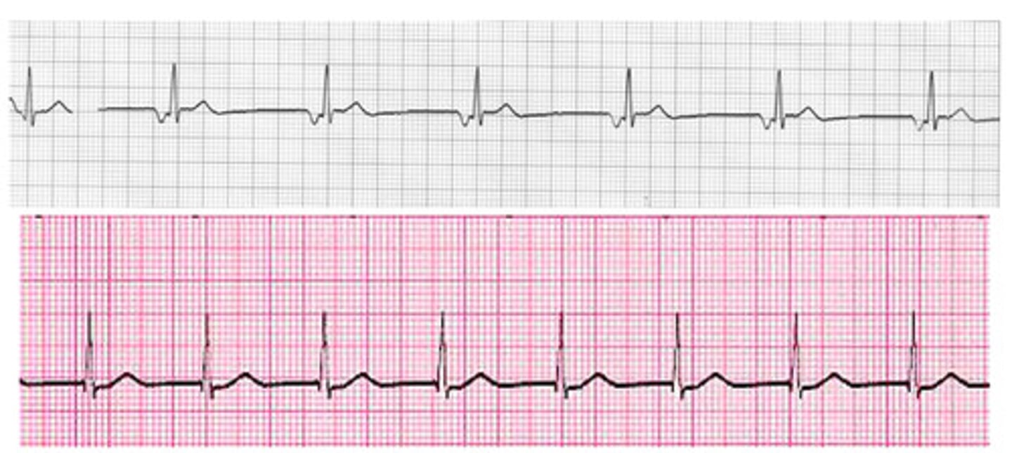
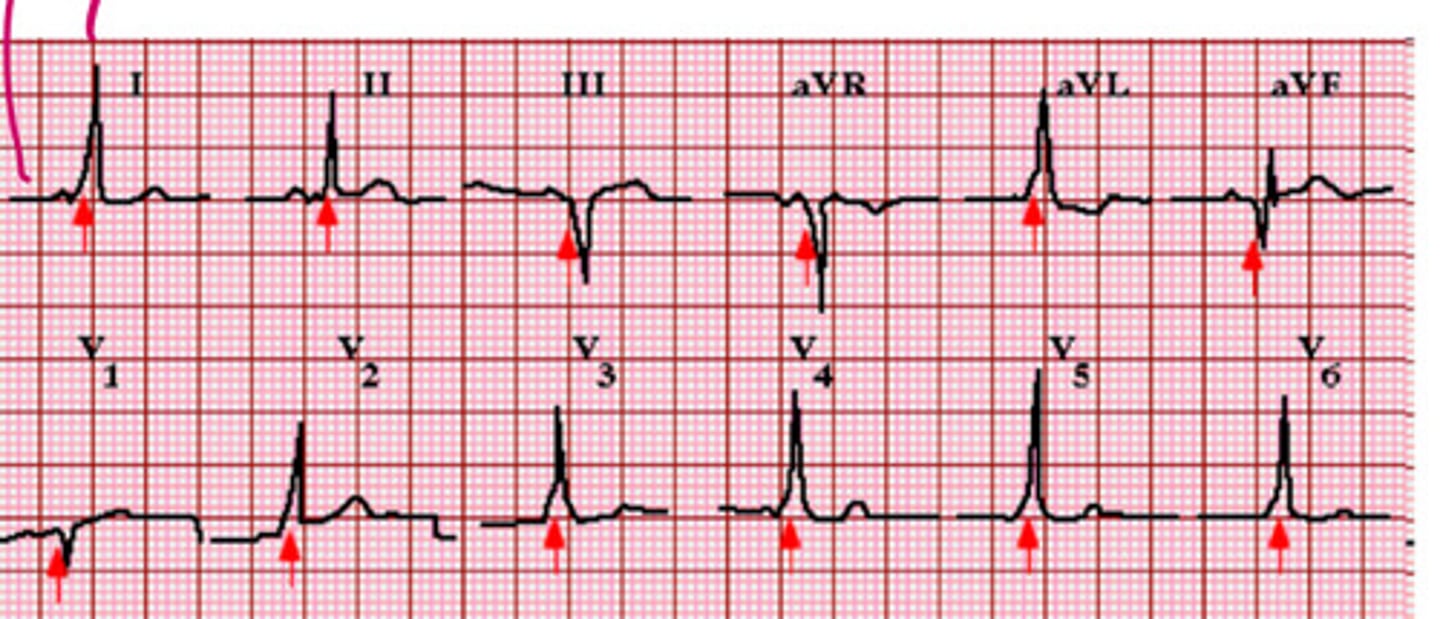
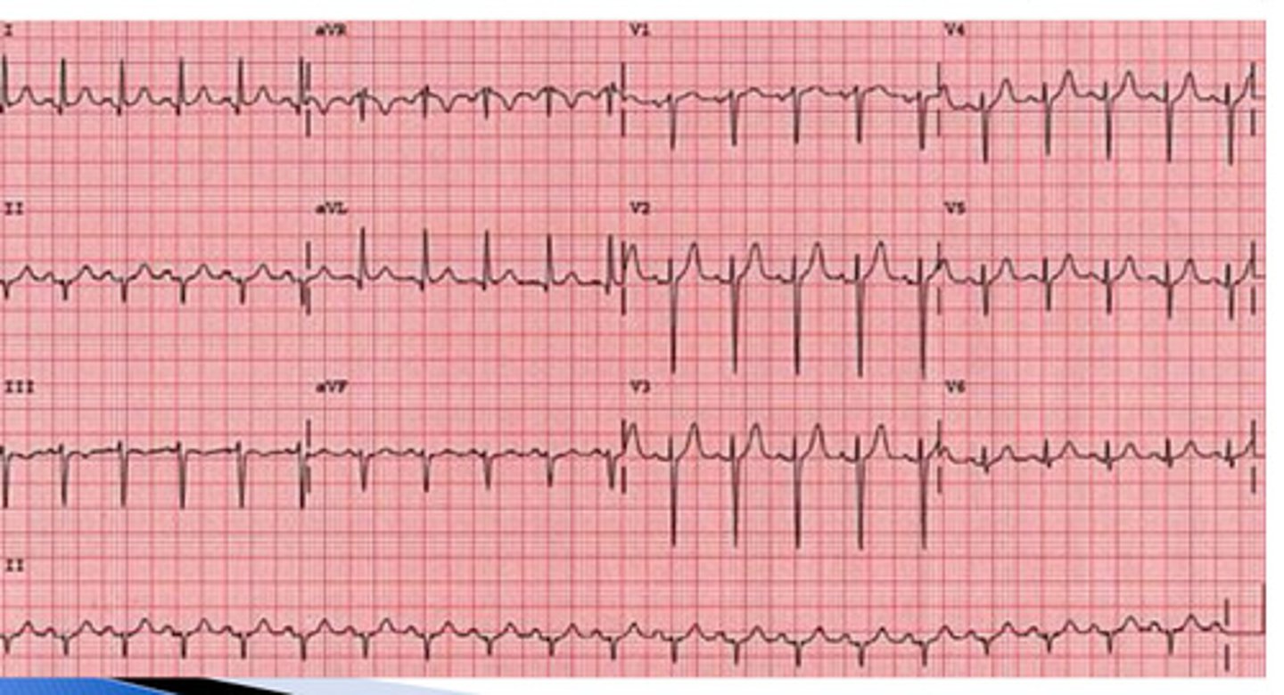
normal ECG
what kind of ECG is this?
normal axis deviation; upright in I and AVF
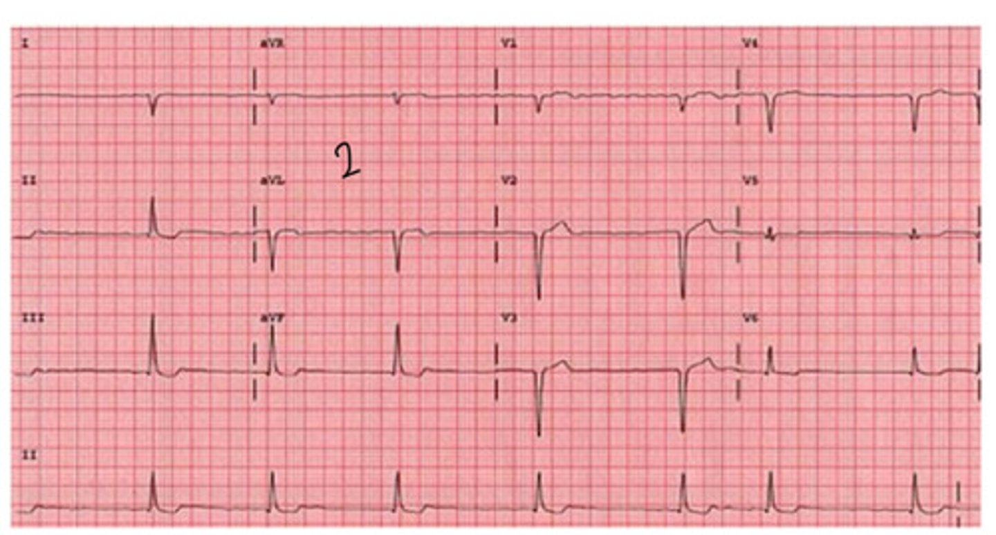
is this normal, left, or right axis deviation? why
right axis deviation; down in 1, upright in AVF
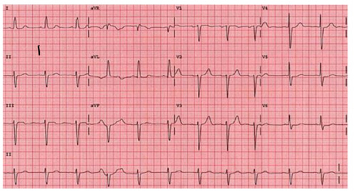
is this normal, left, or right axis deviation? why
left axis deviation; up in 1, down in AVF
is this normal, left, or right axis deviation? why
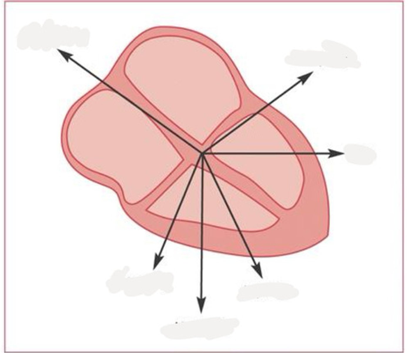
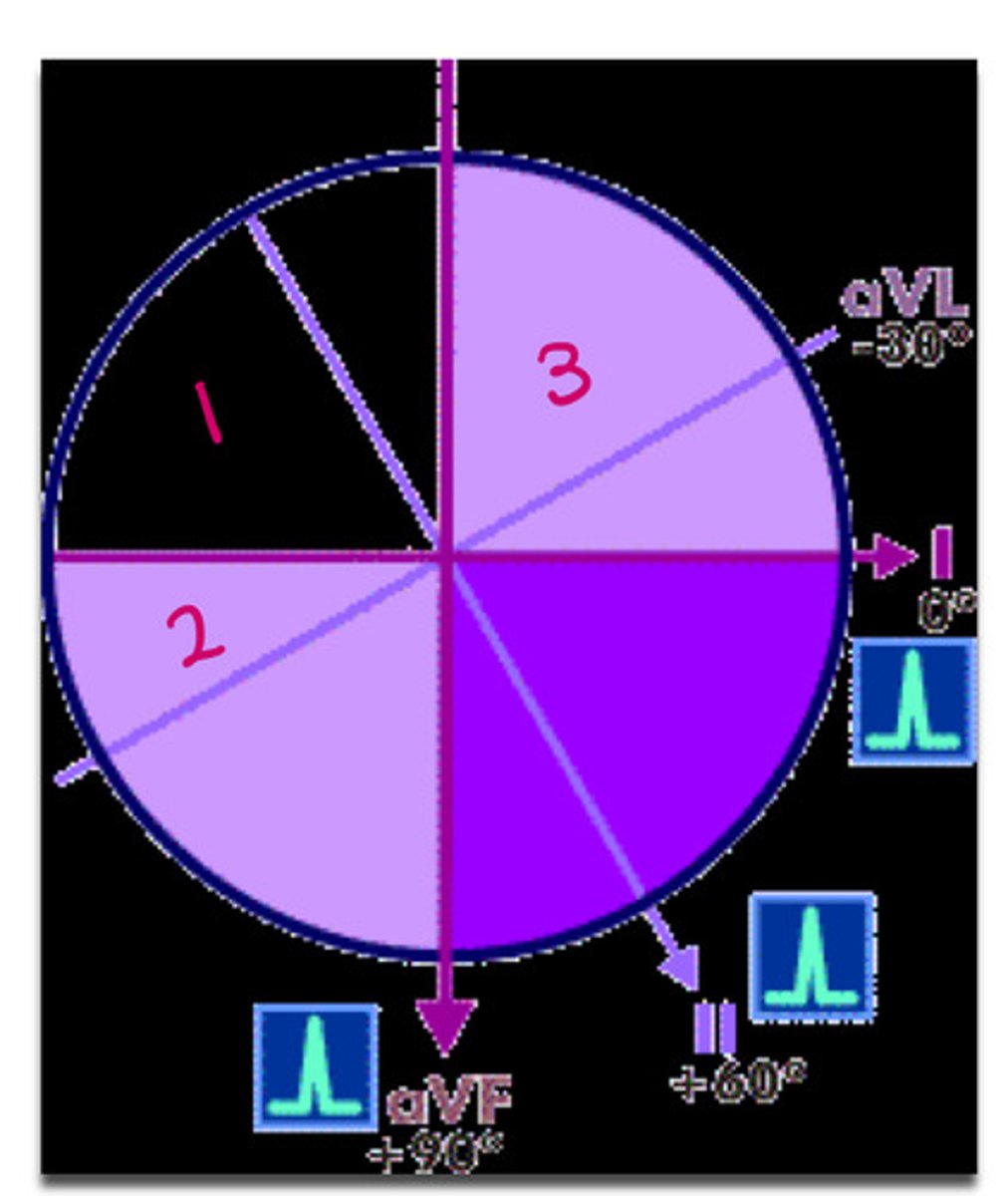
AVR -150
AVL -30
I 0
II +60
AVF +90
III +120
what are the angles from top left to clock wise
*
be able to determine precise axis
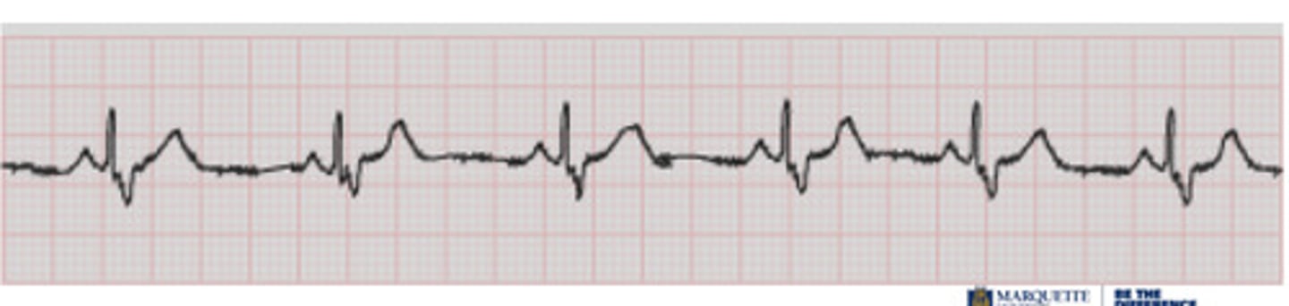
LVH w/ strain pattern
what is this
right ventricular hypertrophy
what is this
left atrial enlargement
what is this
right atrial enlargement
what is this
1: extreme R axis deviation
2: R axis deviation
3: left axis deviation
what do the numbers correlate to
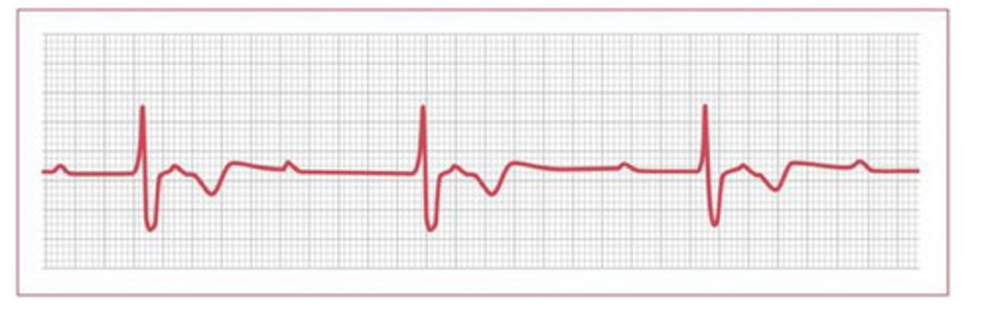
sinus arrest/exit block
what is this
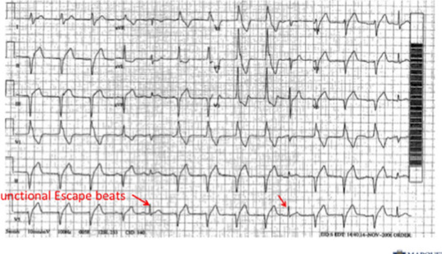
junctional rhythm
what is this
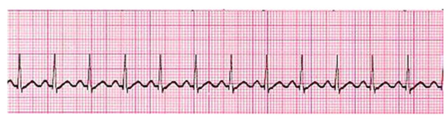
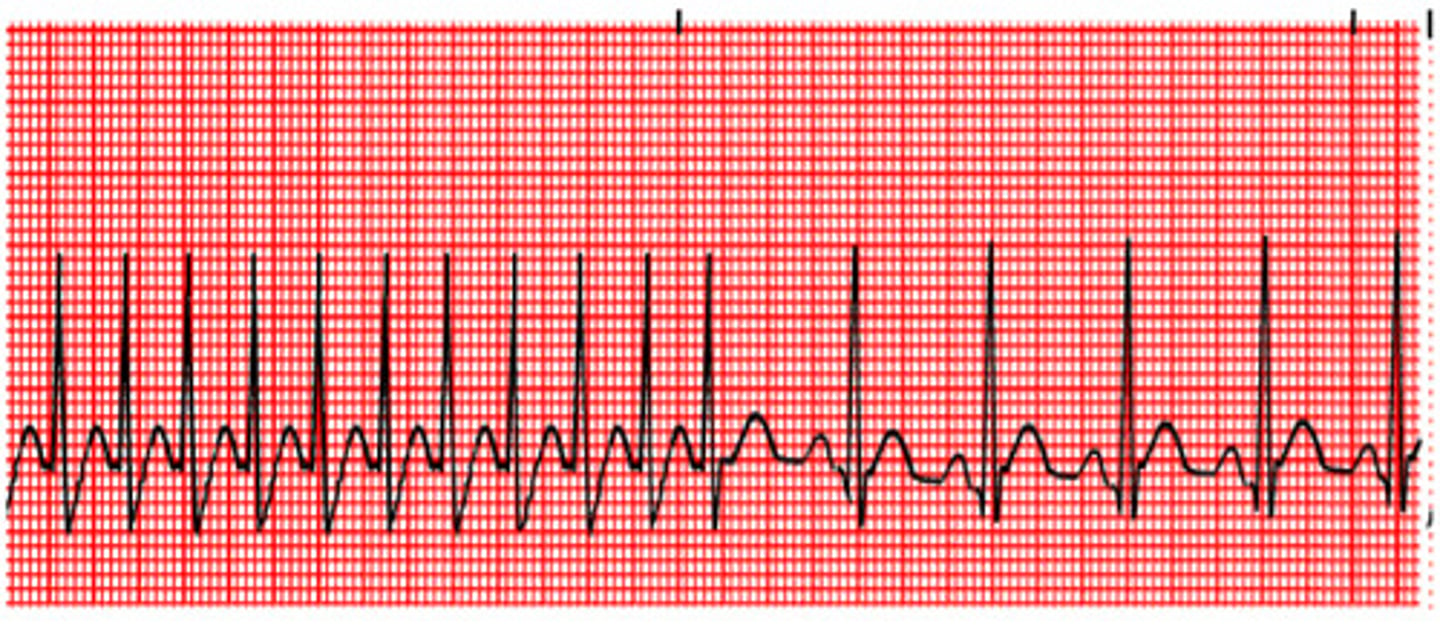
sinus tachycardia
what is this
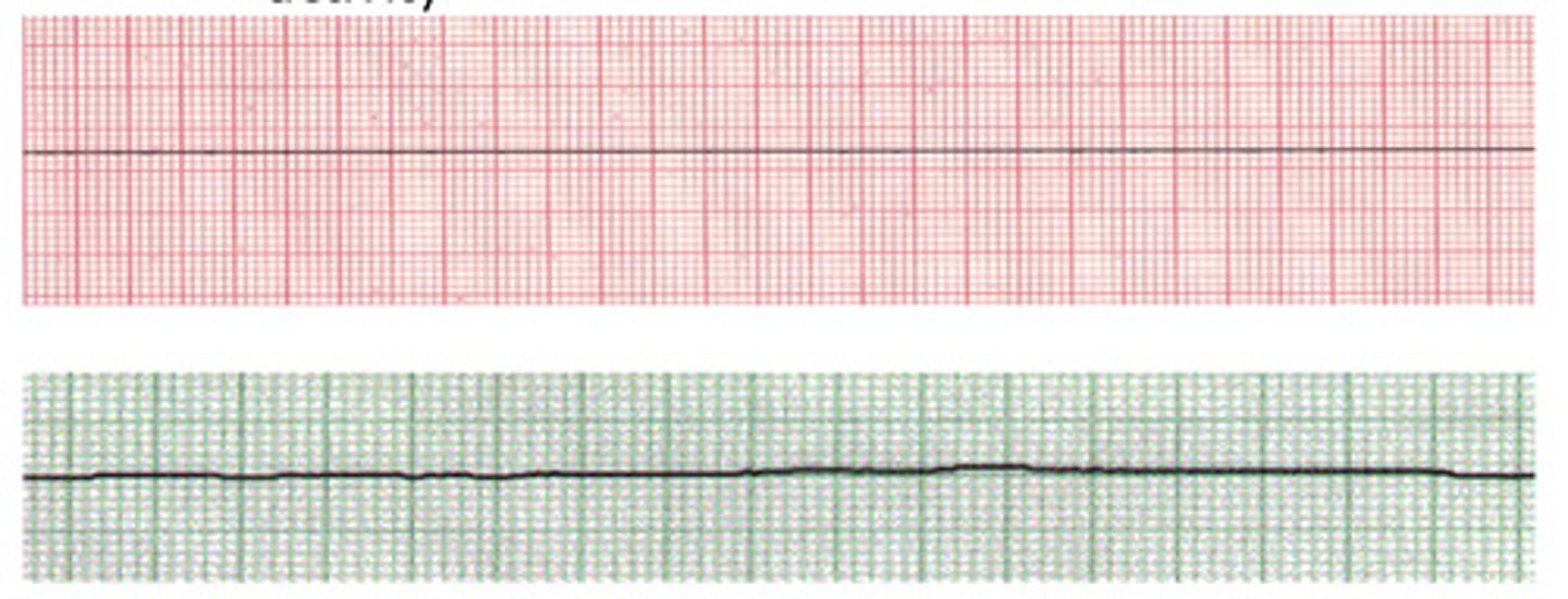
asystole
what is this
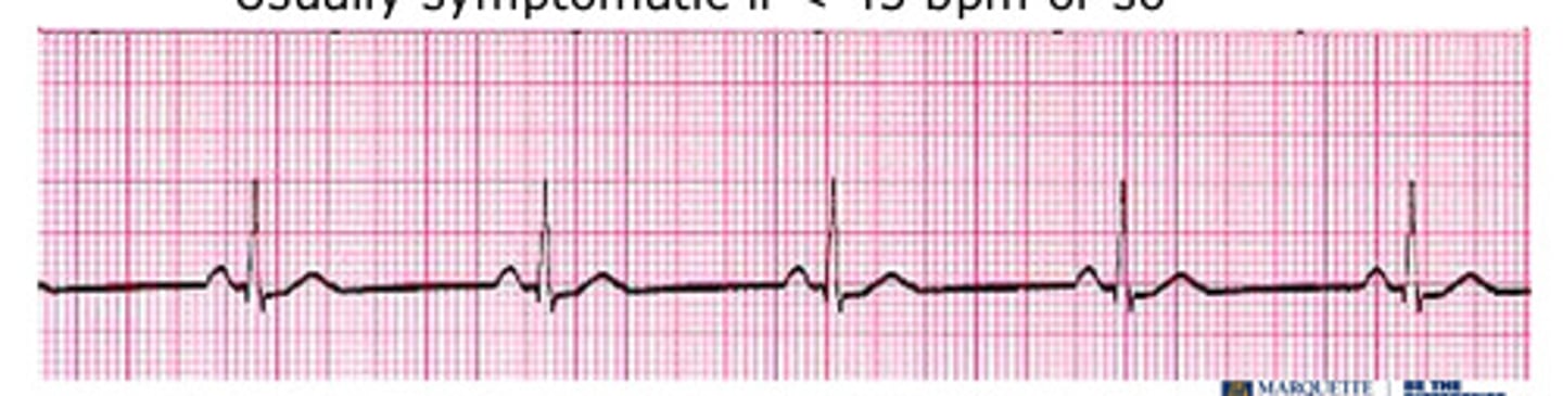
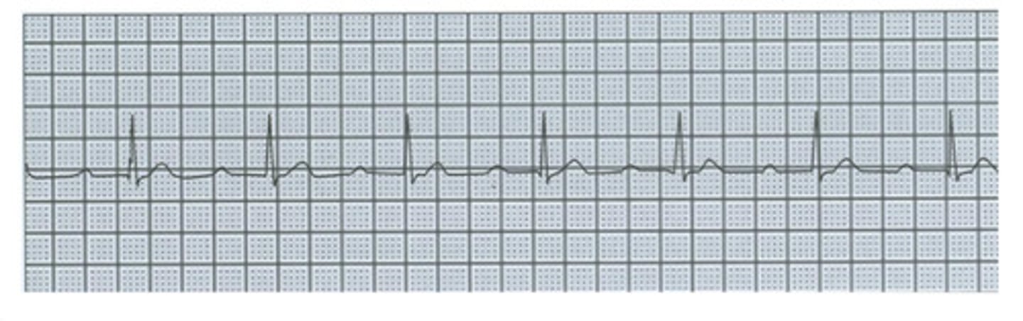
sinus bradycardia
what is this
general sinus arrhythmia
what is this
PSVT (paroxysmal supraventricular tachycardia)
what is this
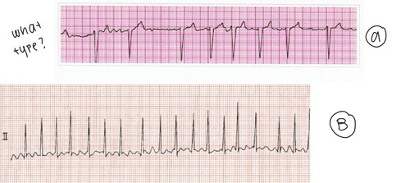
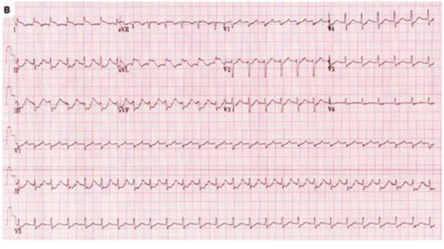
a fib
- a: fine
- b: coarse
what is this & what types are they?
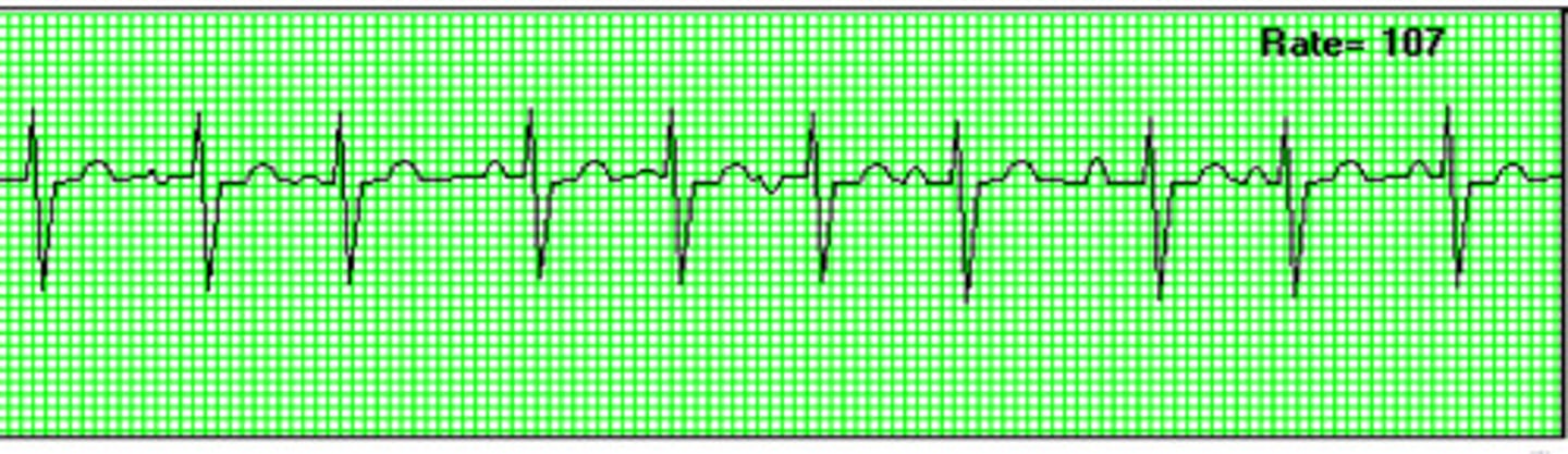
multifocal atrial tachycardia
what is this
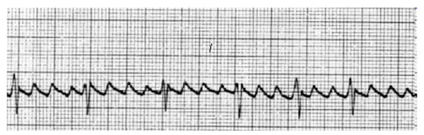
atrial flutter
what is this
premature junctional beat
what is this
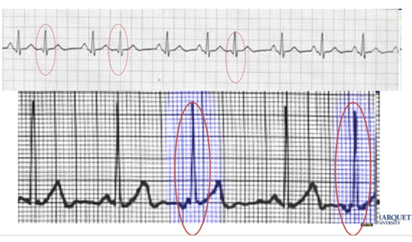
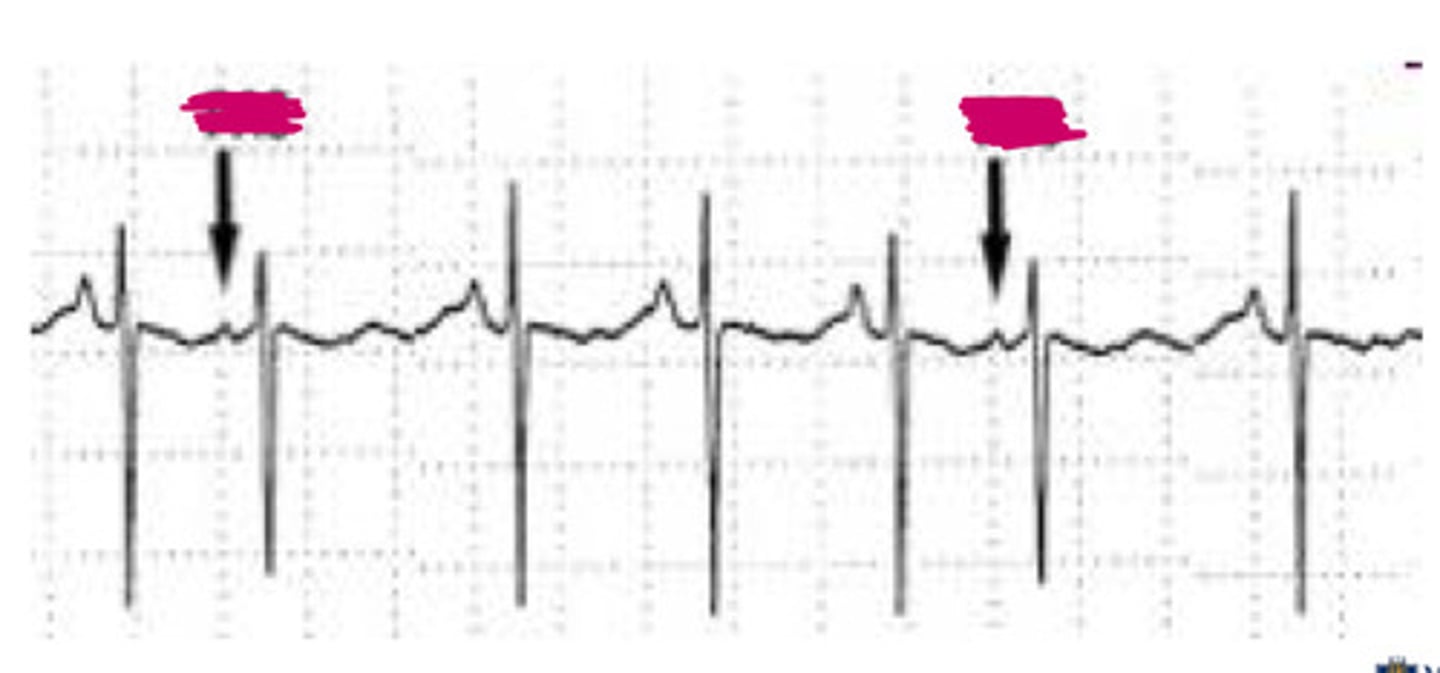
premature atrial contraction (PAC)
what is this
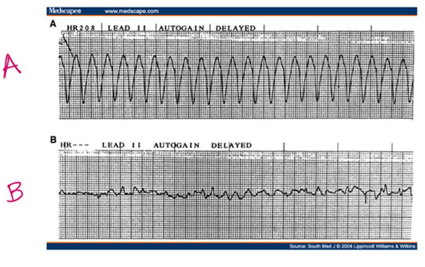
- a: vtach
- b: vfib
what is this
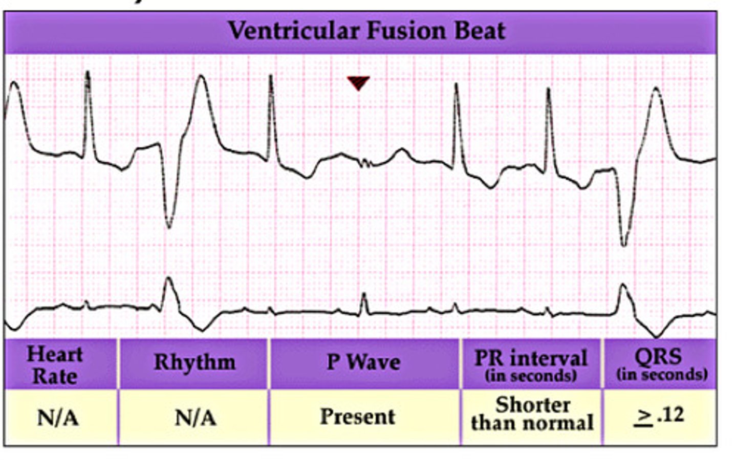
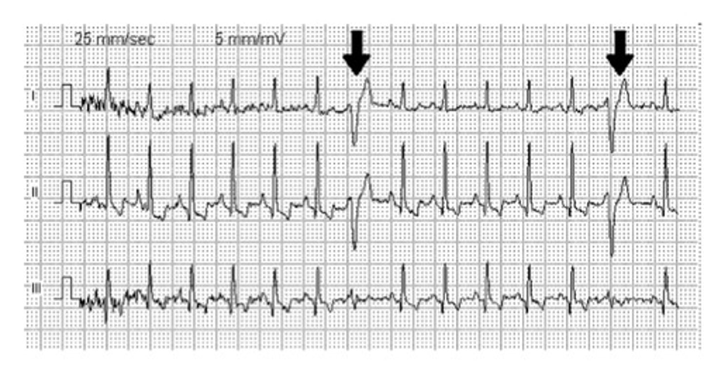
ventricular fusion beat lol
what is this
accelerated idioventricular
what is this
R on T phenomenon
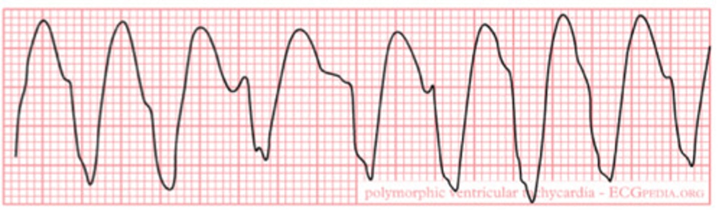
what is this
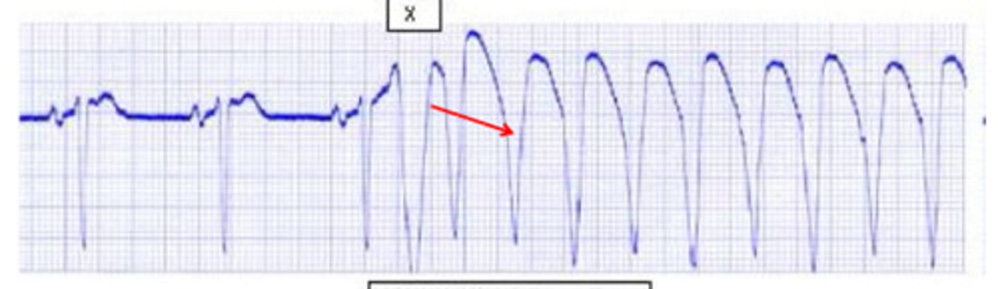
polymorphic v tach
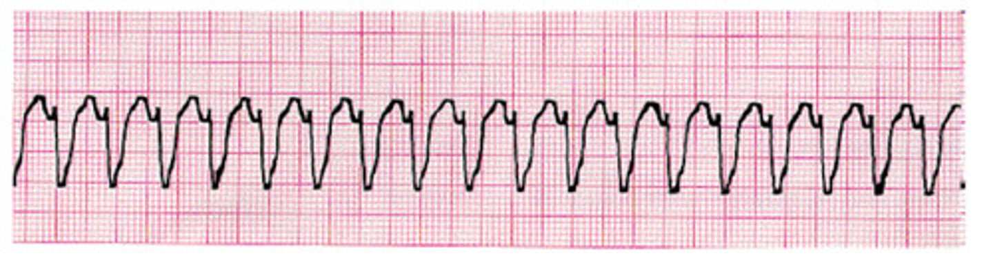
what is this
monomorphic v tach
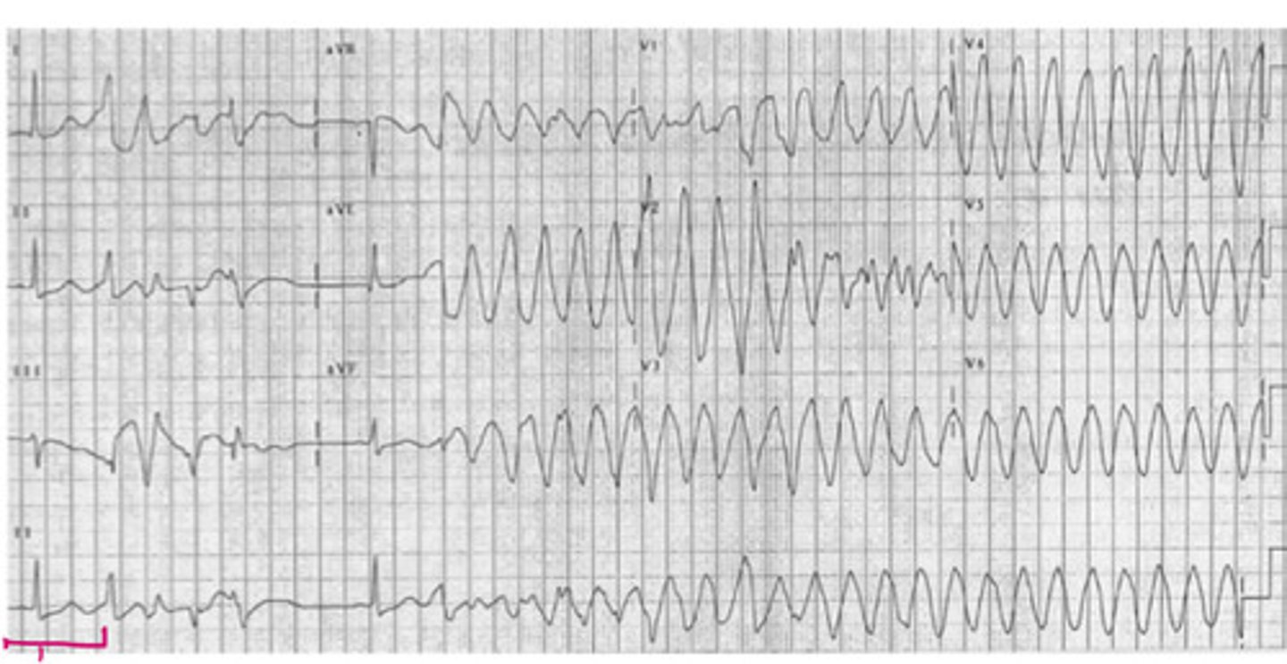
what is this
torsades de pointes
what is this
unifocal PVC
what is this
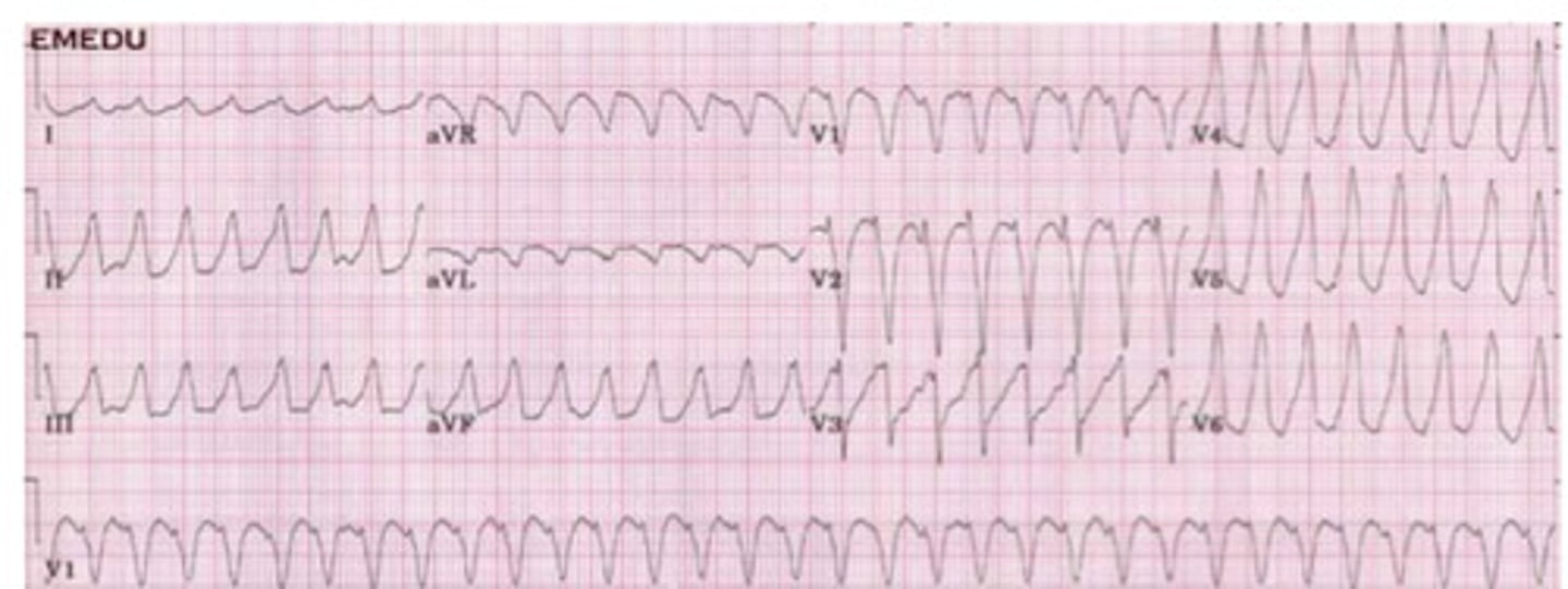
ventricular tachycardia
what is this
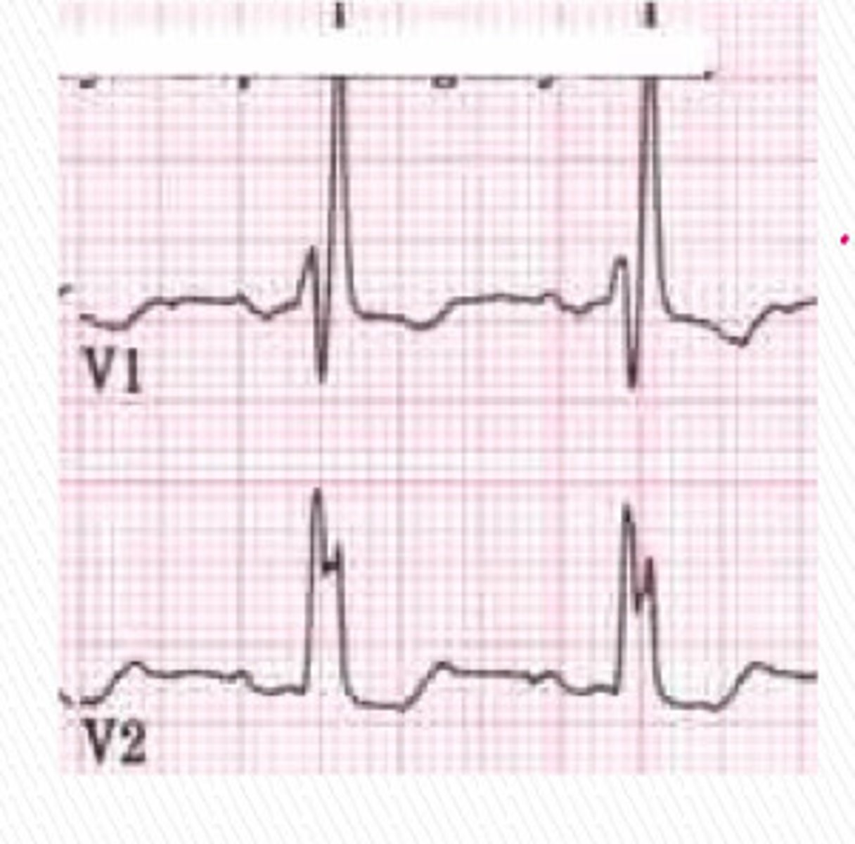
Wolf-Parkinson-White Syndrome
what is this
left bundle branch block
what is this
left posterior fascicular block
what is this
right bundle branch block
what is this
3rd degree heart block
what is this
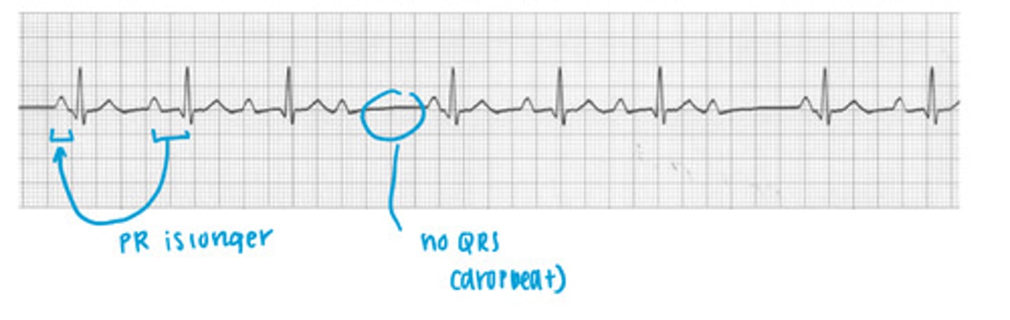
wenckebach
what is this
left anterior fascicular block
what is this
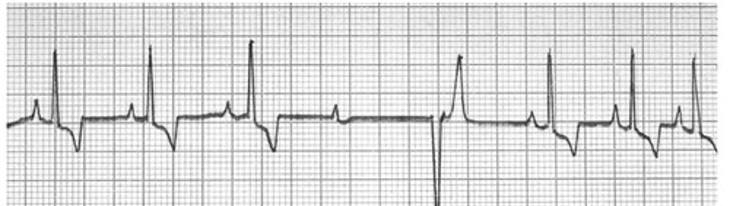
first degree av block
what is this
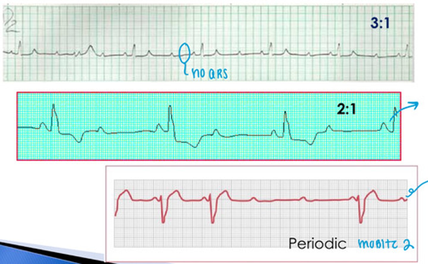
mobitz 2
what is this
mobitz type II block
what are these examples of
sinus node block
what is this
wenckebach's
what is this
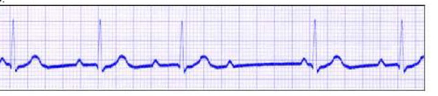
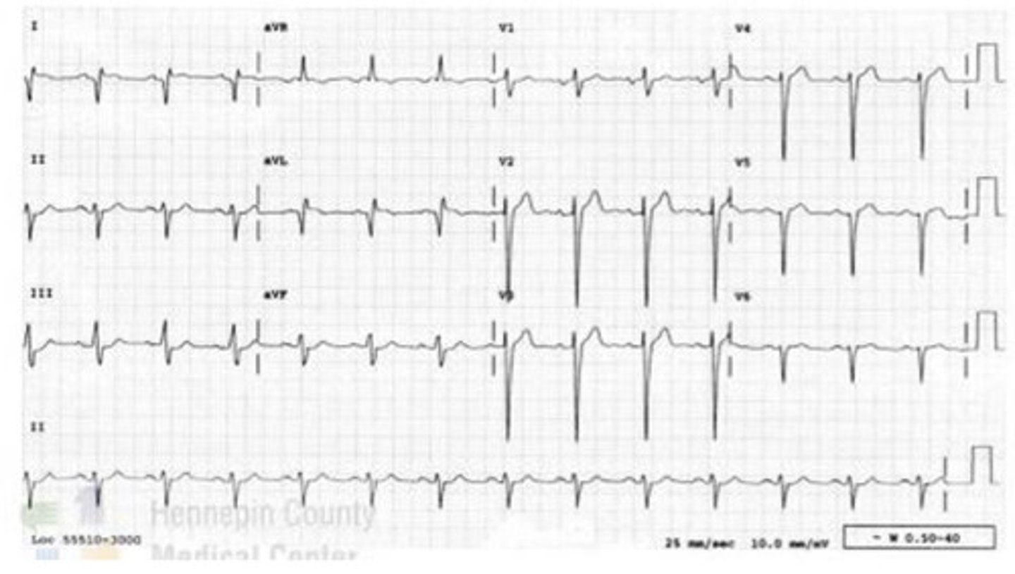
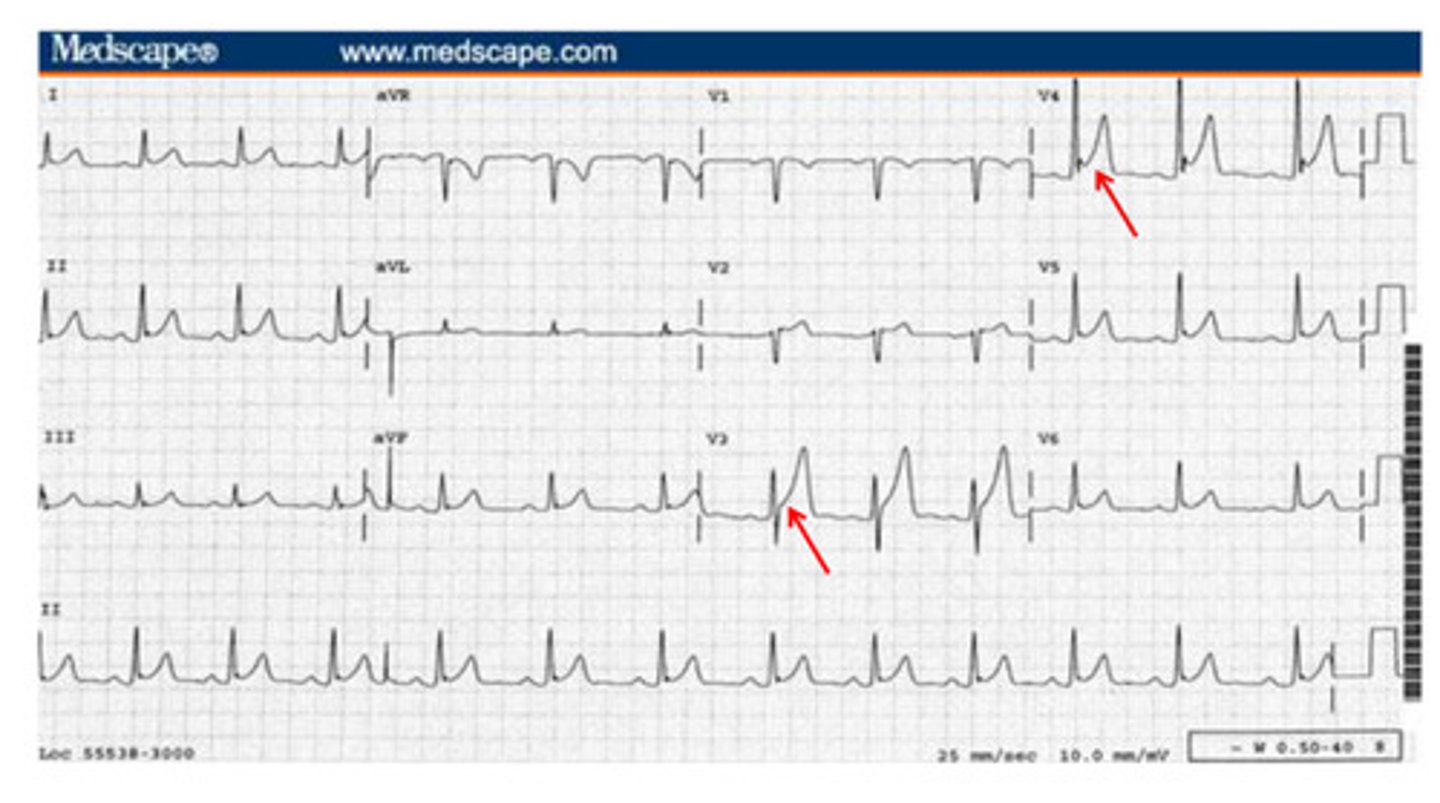
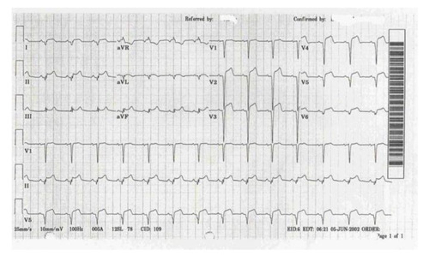
acute anterior mi
what is this

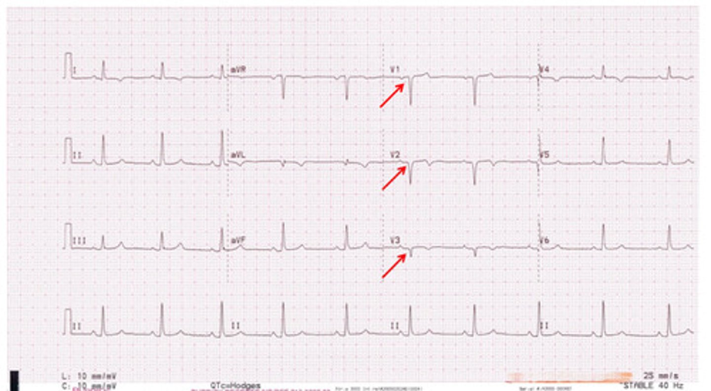
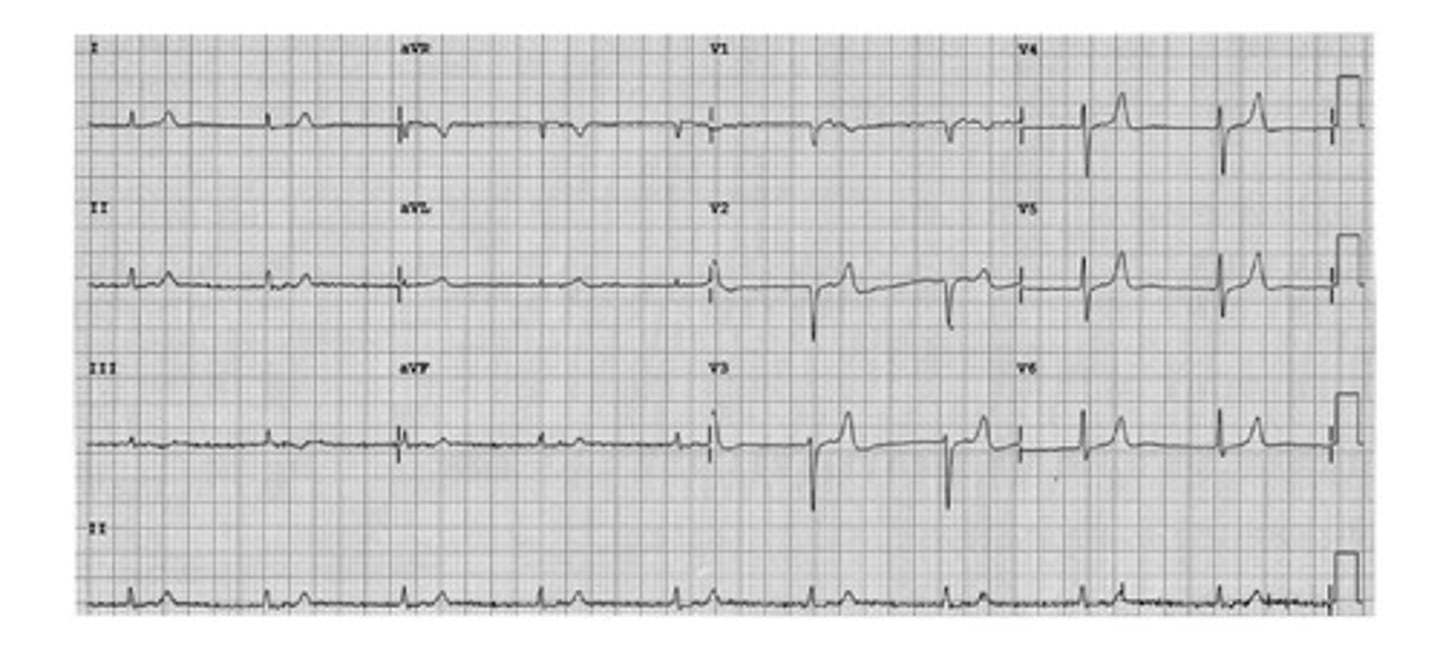
acute lateral mi
what is this
old lateral mi
what is this
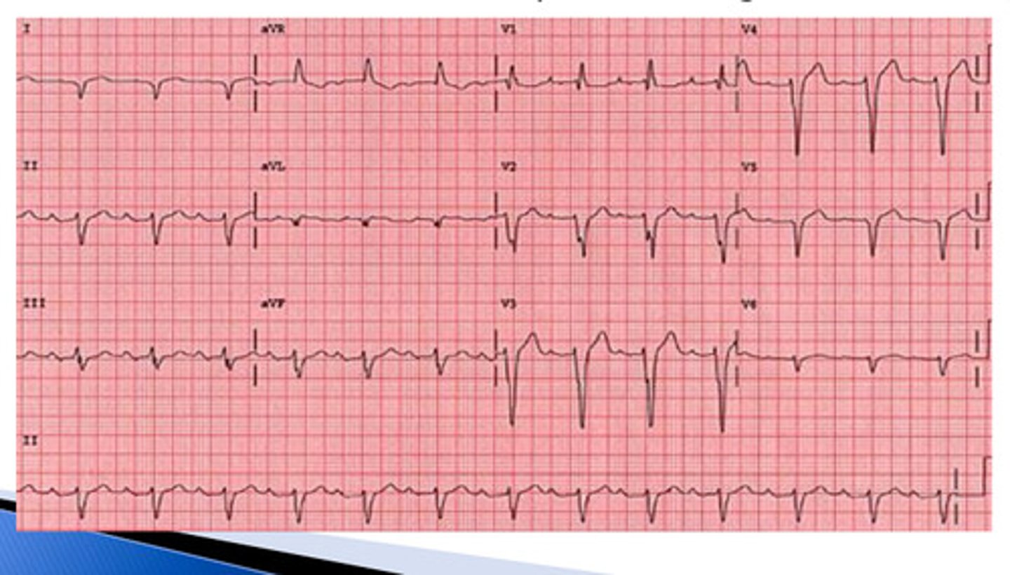
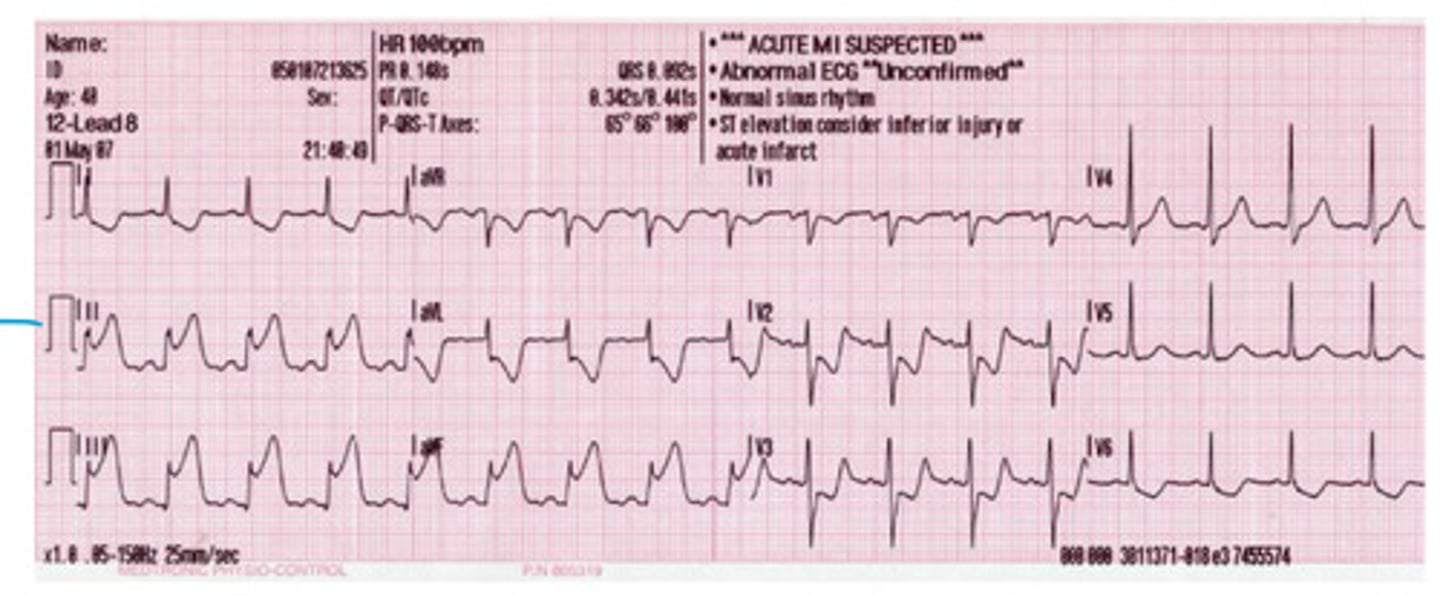
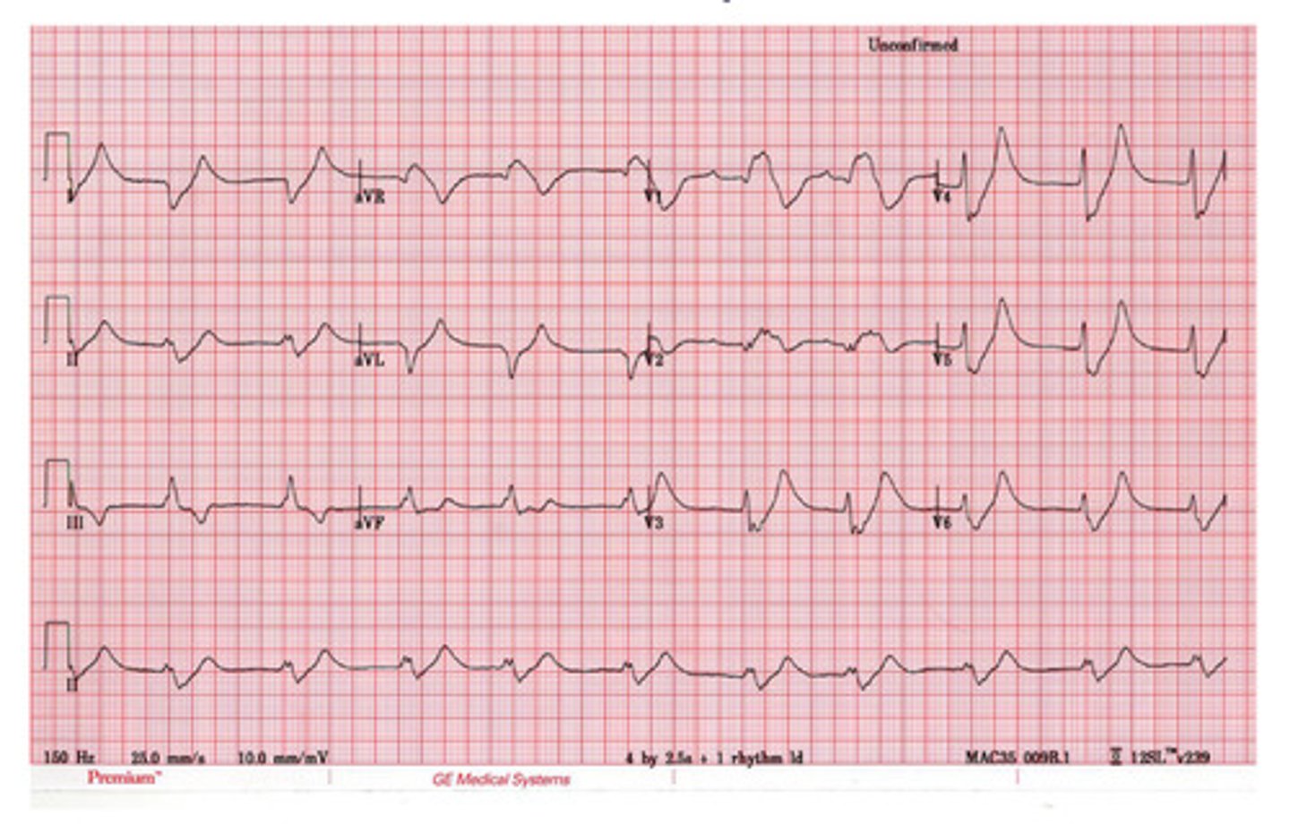
acute inferior MI (STEMI)
what is this
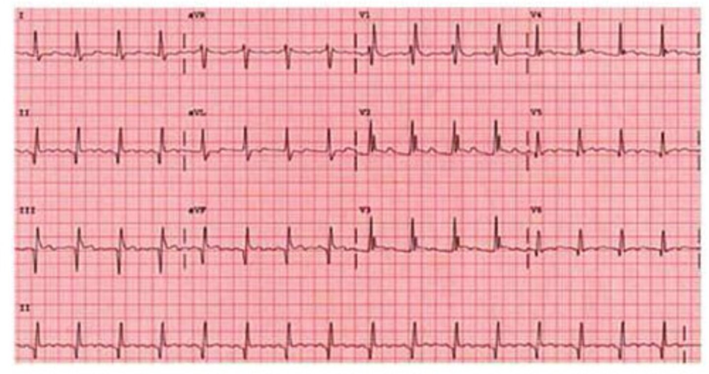
old inferior MI
what is this
early repolarization
what is this
pathologic
this ekg shows ______ Q waves
ST segment elevation
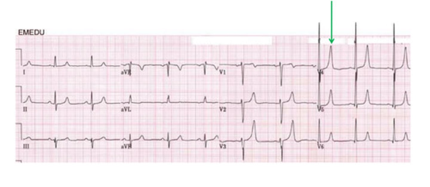
what is this
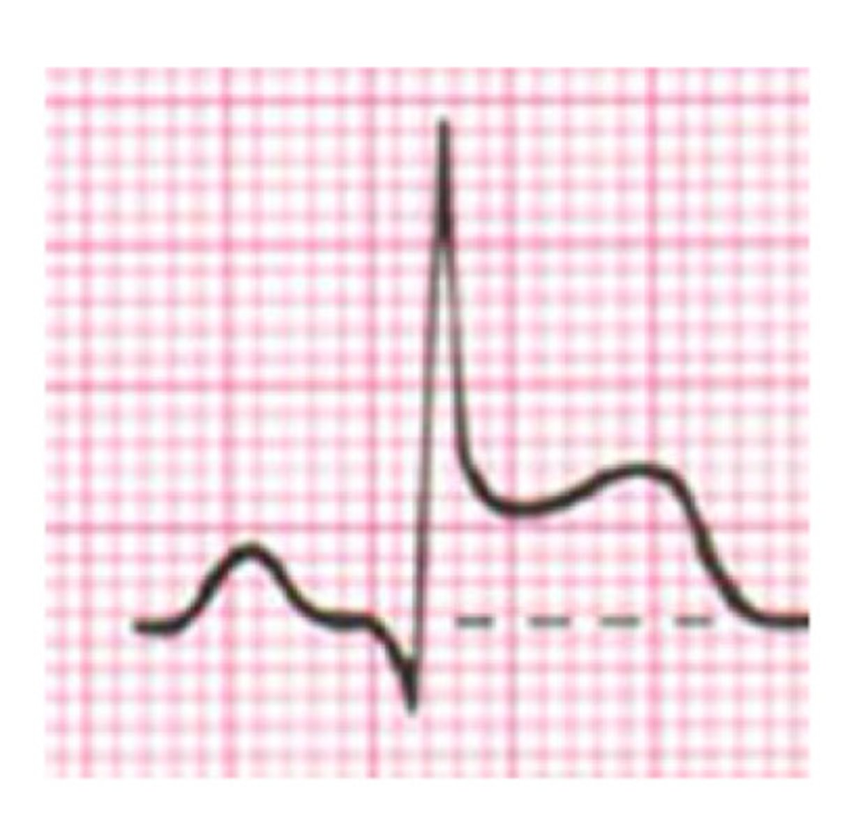
T wave peaking
what is this
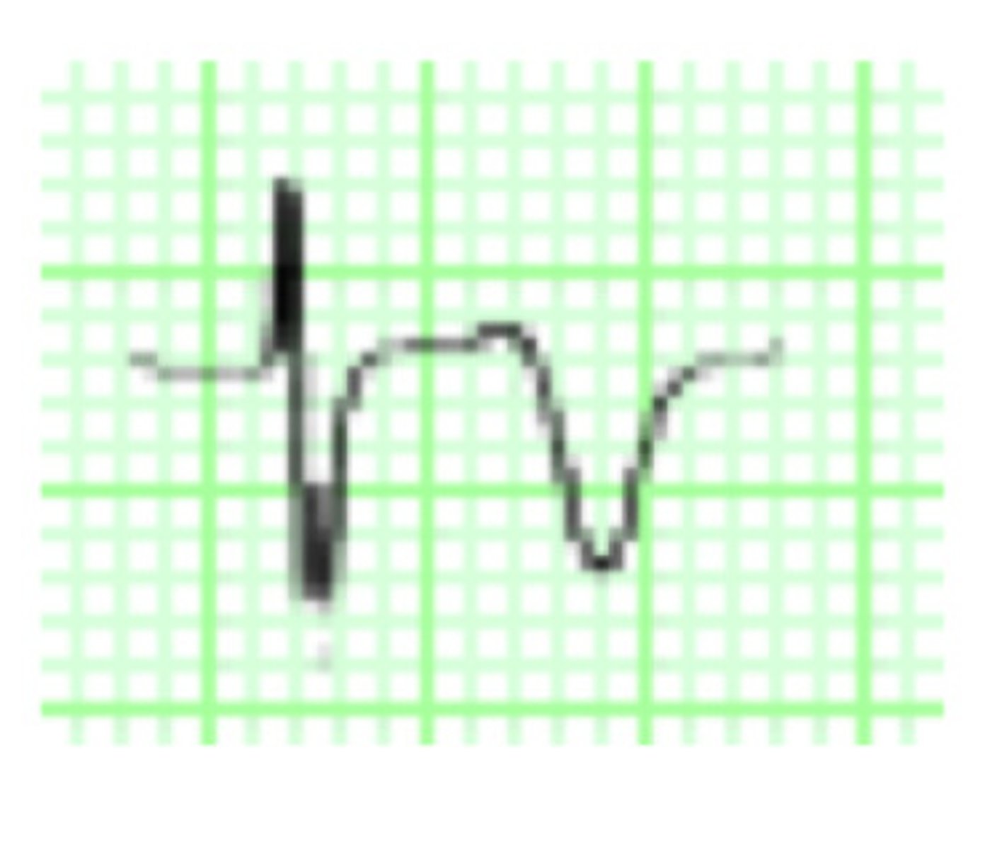
T wave inversion
what is this
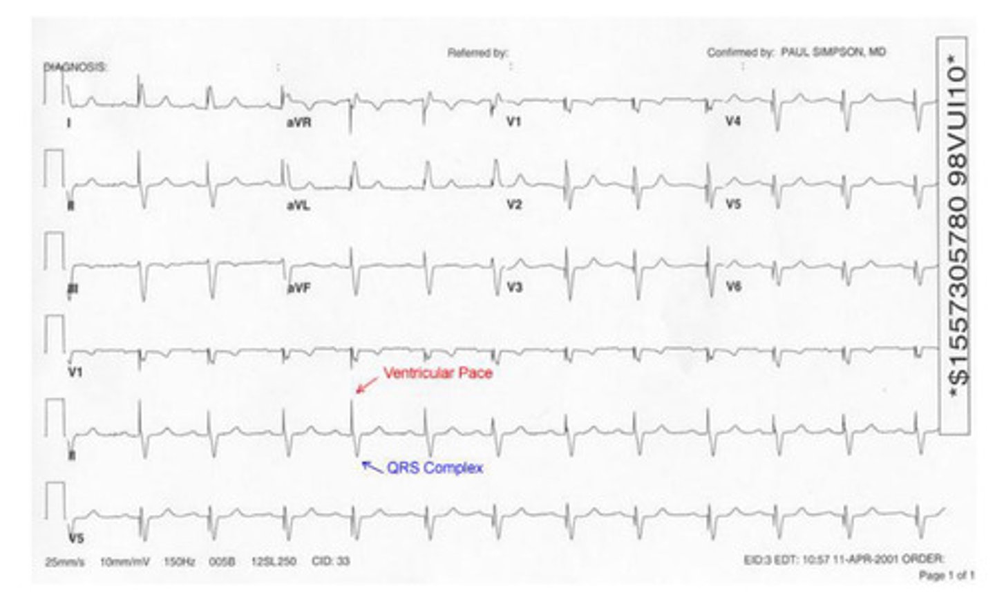
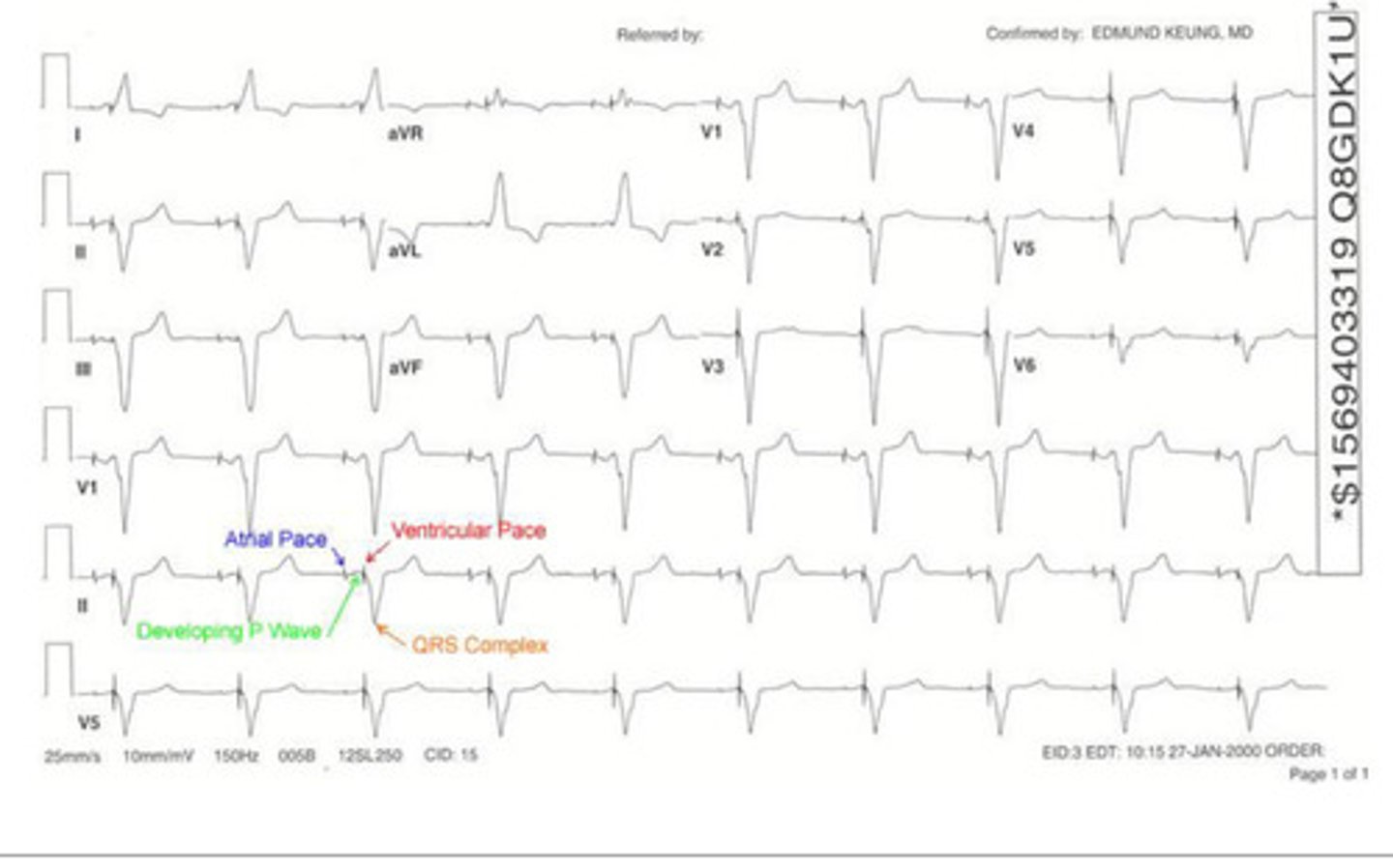
ventricular pacemaker
this is an example of a _____ _____
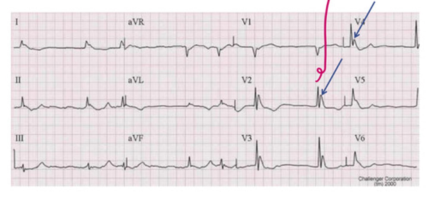
dual chamber
this is an example of a ____ _____ pacemaker
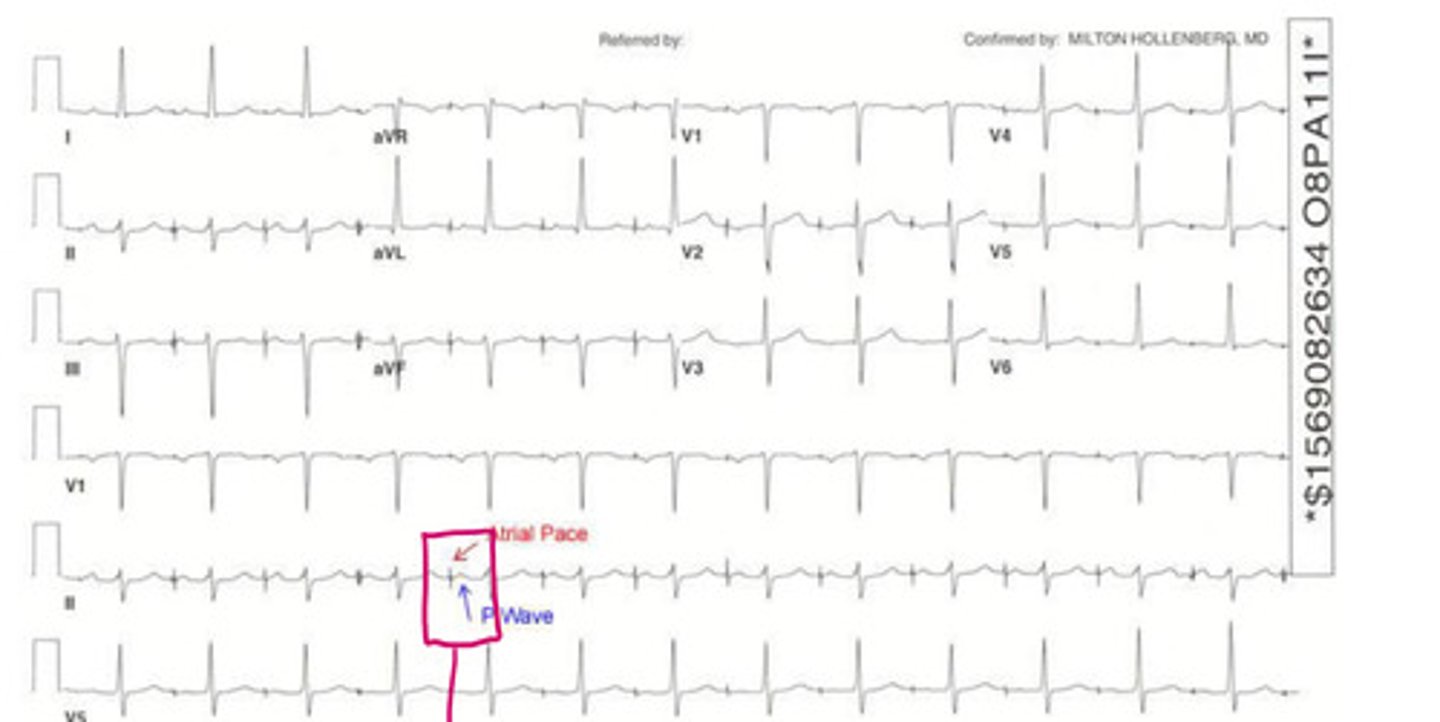
atrial pacemaker
this is an example of an _____ ______
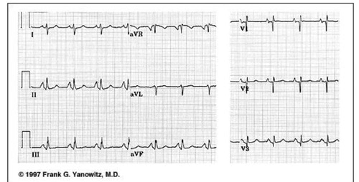
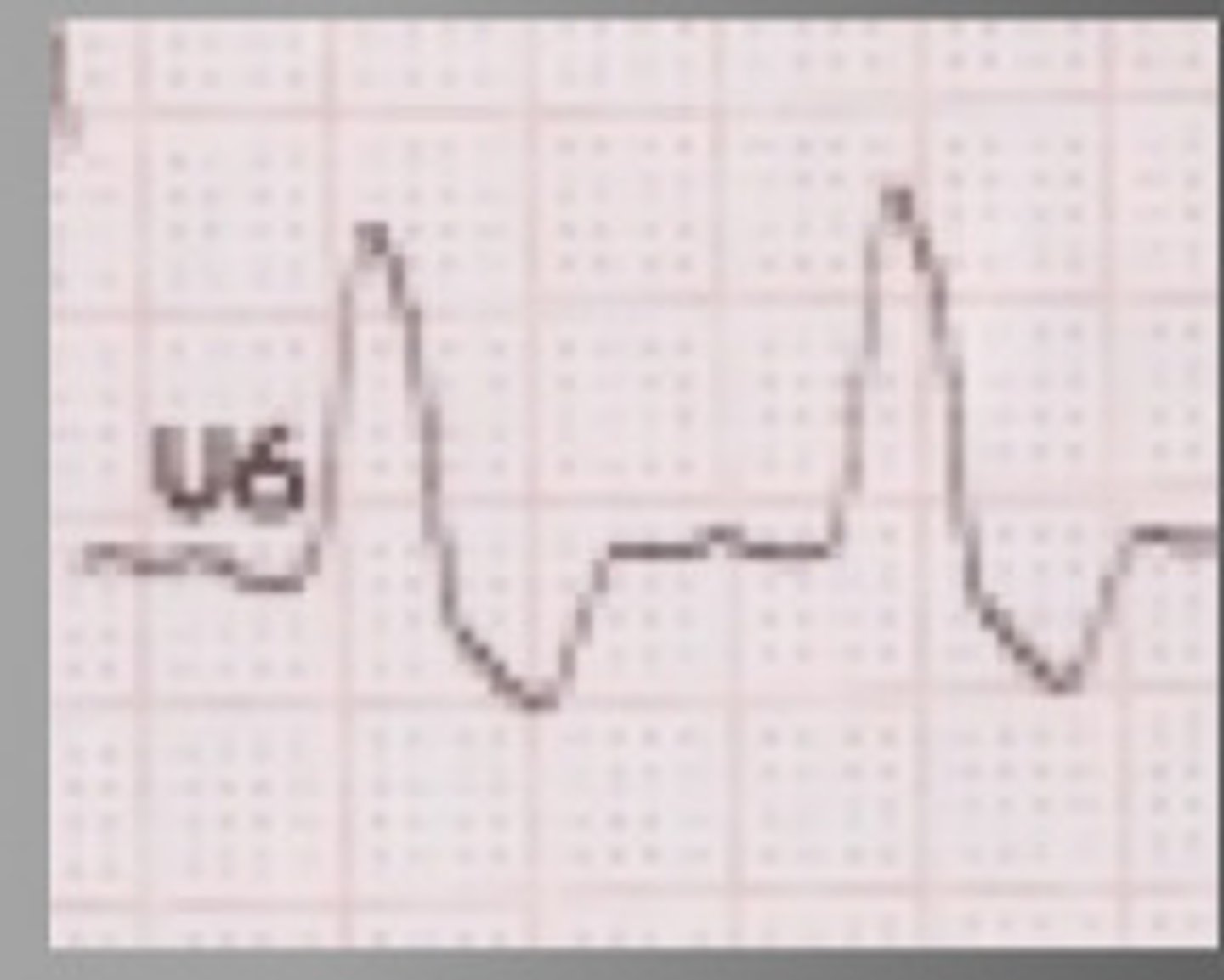
hypothermia; osborne
this is an example of ______, which is characterized by _____ waves
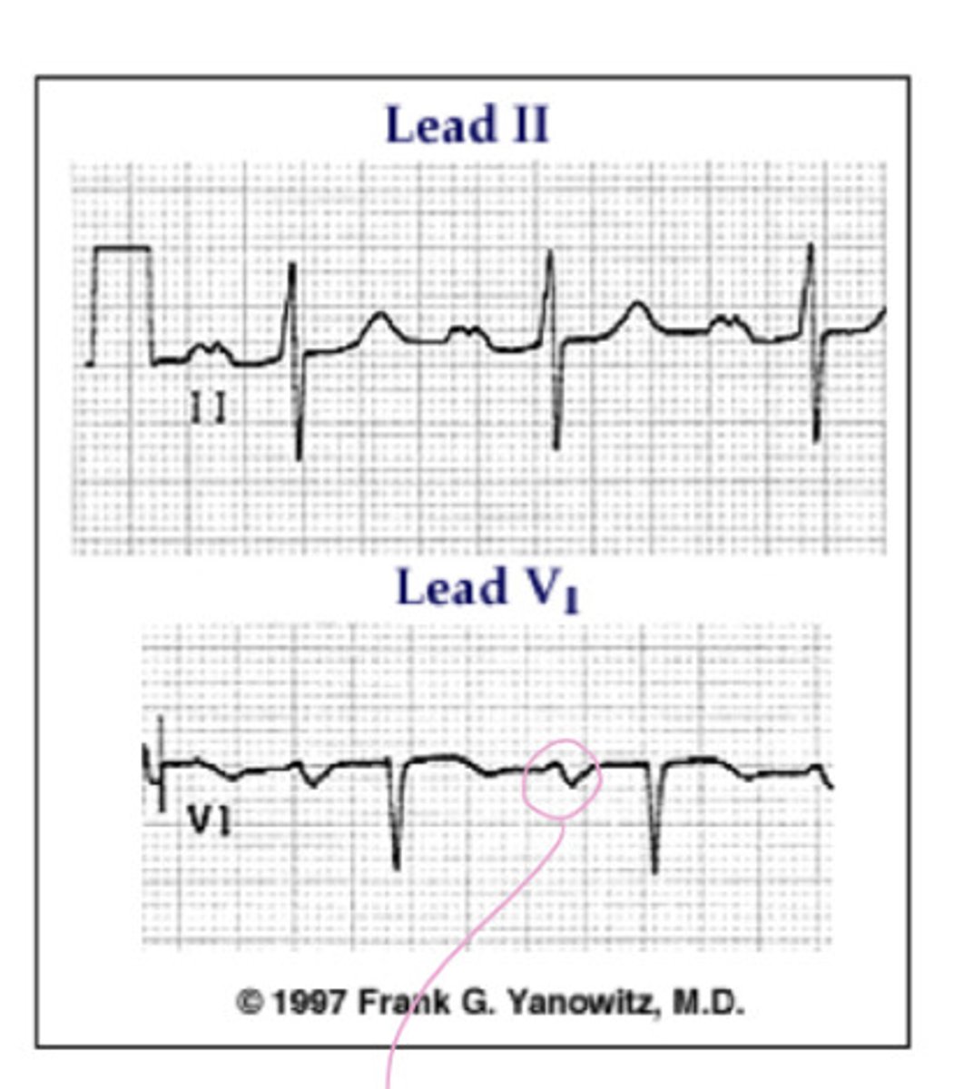
S1Q3 pattern in pulmonary embolisim
this is an example of ________ pattern in ______ _______
pericardial effusion
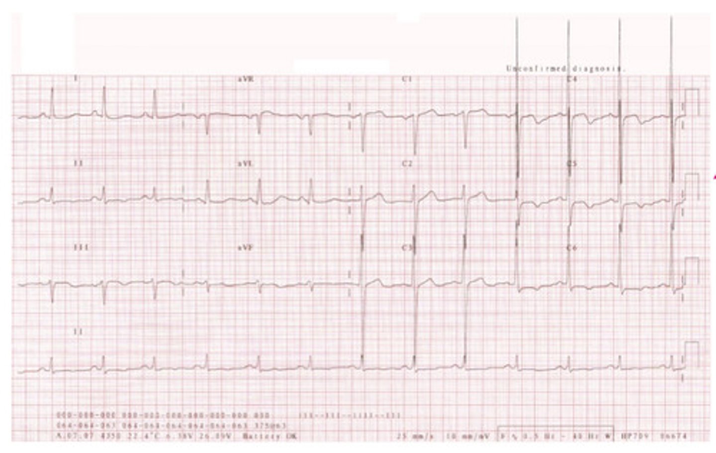
what is this? hint: low voltage & electrical alternans
pericarditis; ST
this is an example of _____, which has a key characteristic of diffuse ___ elevation
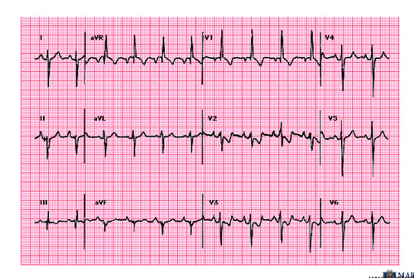
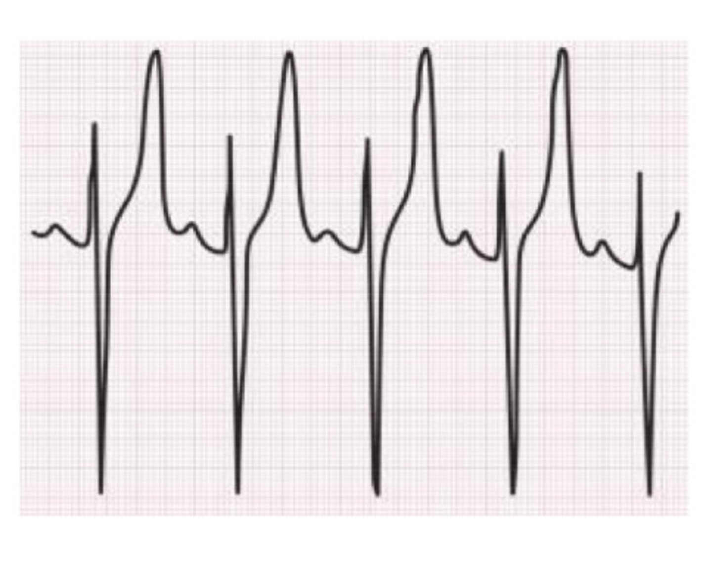
hyperkalemia (absent P waves, peaked T waves)
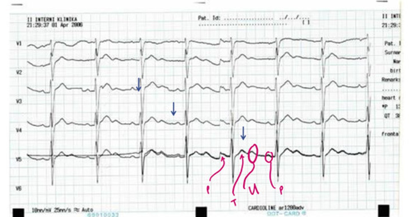
what is this
hypokalemia; u waves
this is an example of _______, which has characteristic ___ waves
hypokalemia (flat T waves, ST depression)
this is an example of ______, why
hyperkalemia =
this is an example of _____, characterized by sine wave pattern
hyperkalemia
this shows T wave peaking in _______
- calculate HR
- check intervals (PR, QTc, QRS width)
- check axis (normal, left, right)
- rhythm (ask these questions if not obvious): P wave before every QRS (& are they upright in I, II, AVR?), is QRS wide or narrow, is it regular or irregular
- blocks?
- check for evidence of infarct, injury, ischemia (ST segments, T waves, pathologic Q waves); remember LBBB, pacemaker, and WPW obscure findings
- check for chamber enlargement
- overall impression?
stepwise approach to interpreting EKG's