Developmental Biology Exam 1
 Call Kai
 Learn
Learn Practice Test
 Spaced Repetition
 Match
Match Flashcards
 Knowt Play
1/80

There's no tags or description
Looks like no tags are added yet.
Name | Mastery | Learn | Test | Matching | Spaced | Call with Kai | Chat |
|---|
No analytics yet
Send a link to your students to track their progress
81 Terms
Even though invertebrate embryos are morphologically different from humans, why are they still important model organisms? Give an example
Invertebrate research has applications to human/vertebrate development, e.g., the hedgehog protein
In Drosophila, the hedgehog protein is a secreted signaling molecule involved in pattern formation
Drosophila larvae with mutated hedgehog genes have disrupted ventral pattern formation
The hedgehog protein has been evolutionarily conserved in vertebrates as well (Sonic hedgehog)
In humans, it can result in loss of symmetry along the midline (consequences may not be as major due to gene redundancy)
What is fertilization and what is the purpose?
Union of male and female gametes
Restores diploid condition
Activates development
When was the discovery of sperm? Who discovered it? What were they originally though to be?
Discovered by Antoni van Leeuwenhoek in 1678
Originally thought to be parasites
What were preformationsists?
Before sperm entry into the egg was documented, there were two categories of preformationists:
Spermists: believed life comes from male sperm, and that a tiny preformed human lived inside the head of the sperm cell, and that the egg just provided nutrients
Ovists: believed life comes from the egg, and that the maternal egg contained the preformed miniature offspring
What makes a good model organism?
Relevant to the question under study
Ease of growth in laboratory
Large brood size
Cheap
Accessibility of embryos
Size of eggs
Optical clarity
Genetics
Relevance to humans
Why are sea urchins good model organisms?
They are useful for studies of fertilization and the cell cycle
Produce many gametes
Cyclins
What is the difference between G and F actin?
G-actin is the free, single spherical monomer
F-actin is the long, linear polymer made of those monomers twisted into a double helix
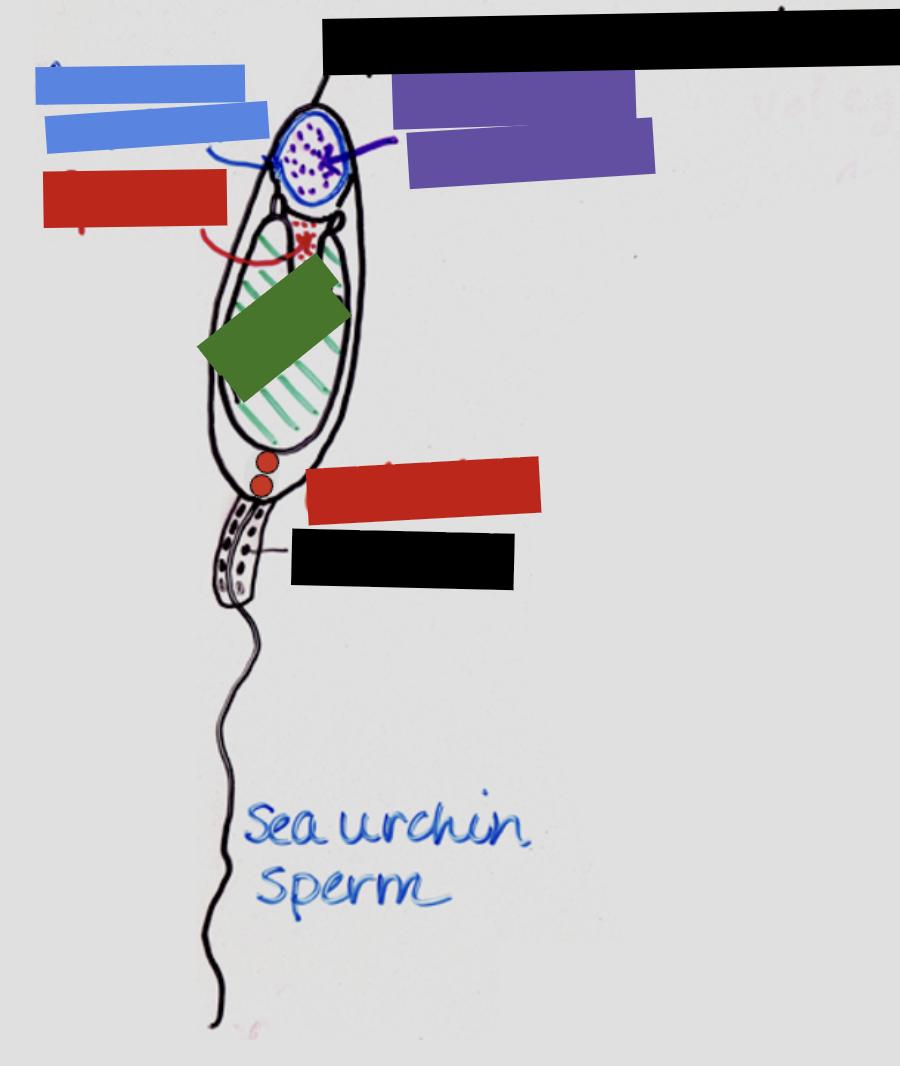
Identify the structures of sea urchin sperm


Identify the structures of sea urchin eggs

What are the four fertilization events?
Contact and recognition
Regulation of sperm entry
Fusion of genetic material
Activation of egg metabolism
What are the elements of recognition and contact?
Chemotaxis: contact, finding egg
Penetration of jelly layer
Sperm-egg recognition
What is chemotaxis? What are the characteristics?
Oriented movement of a cell/organism in response to an external chemical signal
Species-specific
Small and able to diffuse rapidly
Receptor located on sperm
Low concentration of chemoattractant: cells require high sensitivity to detect faint chemical trails and avoid receptor saturation
What are the chemoattractants used by sea urchins (Arbacia punctulata and Strongylocentrotus purpuratus)
Use sperm activating peptides (SAPs)
A. punctulata: resact
S. purpuratus: speract
How do SAPs work?
They:
Give orientation signal
Activate sperm motility
Provide species specificity
(RGC = receptor guanylyl cyclase)

What genes encode calcium channels?
CatSper genes
What happens if the chemoattractant concentration is too high?
Would result in no gradient
Saturates sperm receptors, therefore giving no directional cue
How does a sperm cell penetrate the jelly of the egg?
The acrosome reaction
Describe the acrosome reaction (in sea urchins)
It is triggered by contact with specific glycoproteins in the jelly layer (species-specific)
This causes protein-digesting enzymes released from the acrosome to digest a path through the jelly coat to the egg cell surface
Formation of the acrosomal process then occurs in sea urchins
Changes in pH allow the globular actin proteins between the nucleus and the acrosome to organize into fibrous actin microfilaments and extend forward to create the acrosomal process
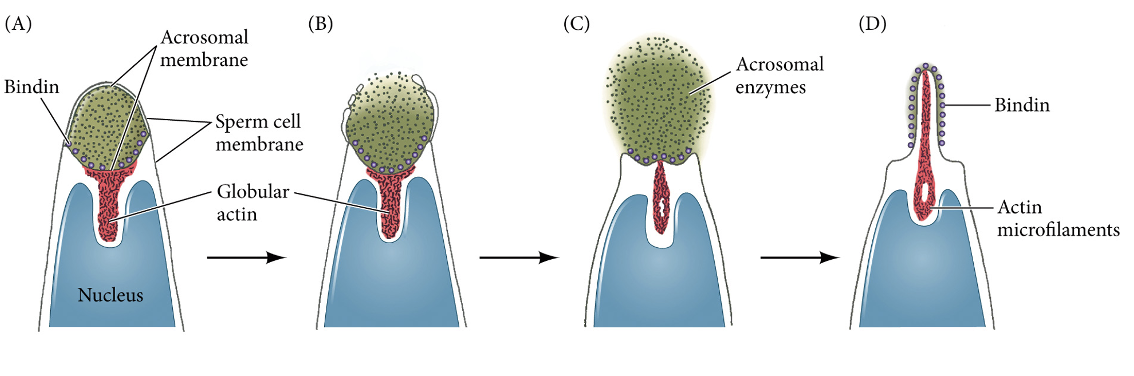
How is Ca++ concentration increased in the acrosome reaction? How is pH raised?
Jelly-sperm interaction activates:
Ca++ transporters (Ca++ moves into the sperm head)
Na+/H+ exchanger (Na+ moves in, H+ moved out, making cytoplasm more basic)
A phospholipase that makes IP3, which causes Ca++ release from internal stores (ER)
What does the increase in Na++ in the increasingly basic cytoplasm cause (in acrosome reaction)?
Triggers the fusion of the sperm plasma membrane and the outer acrosomal membrane, which allows the exocytosis of acrosomal enzymes
Activates the GTP-binding protein (RhoB) that organizes the actin cytoskeleton, leading to the formation of the acrosomal process

What is the function of the acrosomal process?
Penetrate the jelly layer
Promote adhesion to the vitelline envelope
Allows for fusion to happen
What is the GCD4161 Mantra?
Find it, lose it, move it:
Find it: observations to establish correlation
Lose it: loss-of-function experiment to establish necessity
Move it: gain-of-function experiment to establish sufficiency
Define “find it” (what questions are you trying to answer)
Look for:
Where within an embryo (or cell) does a particular protein accumulate
When during development does the protein accumulate
What method can be used to “find it”? Name an example of this being used
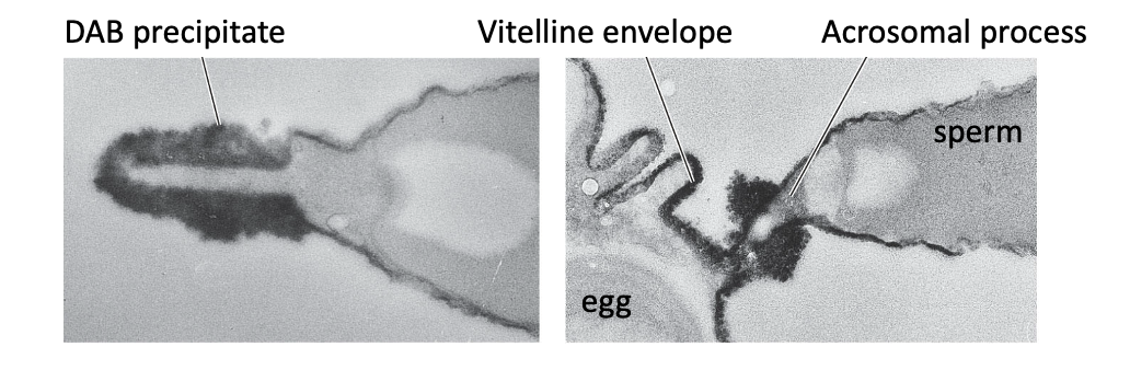
Immunohistochemical localization (aka immunolocalization):
a laboratory technique that uses specific antibodies to detect, image, and map the precise spatial location of target proteins or other macromolecules within preserved tissue sections
Ex: put bindin into a rabbit for it to make specific antibodies for it through immune response. Attach it to DAB, which forms a precipitate

Where is bindin found?
Inside the acrosome of intact sperm
On the outer surface of the acrosomal process following the acrosome reaction
Spreads onto the egg during binding
Define “lose it’ (what question are you trying to answer)?
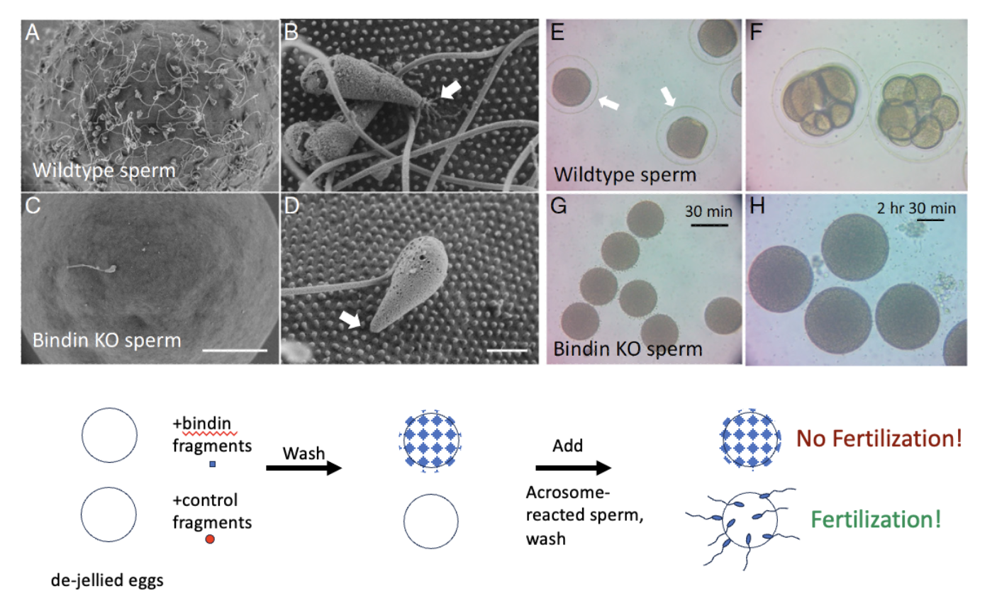
What is the molecule required for?
What methods can be used to “lose it”? Name an example
Knock out a gene
Ex: When bindin gene was knocked out,
Utilize an inhibitor of the molecule
Ex: mixing a de-jellied egg with peptide fragments of bindin, there was no fertilization
This is because binding sites have been pre-bound due to washing the egg with binding fragments
Define “move it” (what questions are you trying to answer)?
What is the power of the molecule?
Can the molecule cause the process to occur in a new location/at a new time?
Name an example for “move it”
The isolated bindin protein was found it to be capable of binding to dejellied S. purpuratus eggs, which indicated that the receptor for bindin was on the vitelline layer
Furthermore, both sperm bindin and egg jelly polysaccharides were species-specific—bindin isolated from the acrosomes of S. purpuratus bound to its own dejellied eggs but not to those of S. franciscanus

What does bindin do?
Sperm-egg recognition
Bind to egg binding receptors (EBR) extend through the egg plasma membrane and vitelline envelope and attach bindin protein on the acrosomal process
This is another step for species specificity
What does it mean that bindin is fusogenic in some species?
It can promote membrane fusion
Egg-binding receptors (EBRs) also have fusion domains
What is a fertilization cone?
A temporary, cone-like cytoplasmic projection that forms on the surface of an egg's plasma membrane when a sperm makes contact and fuses with it
Actin polymerization in egg makes fertilization cone, which makes bridge with acrosomal process
Why is polyspermy bad?
It messes with the diploidy
Centrioles help with early cell divisions; multiple centrioles would result in the cell trying to divide in multiple ways at the same time, often leading to issues with chromosome segregation and death
What are the characteristics of the fast and slow blocks to polyspermy?
Fast block:
Electrical
Fast (1-3 sec)
Transient (60s)
Slow block:
Mechanical
Slow (30-60 sec)
Permanent
How does the fast block to polyspermy work?
Initially, the egg is at a resting potential of -70mV
Chemicals from the fusing sperm cytoplasm open voltage-gated Na+ channels, which allows Na+ to enter the egg, raising the membrane potential to +20 mV
When the potential is negative, sperm can bind and fuse
When the potential is positive, sperm can bind, but fusion is blocked, preventing more sperm from binding
What could a voltage clamp be used for in polyspermy-blocking experiments?
To maintain a sea urchin egg at a specific electrical potential for experimentation
Why is fast blocking alone not enough?
The effect wears off
After roughly 70 seconds, the potential becomes negative again, meaning more sperm could bind and fuse again
How does the slow block to polyspermy work? What is the other name for it?
Also called the cortical granule reaction
Upon sperm entry, cortical granules fuse with the egg cell membrane, releasing their contents
This results in the vitelline enveleope and plasma membrane to separate (after release, the VE become the fertilization envelope/FE)
What are cortical granules?
Effectors of the slow block
They lie just beneath the sea urchin egg cell membrane: about 15,000, each about 1 μm in diameter
In close proximity to the plasma membrane, as they will fuse with the plasma membrane to exocytose their contents for the slow block to polyspermy
What is in a cortical granule? What does each one do?
Proteases, such as cortical granule serine proteases (CGSP)
Cleave the protein tethers between the PM and VE
Mucopolysaccharides / glycosaminoglycans
Absorb water to expand the space between the cell membrane and the fertilization envelope so that the envelope moves radially away from the egg
Hyaline
Form the hyaline layer, which provides support for the blastomeres during cleavage
Peroxidases
Stabilize the fertilization envelope by crosslinking adjacent proteins
What is the cortical flash?
When fertilization happens and the membrane potential becomes depolarized, Ca++ channels throughout the egg membrane open (not to be confused with the Na+ channels also opening)
Leads to Ca++ entering from the outside, creating a cortical flash: a brief rise in Ca++ levels throughout the cortical cytoplasm (outer layer of cytoplasm)
May contribute to the fast block to polyspermy
What is the calcium wave?
The second flux of Ca++
Starts where the sperm enters and transverses the entire egg
In this case, the Ca++ comes from inside the oocyte, from the ER

What surrounds cortical granules in unfertilized eggs? Why?
Endoplasmic reticulum
Since Ca++ is released from the ER after fertilization, the ER must be near the CGs, as the Ca++ promotes cortical granule fusion with the plasma membrane
What is the pathway of the slow block to polyspermy?
Note: Ca++ release triggers Ca++ release channels on neighboring ER, wave propagates around egg as additional ER releases Ca++

Are unfertilized sea urchin eggs translationally active?
No, they are translationally inactive, but translationally competent
Endogenous mRNAs are present, but not translated

What does this show?
An unfertilized egg is translationally inactive
It contains endogenous mRNAs, but they’re not being translated
If exogenous mRNA is added to an unfertilized egg, it is translated
Translational machinery is functional, but stored mRNAs are untranslatable
How are stored mRNA kept inactive in unfertilized eggs?
Prevent polyadenylation (polyA tail) of an mRNA
Mask the mRNA with RNA-binding proteins that block the translational machinery
Keep an mRNA out of the cytoplasm (such as histones that pack DNA into heterochromatin that can’t be transcribed)
Bind the mRNA with miRNAs
What are ways to activate mRNA translation at fertilization?
Activate polyA polymerase, unmask, break down the nuclear envelope
pH changes (increase)
Ion concentration changes
Change activity of kinases/phosphatases
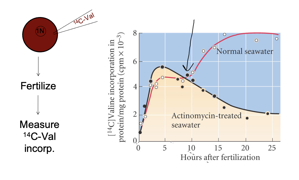
Explain what this means. What is the arrow pointing at?
Fertilized eggs were placed into either regular seawater or seawater treated with actinomycin (which inhibits transcription of zygotic DNA
In normal seawater, tranlation of mRNAs then transitioned into transription of zygotic DNA
In the actinomycin-treated water, protein levels eventually dropped as zygotic DNA could not be transcribed
The switch from mRNA to zygotic DNA is called the mid-blastula transition (MBT)
What are the differences between early and late cleavage stages? What marks the transition?
The transition from early to late cleavage stages is marked by the MBT/MZT
Early cleavage: M → S
Fast
Synchronous
Cells decrease in size (reductive divisions)
Later cleavage: M → G1 → S → G2
Slower
Asynchronous
Cells maintain size (grow back)

Identify
Note:
Spindle orientation controls the plane of cytokinesis
Cytokinesis is perpendicular to axis of the mitotic spindle
Cytokinesis always occurs equidistant from the two spindle poles

What are the two types of cleavage? What are the subtypes?
Holoblastic: complete or total cleavage where the division furrow passes entirely through the egg
Meroblastic: partial or incomplete cleavage where the division furrow does not penetrate through
Discoidal: a type of partial embryonic cell division where cell division happens only in a small disc of cytoplasm at the top of a heavy yolk
Superficial: occurs without immediate cytoplasmic division (cytokinesis), producing a multinucleated cell

What are the yolk types for the two types of cleavage (holo- and meroblastic)?
Holoblastic:
Isolecithal: sparse, evenly distributed yolk
Mesolecithal: moderate vegetal yolk disposition
Meroblastic:
Telolecithal: dense yolk throughout most of the cell
Centrolecithal: yolk in center of the egg

What is a blastomere?
types of embryonic cells produced by the division (cleavage) of a zygote right after fertilization
What is the difference between radial and spiral cleavage? Which do sea urchins have?
While they are both types of holoblastic cleavage:
Radial cleavage results in daughter cells that stack directly on top of each other, forming neat, symmetrical vertical tiers
Spiral cleavage results in daughter cells that sit at oblique angles, creating a twisted, interlocking, and diagonal spiral pattern
Sea urchins exhibit radial
What are meridional and equatorial divisions?
Meridional runs north-south (along lines of longitude or pole-to-pole), while equatorial runs east-west (parallel to the equator)

What is the difference between the animal and vegetal poles?
Animal pole: consists of fast-dividing, cytoplasm-rich cells with little yolk
Vegetal pole: consists of slow-dividing, large cells packed with dense yolk nutrients
What is unique about the 4th cleavage in sea urchin development? What other cells does this happen in?
The vegetal half begins to cleave asymmetrically
It does so by positioning the mitotic spindle asymmetrically in the cell (since the cleavage furrow is always in the center of the spindle)
Also happens in oocytes and stem cells
Add the diagram from the slides once they are posted
Why are primordial germ cells set aside early in development?
To protect the germline from the accumulation of somatic mutations and to preserve the totipotency or pluripotency needed to transmit clean genetic information to the next generation
What does a sea urchin embryo look like before gastrulation?
Vegetal pole will be wider (hatched blastula stage)

What are the types of cell movements during gastrulation?
Invagination
Ingression
Epiboly
Involution
Delamination
Convergent extension
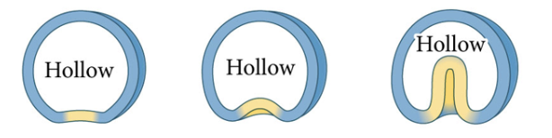
What is invagination (cell movement during gastrulation)?
Infolding of a sheet (epithelium) of cells
What is ingression (cell movement during gastrulation)?
Migration of individual cells from the surface into the embryo’s interior
Individual cells become mesenchymal and migrate independently

What is epiboly (cell movement during gastrulation)?
Movement of epithelial sheets, spreading as a unit to enclose deeper layers of the embryo
Can occur by cells dividing, by cells changing their shape, or by several layers of cells radially intercalating into fewer layers

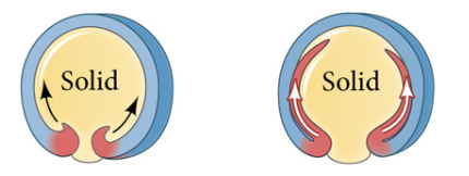
What is involution (cell movement during gastrulation)?
Inward movement of an expanding outer layer so that it spreads over the internal surface of the remaining external cells
What is delamination (cell movement during gastrulation)?
Splitting of one cellular sheet into two more or less parallel sheets
While on a cellular basis it resembles ingression, the result is the formation of a new epithelial sheet of cells

What is convergent extension (cell movement during gastrulation)?
Movement of more lateral cells of all germ layers toward the midline
This leads to a convergence of those cells, causing their intercalation along the medial-to-lateral axis
As these cells converge en masse, the embryo extends along the anterior-to-posterior axis

What is gastrulation?
Movement and rearrangement of blastomeres to form the three germ layers that will comprise the embryo: ectoderm, endoderm, and mesoderm
What is the first step of gastrulation in sea urchins?
The floor plate thickens (the vegetal pole cells thicken)
What is the second step of gastrulation in sea urchins? What processes are involved?
Ingresssion: large micromeres (primary mesenchyme cells/PMCs/skeletogenic/large micromeres) move inwards from the vegetal region
This is an epithelial-mesenchymal transition (EMT)
Involves:
Apical-basal polarity: the dynamic reorganization and eventual loss or remodeling of a cell's distinct top (apical) and bottom/side (basal/lateral) structural domains to allow an epithelial cell to detach, change shape, and move inward into a tissue
Apical constriction
Basal lamina remodels
De-adhesion
Cell motility
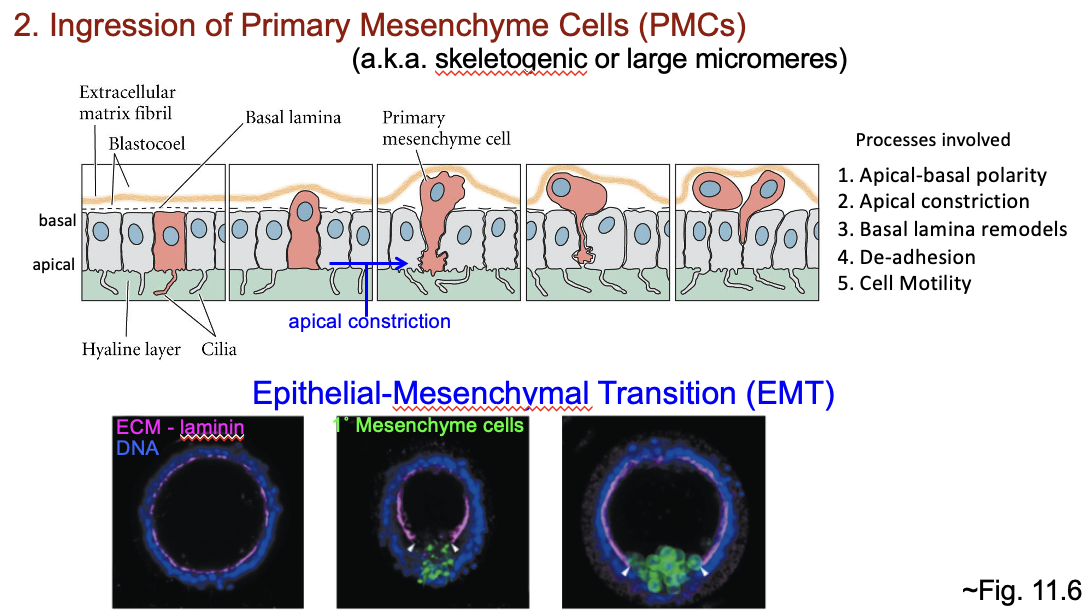
What happens after the epithelial-mesenchymal transition?
The primary mesenchyme cells begin extending and contracting long, thin, filopodia to the ectodermal wall
Allows the cells to move by extending, attaching, and then contracting their filopodia
The cells migrate to the points of FGF synthesis and arrange themselves in a ring along the animal-vegetal axis
The primary mesenchyme cells eventually make spicules (skeleton)

How do we know that spicules are micromere-derived? What does this technique allow for us to create?
Lineage tracing: by injecting dye into the micromere, progeny of the cell maintain the dye, allowing you to see where they are in the blastula
Allows you to make a fate map: a map of the early embryo that indicates what cell types/structures will arise from a given blastomere
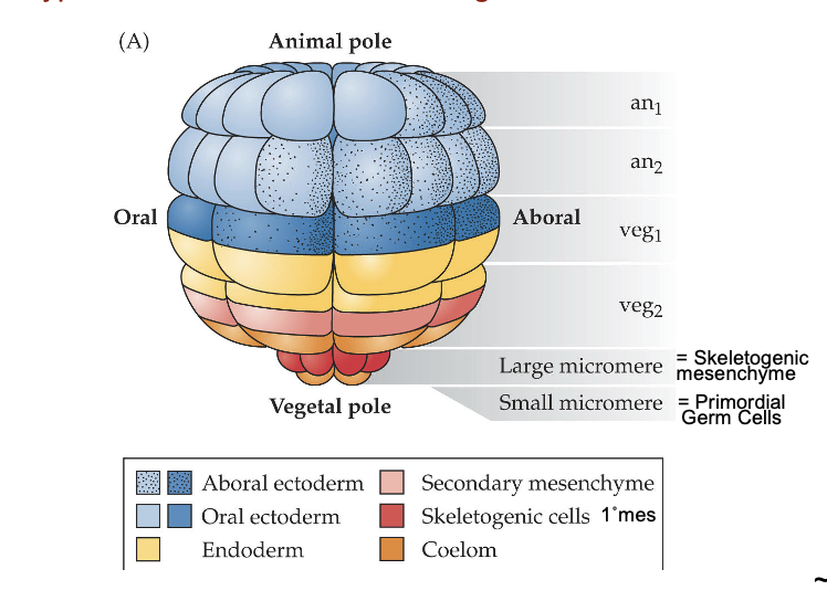
How do we know that micromeres know to make spicules, rather than relying on signals from other cells?
By isolating micromeres and putting them on a petri dish, it was found that they make spicules, meaning spicule production is not signal dependent
What is the third step of gastrulation in sea urchins?
Invagination to initiate archenteron formation
As the skeletogenic mesenchyme cells leave the vegetal region of the spherical embryo, important changes are occurring in the cells that remain there:
These cells thicken and flatten to form a vegetal plate, changing the shape of the blastula
The vegetal plate involutes inward by altering its cell shape, then invaginates about one-fourth to one-half of the way into the blastocoel before invagination suddenly ceases
The invaginated region is called the archenteron (primitive gut), and the opening of the archenteron at the vegetal pole is the blastopore
What is the fourth step of gastrulation in sea urchins?
Archenteron extension via:
Convergent extension: tissue narrows (converges) along one axis while lengthening (extends) along a perpendicular axis
Cell division
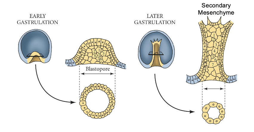
What is the fifth step of gastrulation in sea urchins?
Secondary mesenchyme cells form from the tip of the archenteron during and after the elongation phase of gastrulation and extend filopodia to where the mouth eventually forms


What does this experiment show?
Induction:
The autonomously specified large micromeres produce paracrine and juxtacrine factors that conditionally specify the fates of their neighbors
These factors signal the cells above the micromeres to become endomesoderm (the endoderm and the non-skeletogenic secondary mesenchyme cells) and to invaginate into the embryo
Animal cap cells were induced by micromeres to change their fate, resulting in endoderm
Micromeres retained their fate and made spicules
What is specification?
The first stage of commitment to a fate
The cell/tissue is capable of differentiating autonomously (by itself) when placed in a neutral environment. Fates of cells at this stage are capable of being reversed
Requires no signals to differentiate
Animal pole cells are specified to make ectoderm
What is conditional specification?
When a cell’s fate can be altered by its environment. Usually achieved by interactions with other cells, often through cell signaling molecules
Requires signals to differentiate
What is determination?
The stage of commitment following specification
The cell/tissue is committed to a fate and will retain that fate when placed in a new location, even if it is a non-neutral environment
Considered irreversible

What does this experiment show?
A “move it” transplantation experiment to test micromere function