7. Signal Transmission at Synapses
 Call Kai
 Learn
Learn Practice Test
 Spaced Repetition
 Match
Match Flashcards
 Knowt Play
1/62

There's no tags or description
Looks like no tags are added yet.
Name | Mastery | Learn | Test | Matching | Spaced | Call with Kai | Chat |
|---|
No analytics yet
Send a link to your students to track their progress
63 Terms
Synapse
A region where communication occurs between two neurons or between a neuron and an effector cell
Synapse: Function
Allows information to be filtered and integrated that are essential for homeostasis
Synapse: Presynaptic Neuron
The neuron that carries a nerve impulse toward a synapse and sends the signa
Synapse: Postsynaptic Cell
A cell that receives the action potential, whether it be a postsynaptic neuron or an effector cell
Synapse: Postsynaptic Neuron
It receives a signal at the synapse and carries a nerve impulse away from the synapse.
Synapse: Effector Cell
It responds to the signal at the synapse
Axodendritic
Refers to synapses between neurons
Axosomatic
Refers to synapses between axon to dendrite
Axoaxonal
Refers to synapses between axons and cell body
Electrical Synapses
A region where action potentials conduct directly between the plasma membranes of adjacent neurons through structures called gap junctions, most common in smooth muscle, cardiac muscle, and the developing embryo.
Electrical Synapses: Advantages
Faster Communication and Synchronization
Electrical Synapses: Advantages: Communication
Action potential passes directly from the presynaptic cell to the postsynaptic cell, unlike the delay communication at the chemical synapse
Electrical Synapses: Advantages: Synchronization
A large number of neurons or muscle fibers can produce action potentials in unison if they are connected by gap junctions
Chemical Synapses
A region where neurons indirectly sends a message to another cell using chemical messengers called neurotransmitters, as they are separated by a synaptic cleft
Chemical Synapses: Synaptic Cleft
The small space (about 20–50 nm wide) separating the presynaptic and postsynaptic membranes at a chemical synapse that is filled with interstitial fluid
Because the nerve impulse cannot cross the synaptic cleft, the presynaptic neuron converts the electrical signal into a chemical signal using a neurotransmitter across the synaptic cleft
Chemical Synapse: Movement
The neurotransmitter diffuses across the synaptic cleft through the interstitial fluid to eventually bind to receptors on the postsynaptic neuron.
Chemical Synapse: Postsynaptic Potential
A type of graded potential produced in the postsynaptic neuron in response to a neurotransmitter.
Synaptic Delay
The short delay (about 0.5 msec) occurs while a neurotransmitter is released, crosses the synaptic cleft, and produces a response in the postsynaptic neuron.
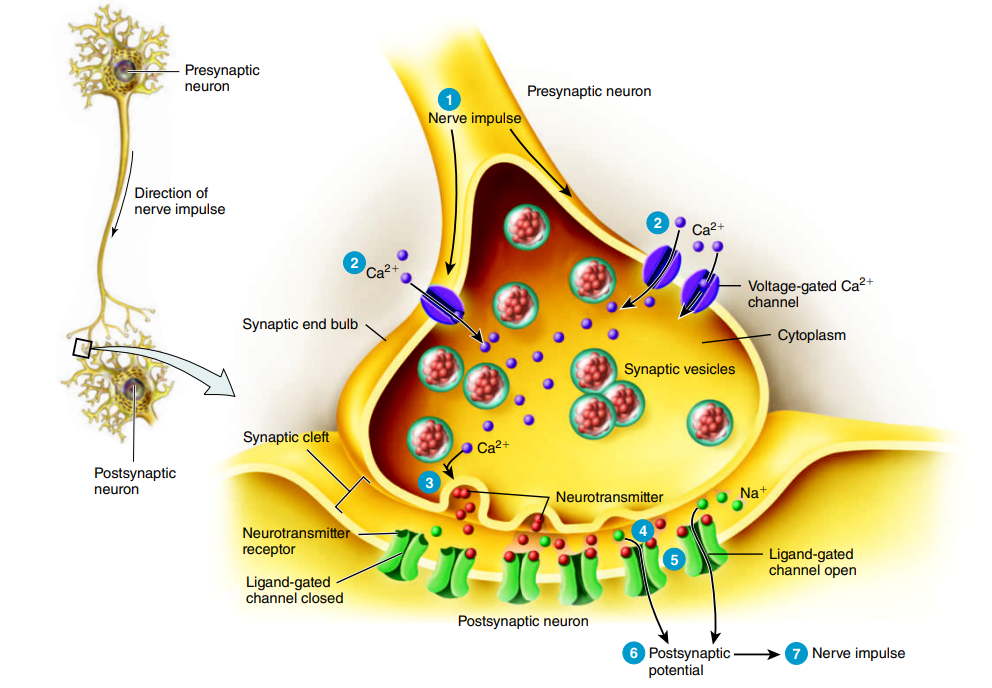
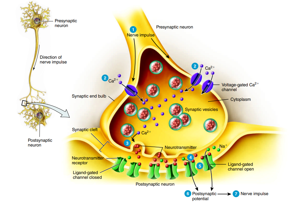
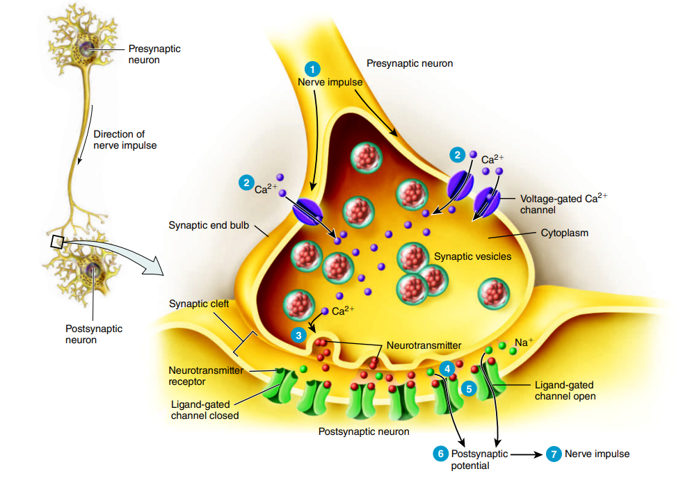
Chemical Synapse: Example Pathway: Step 1
A nerve impulse arrives at the synaptic end bulb or varicosity of the presynaptic axon.
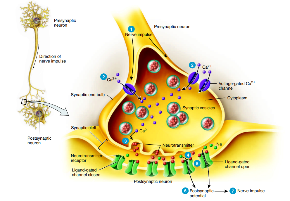
Chemical Synapse: Example Pathway: Step 2
The depolarizing phase of the nerve impulse opens voltage-gated Ca²⁺ channels in the synaptic end bulb to let Ca²⁺ flow into the presynaptic neuron due to the concentration gradient
Chemical Synapse: Example Pathway: Step 3
The increase in Ca²⁺ inside the presynaptic neuron triggers exocytosis of synaptic vesicles, which releases neurotransmitter molecules into the synaptic cleft.

Chemical Synapse: Example Pathway: Step 4
Neurotransmitters diffuse across the synaptic cleft and bind to receptors on the postsynaptic neuron.
Chemical Synapse: Example Pathway: Step 5
Neurotransmitter binding to ligand-gated channels opens the channels, allowing specific ions to flow across the postsynaptic membrane.
Chemical Synapse: Example Pathway: Step 6
Ion movement through the opened channels changes the voltage across the postsynaptic membrane, producing a postsynaptic potential.

Chemical Synapse: Step 7
When a depolarizing postsynaptic potential reaches threshold, it triggers a nerve impulse in the axon of the postsynaptic neuron.

Neurotransmitter: Types of Graded Potential
Either produces an excitatory or an inhibitory graded potential
Neurotransmitter: Excitatory Postsynaptic Potential
A temporary depolarization of a postsynaptic membrane caused by the influx of positively charged ions into the cell, but it does not initiate an action potential
Neurotransmitter: Inhibitory Postsynaptic Potential
A temporary hyperpolarization of a postsynaptic membrane caused by the efflux of positively charged ions out of the cell, making it less likely to fire an action potential
Neurotransmitter Receptors: Types
Based on whether the neurotransmitter binding site and the ion channel are components of the same protein or are components of different proteins: ionotropic receptors or metabotropic receptors
Ionotropic Receptors
A neurotransmitter receptor that contains the neurotransmitter binding site and the ion channel, which are components of the same protein, is also known as a ligand-gated channel
Ionotropic Receptor: Neurotransmitter Binding
When the correct neurotransmitter binds to an ionotropic receptor, its ion channel opens, producing either an EPSP or an IPSP
Ionotropic Receptor: Excitatory Postsynaptic Potential
Many excitatory neurotransmitters bind to ionotropic receptors containing cation channels that allow Na⁺, K⁺, and Ca²⁺ to cross the postsynaptic membrane
Ionotropic Receptor: Excitatory Postsynaptic Potential: Effect
An EPSP depolarizes the postsynaptic cell, making it more likely to reach threshold and generate a nerve impulse.
Ionotropic Receptor: Inhibitory Postsynaptic Potential
Many inhibitory neurotransmitters bind to ionotropic receptors containing Cl⁻ channels that allow Cl⁻ ions to diffuse into the postsynaptic cell
Ionotropic Receptor: Inhibitory Postsynaptic Potential: Effect
Metabotropic Receptors
A ligand-gated channel that contains the neurotransmitter binding site but lacks an ion channel, yet coupled by
Metabotropic Receptor
A neurotransmitter receptor that has a neurotransmitter-binding site but does not contain an ion channel as part of its structure, but is connected to a separate ion channel through a membrane protein called a G protein.
Metabotropic Receptor: G Protein
A G protein is a membrane protein that connects a metabotropic receptor to a separate ion channel.
Metabotropic Receptor: G Protein: Function
When a neurotransmitter binds to the receptor, the G protein can either directly open or close the ion channel, or indirectly activate a “second messenger” in the cytosol, which then opens or closes the ion channel.
Metabotropic Receptor: Direct Pathway
Neurotransmitter binds → G protein activates → G protein directly opens/closes ion channel.
Neurotransmitter Removal
Neurotransmitters must be removed from the synaptic cleft so their effects on the postsynaptic cell do not continue indefinitely to prepare the synapse for another signal.
Neurotransmitter Removal: Methods
Diffusion, Enzymatic Degradation, and Uptake by Cells.
Neurotransmitter Removal: Diffusion
Once a neurotransmitter molecule is out of reach of its receptors, it can no longer exert an effect.
Neurotransmitter Removal: Enzymatic Degradation
Some enzymes could break down certain neurotransmitters
Neurotransmitter Removal: Uptake
Neurotransmitters can be removed from the synaptic cleft by being actively transported back into the neuron that released them (reuptake) or into neighboring neuroglia (uptake) by neurotransmitter transporters.
Synaptic Integration
The process by which a postsynaptic neuron combines (sums) the postsynaptic potentials it receives from many synapses.
Summation is the process in which graded potentials, including EPSPs and IPSPs, add together to increase the likelihood of meeting the treshold.
Summation: Types
Spatial and Temporal Summation
Summation: Spatial
Process in which several presynaptic end bulbs release neurotransmitters simultaneously, producing postsynaptic potentials at different locations at the same time.
Summation: Temporal
Process in which one presynaptic neuron repeatedly releases neurotransmitter, producing postsynaptic potentials at same locations at different time.
If excitatory effects are greater than inhibitory effects but still below threshold, an EPSP occurs without triggering a nerve impulse because the neuron is partially depolarized; subsequent stimuli can more easily reach threshold through summation.
If total excitatory effects are greater than inhibitory effects and reach threshold, one or more nerve impulses are triggered and continue as long as the EPSP remains at or above threshold.
If total inhibitory effects are greater than excitatory effects, the postsynaptic membrane hyperpolarizes (IPSP), making the neuron less likely or unable to reach threshold and generate a nerve impulse.