MBIO 1010 / Lab 1
1/23
There's no tags or description
Looks like no tags are added yet.
Name | Mastery | Learn | Test | Matching | Spaced |
|---|
No study sessions yet.
24 Terms
What are the eight microscope guidelines when wrapping up a lab?
Clean ocular lenses.
Wipe off any oil from objectives and stage.
No objectives sticking out of microscope.
Move slide holders so that they are centred on stage.
Move stage down to the max.
Wrap power cord around cord holder or base.
Check microscope with demonstrator.
Place microscope back with both hands.
What are the six things to write on your Petri plate?
Full name.
Seat number.
Bench number.
Name of the culture.
Medium of the culture.
Date of the culture.
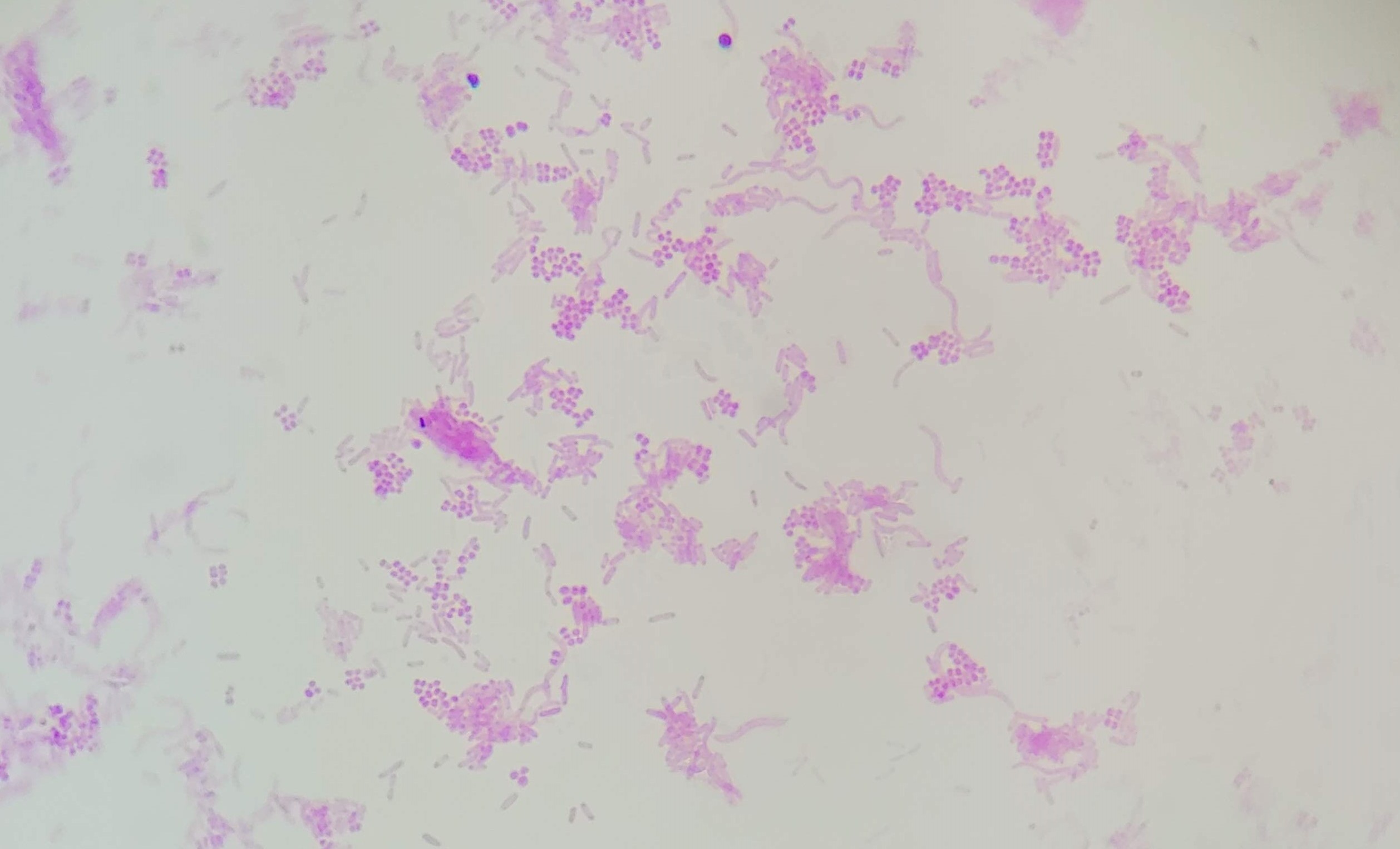
What is the prokaryote shown? What’s its domain?
Mixed bacteria of Bacteria.
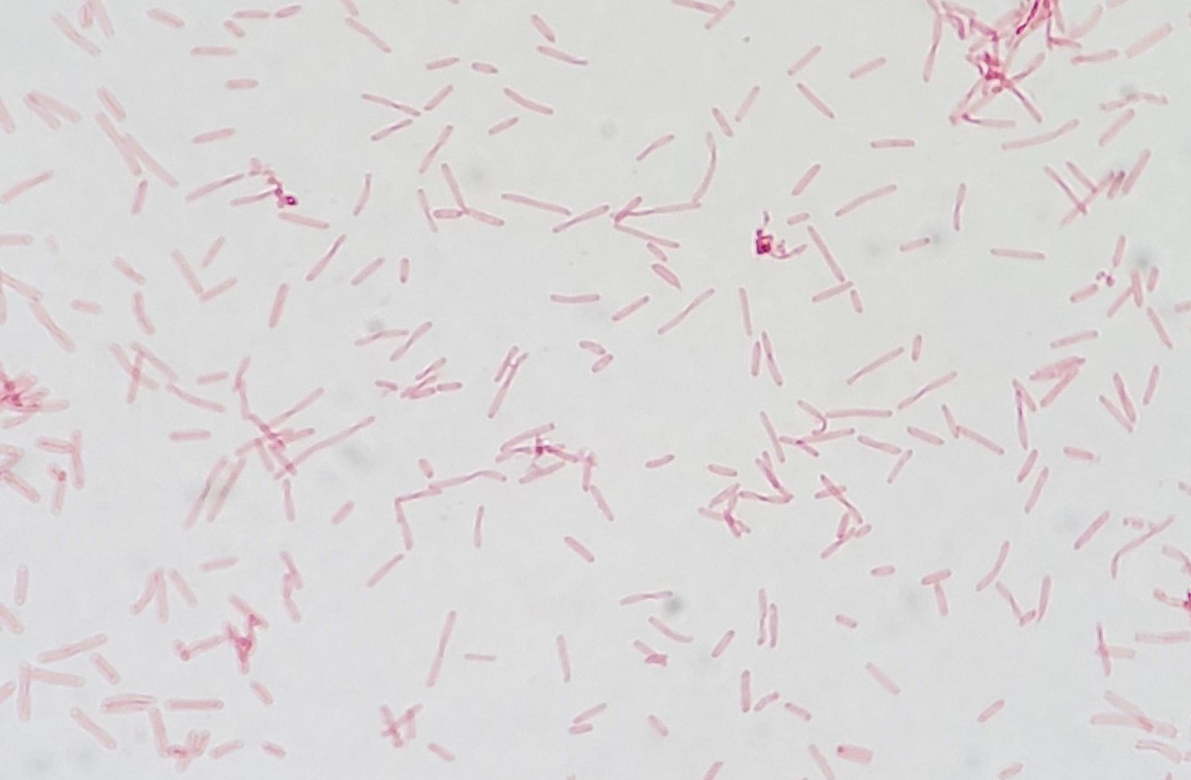
What is the prokaryote shown? What’s its domain?
Halobacterium salinarum of Archaea.
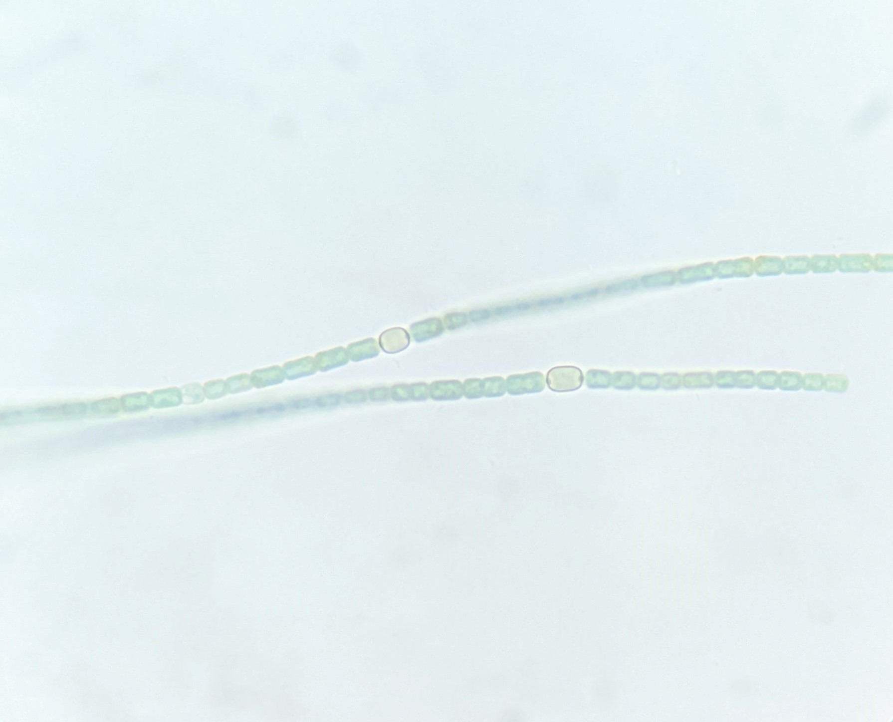
What is the prokaryote shown? What’s its domain?
genus Anabaena of Bacteria.
What is the eukaryote shown?
genus Euglena.
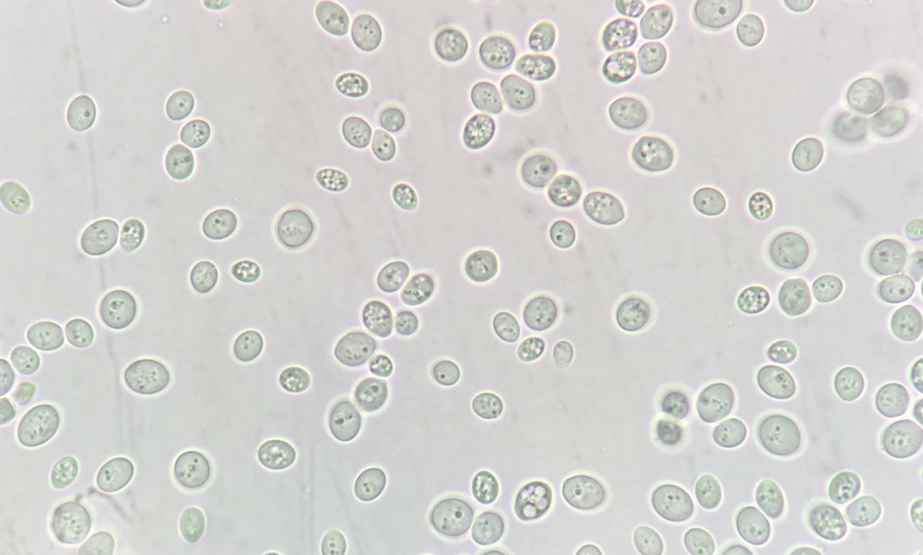
What is the eukaryote shown?
Saccharomyces cerevisiae. (Baker’s yeast or brewer’s yeast)
What is the eukaryote shown?
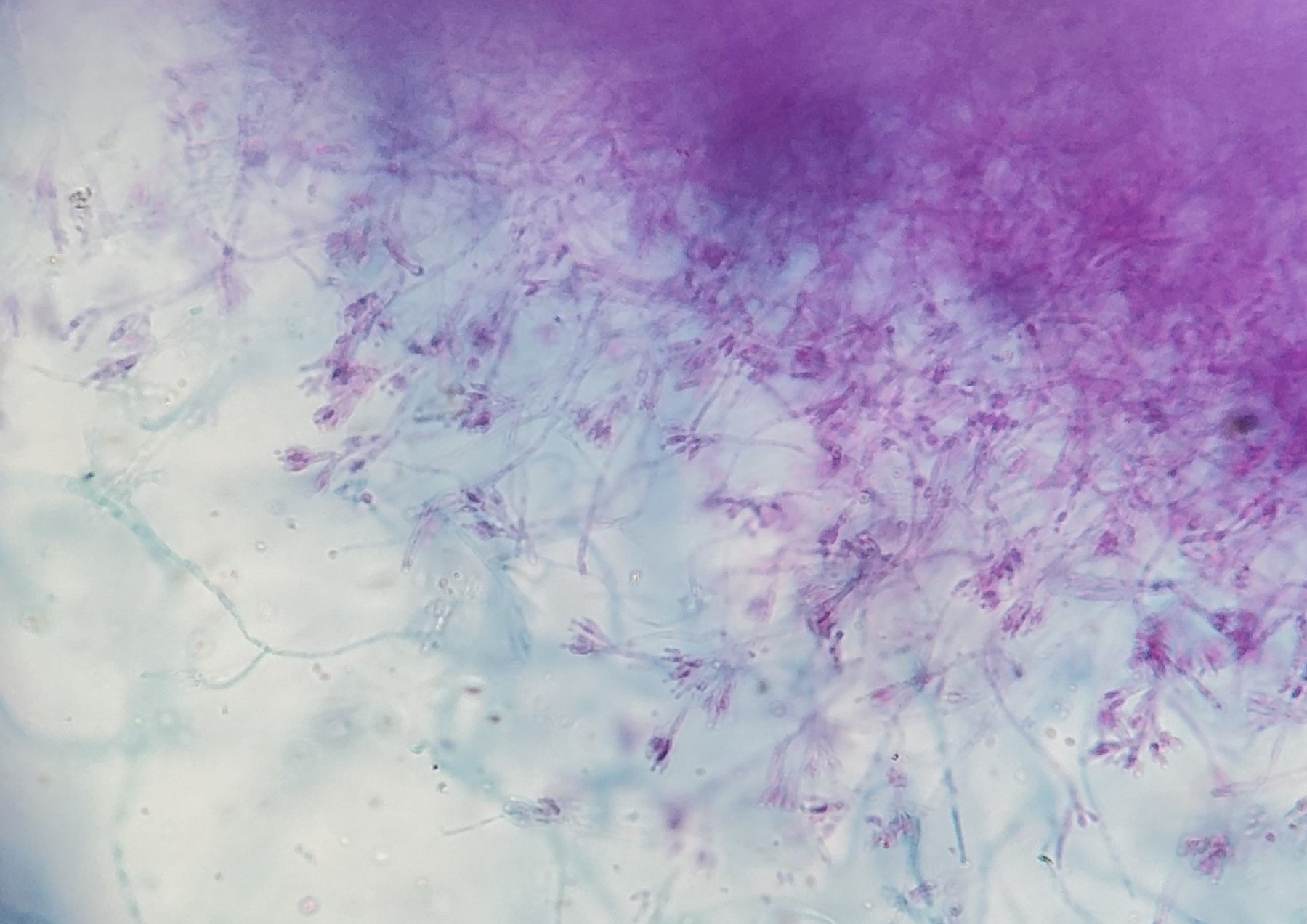
genus Penicillium. (common blue mold to produce penicillin and make blue cheese)
Where will you find your microscope in the microbiology lab room?
In the cupboard to the right of my work seat.
When disinfecting the workbench, you should…
Spread a thin layer of disinfectant on the counter and then let it dry down into a film on the surface of the work bench.
Where can you find microscope slides and cover slips?
On the shelf right above the work space.
Where can you find media supplies, like Petri plates for growing bacteria?
On the supply bench at the side of the room.
If you find you're having trouble looking into the microscope with both eyes, what should you do?
Grab the oculars and move them closer together, or further apart, until you can look into them comfortably.
Where is the condenser located on the microscope, and how is it controlled?
It's located directly under the stage, and it is controlled by a lever on the left side of the microscope.
How should the iris diaphragm slider be positioned in you want to look at a colourful specimen, like blue-green algae, or bacteria that have been stained with a dye?
The iris slider should be positioned over to the left, so that the iris is wide open to improve resolution.
Where are coarse and fine focus knobs on our microscopes?
There is both a coarse and fine focus knob on the left side of the microscope, while there is only a fine focus knob on the right.
When trying to find a specimen with my microscope, I should…
Start with the 10x objective, and the stage raised all the way to the top, so that I only need to turn my coarse focus knob one direction, until the specimen comes into view.
When moving up to the 100x oil immersion objective, I should…
Turn the nosepiece on the microscope to move the objectives out of the way, add a drop of oil to the slide, and then rotate the 100x objective into place, all without touching either of the focus knobs.
After using the microscope, you'll need to clean the oil from the 100x objective, before you put the microscope away. When cleaning the objective, you should…
Push the spring loaded objective in, and give the objective a good wipe with a Kim wipe to remove all the oil.
If you need to mix a culture of bacteria in a test tube, what is the best way to do that?
Hold the test tube by the glass, and use a finger on your other hand to tap the tube at the bottom.
In this lab period, you will make a wet mount of baker's yeast to look at it with your microscope. And the baker's yeast will be in a test tube (with a loose fitting metal cap). How will this test tube be labeled?
The test tube should be labeled with a short abbreviation, S. ce, because that saves the instructor time and space writing on test tubes!
When making a wet mount for microscopy, you should…
Draw a small circle on your microscope slide with a grease pencil or permanent marker, then place a single drop of culture in the middle of the circle, and then carefully place a cover slip on top.
What are the two reasons why we always label our plates on the back (or bottom) instead of on the lid?
Because then if the lid gets lost, we still know what's on the plate!
Because then we can still see through the lid to observe any colonies that grow.
What temperature do we incubate our plates at, when we have lab period once per week?
We incubate them at 25°C so the colonies grow slowly.