hematology exam 1 Diagram | Quizlet
 Call Kai
 Learn
Learn Practice Test
 Spaced Repetition
 Match
Match Flashcards
 Knowt Play
1/283

There's no tags or description
Looks like no tags are added yet.
Name | Mastery | Learn | Test | Matching | Spaced | Call with Kai | Chat |
|---|
No analytics yet
Send a link to your students to track their progress
284 Terms
major components of blood
plasma, red blood cells (erythrocytes), white blood cells (leukocytes), platelets
fluid phase
plasma
-fluid portion of blood proteins
-transport cells and molecules
cell phase
erythrocytes (RBCs)
-O2 and CO2 transport
-H+ buffering
leukocytes (WBCs)
-body defenses
platelets (thrombocytes in some species)
-hemostasis
-role in inflammation & wound healing
Blood cells have precursors in the
bone marrow
plasma and plasma proteins are mostly made in the
liver
blood volumes as % of body weight for horses
10-11% for "hot blooded" (aka race, muscular)
6-7% in "cold blooded" (aka older, work horses)
blood volume as % of body weight for dogs
8-9%
blood volume as % of body weight for cats and rodents
6-7%
blood volume as % of body weight for pigs
5-6%
what is the maximum amount of blood (in mL) that can be drawn from an 8.8 lb donor cat if you don't want to exceed 20% of its total blood volume?
(assuming cat's blood volume is 7% of body weight) ***
8.8 lb x 0.4536 kg/lb = 3.99 kg
3.99 kg x 0.07 (percent of blood volume) = 0.2793 kg
1 kg = 1000 ml; so.. 0.2793 kg = 279.3 ml
297.3 ml x 0.20 (20% blood volume) = 55.86 ml
the max amount of blood that can be drawn from the 8.8 lb donor cat without exceeding 20% of its blood volume is approximately 55.86 ml.
spun microhematocrit tubes
using a microhematocrit tube and a centrifuge, we can get a spun hematocrit, PCV, and total solids.
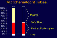
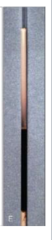
what are the blood components of a spun microhematocrit tube?
top: plasma
middle: buffy coat, WBCs + platelets
bottom: packed erythrocytes (RBCs)
very bottom: clay
mean PCV for dogs
45%
(some breeds i.e greyhounds are routinely > 50%)
mean PCV for horses + pigs
42%
mean PCV for sheep
38%
mean PCV for cats
37%
mean PCV for catle
35%
mean PCV for goats
28%
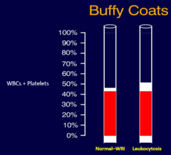
if there is a large buffy coat in a spun microhematocrit tube, it can indicate...
leukocytosis
i.e. infection or neoplasia
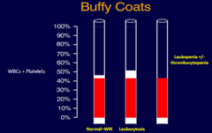
if there is an absence of a buffy coat in a spun microhematocrit tube, it can indicate...
leukopenia +/- thrombocytopenia
in dogs and cats, plasma will appear what color in a normal spun microhematocrit tube?
colorless
in horses and cattle, plasma will appear what color in a normal spun microhematocrit tube?
light yellow
due to the carotenoids in their diet.
an icteric plasma appears what color?
yellow
may indicate liver disease

a hemolytic plasma appears what color?
red
indicates breakdown of RBCs, most commonly a result of damage to blood during collection
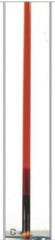
a lipemic plasma appears what color?
white
could indicate a lot of triglycerides, neoplasia, metabolic diseases that cause an imbalance of hormones.
plasma
functions:
-transport nutrients, by-products and waste, cells
-maintains homeostasis (pH, temperature, etc.)
92% water
8% solids
-nutrients: glucose, lipids, triglycerides, amino acids
-proteins: albumin & globulin
-hormones and enzymes
-electrolytes (minerals)
plasma proteins
collective properties
-transport nutrients, hormones, waste, drugs
-maintain fluid balance (colloid osmotic effects)
-acid-base balance
-immunity
-hemostasis
most are synthesized in the liver
in a patient with liver failure, what happens to the albumin?
there is a decreased ability to make albumin.
b/c albumin holds water within vasculature, decrease in it can result in edemas and swelling.
also, there are less clotting factors produced which can lead to bleeding out..
protein measurements w/ a refractometer
refractometer measures total solids (NA+, Cl-, glucose, urea, lipids) in plasma or in urine via measuring the light refraction
can measure for total solids (plasma proteins) and when using HPP, can also measure for fibrinogen.
if albumin is measured w/ analyzer, then the globulin can also be calculated (TP - albumin = globulins)
what parameters can lead to a false measurement of total protein when using a refractometer?
if the sample is lipemic (light refracts differently)
if there is markedly increased glucose or urea
if there are scratches on the glass
if there is hemolysis
total protein (g/dL) can be increased when
the patient is dehydrated (total body water loss)
or
protein gain (increased globulins)
total protein (g/dL) can be decreased when
the patient is over-hydrated (total body water gain/ dilution)
or
protein loss or decreased protein production
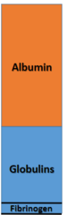
total protein mainly consists of
albumin, globulin, and a small about of fibrinogen
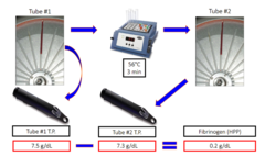
how can we test for fibrinogen using a refractometer?
a HPP (heat-precipitated protein test)
the difference between the TP of a heated microhematocrit and a non-heated microhematocrit yields the fibrinogen amount
Fibrinogen **
coagulation factor 1
-precursor to fibrin in coagulation
-optimal platelet aggregation
-scaffold for inflammatory cells, fibroblasts, and endothelial cells when in tissues (wound healing)
-produced by liver
-not present in serum
a positive acute-phase protein (APP)
-increases w/ inflammation
-most prominent horses, cattle, and goats
if you suspect inflammation in a horse, which protein would you assess?
fibrinogen
what kind of acute-phase protein is fibrinogen?
a positive-acute phase protein (APP) meaning it increases with inflammation
how can you calculate the globulins from a sample if you know the total protein and albumin?
total protein - albumin = globulins
how do you measure albumin?
albumin can only be measured w/ a chemistry analyzer, and not w/ the refractometer.
difference between serum and plasma ***
serum = plasma - clotting factors
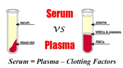
what effect does sample type have on protein measurement?
serum = can measure chemistry, proteins, total globulins, albumin
plasma = can measure WBCs, platelets, clotting factors
albumin ***
single homogenous protein
-small
-made by liver
-negative APP (decreases w/ inflammation)
transports organic and inorganic substances (cations i.e. Ca2+, metabolites, hormones, poorly soluble drugs, toxic substances)
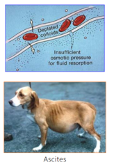
maintains colloidal osmotic pressure (COP) (holds water in vessels)
important for acid/base buffering
which protein is most important for maintaining colloidal osmotic pressure (COP) in the blood?
albumin
-low albumin, liver insufficient, can lead to ascites
globulins ***
all non-albumin proteins
-can be ascertained by a chemistry analyzer
-can be calculated if you know the TP and albumin (TP- albumin = globulins)
-increase w/ inflammatory, neoplastic disease states
-decrease w/ protein loss, immunodeficiency
-can be more accurately measured w/ serum protein electrophoresis (SPE)
why do we use serum when performing a protein electrophoresis?
if we used plasma, the fibrinogen can get in the way.
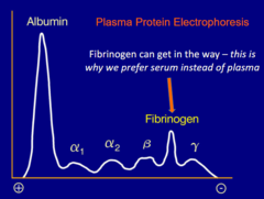
what can a serum protein electrophoresis tell us?
the concentration of different immunoglobulins
i.e. if there is monoclonal or polyclonal gammopathy.
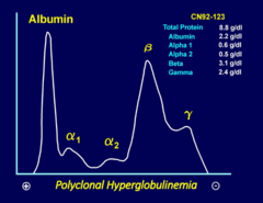
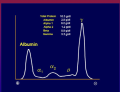
*the albumin is highly concentrated near the left or + side because it is a tiny molecule. smaller molecules travel fastest in gel electrophoresis.
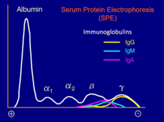
immunoglobulins
antibodies
-glycoproteins
-produced by lymphoid cells (B-lymphocytes, plasma cells)
-recognize antigens (clear infectious agents)
increased concentration of immunoglobulins indicates
gammopathy
monoclonal -> if made from a single cell clone (i.e. increase of just a single kind of immunoglobulin)
polyclonal -> if made from multiple cell clones (i.e. increase of multiple kinds of immunoglobulins)
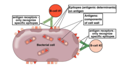
epitopes, antigen receptors, and antigens
epitopes- antigenic determinants on an antigen
antigens - components of the cell wall where epitopes are
antigen receptors - only recognize specific epitopes
polyclonal hyperglobulinemia **
=polyclonal gammopathy
seen w/ inflammation
(also liver disease, autoimmune disorders)
monoclonal hyperglobulinemia **
= monoclonal gammopathy
seen w/ neoplasia (i.e. B-cell lymphoma, multiple myeloma)
what is the acute-phase response?
part of the innate immune response
-modulates production of "acute phase proteins" (APP)
if APPs are positive, then they increase w/ inflammation
if APPs are negative, then they decrease w/ inflammation
positive APPs
positive acute phase proteins
-increase w/ inflammation
fibrinogen
CRP (c-reactive protein, helps immune cells and complement clear pathogens)
haptoglobin (binds free hemoglobin, anti-oxidant and anti-bacterial effects)
serum amyloid A (SAA) (can bind bacteria helping immune system clear organisms)
C3 (part of the complement cascade)
negative APPs
negative acute phase proteins
-decrease w/ inflammation
albumin (Alb)
transferrin (Tf)
what is hematopoiesis?
the formation of blood cells
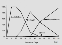
where does hematopoiesis occur in the fetal period? **
it starts in the yolk sac, then the liver, spleen, and finally the bone marrow
what is the primary site for hematopoiesis during the post-natal periods?
the bone marrow
the bone-marrow microenvironment contains .....
stem cells (can't be seen with a light microscope)
-hematopoietic stem cells, hematopoietic progenitor cells, mesenchymal stem cells
stromal elements (provide structure/support)
-endothelial cells, stromal cells, macrophages, adipocytes, extracellular matrix ECM)
hematopoietic precursors
-erythroid, myeloid, lymphoid, megakaryocytic
what are the hematopoietic cells of the bone marrow microenvironment? what are some examples?
the hematopoietic stem cells (HSCs)
- can differentiate in to all blood cell types & some tissue cell types (macrophages, osteoclasts, mast cells)
hematopoietic progenitor cells
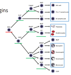
-common lymphoid progenitors (CLP) - differentiate to T-lymphocyte-natural killer cell progenitor and B lymphocyte progenitor
-common myeloid progenitor (CMP) - differentiate in to megakaryocyte-erythroid progenitor (RBCS + platelets) and granulocyte-monocyte progenitor (neutrophil, eosinophil, monocyte, basophil, dendritic cell, mast cell)
what cells are the supporting cells of the bone marrow microenvironment? what are some examples?
the stromal elements
endothelial cells, stromal cells, macrophages, adipocytes, and the extracellular matrix
hematopoietic stem cells
-least differentiated type of hematopoietic cell
-sustained self--replication & slow (@8-10 weeks)
can differentiate into all blood cell types and some tissue cell types (macrophages, dendritic cells, osteoclasts, mast cells)
hematopoietic progenitor cells
-proliferation
-limited self-replication, requires replenishment by HSCs
-more rapid than HSCs
-more restricted lineage potential than HSCs
-1% of bone marrow cells in adults, higher in neonates
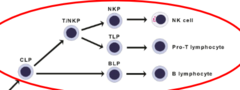
Common lymphoid progenitor (CLP)
Common myeloid progenitor (CMP)
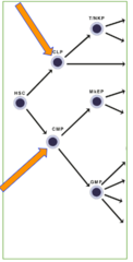
Common lymphoid progenitor (CLP)
hematopoietic progenitor cell
can differentiate into
T/NKP - T lymphocyte and natural killer cell progenitor
BLP - B lymphocyte progenitor
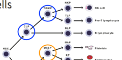
Common myeloid progenitor (CMP)
hematopoietic progenitor cell
can differentiate into
MkEP - megakaryocyte and erythroid progenitor
(RBCs and platelets)
GMP - granulocyte-monocyte progenitor
(neutrophils, eosinophils, monocytes, basophils, dendritic cells, mast cells)
mesenchymal stem cells
produce
-stromal cells and adipocytes
-endothelial cells
-fibroblasts and myoblasts
-osteoblasts and chondrocytes
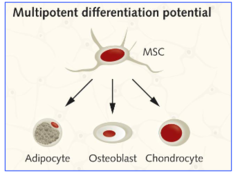
what cell types can arise from mesenchymal stem cells?
stromal cells and adipocytes
endothelial cells
fibroblasts and myoblasts
osteoblasts and chondrocytes
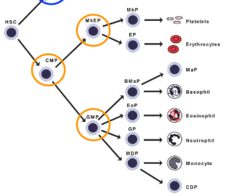
what cell types can arise from hematopoietic stem cells?
HSCs can differentiate into all blood cell types and some tissue cell types
initially, HSCs differentiate into CLP and CMP
all blood cell types:
-NK cells
-Pro-T lymphocytes
-B lymphocytes
-platelets
-erythrocytes
-basophils
-eosinophils
-neutrophils
-monocytes
tissue cell types:
-macrophages
-dendritic cells
-osteoclasts
-mast cells
how is hematopoiesis maintained?
growth factor signaling via
-autocrine action (produced in marrow)
-paracrine action ( produced in marrow)
-endocrine action ( produced in peripheral tissues)
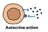
autocrine action
the hormone acts on the same cell that produced it
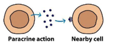
paracrine action
the hormone acts locally by diffusing from its source to target cells in the neighborhood
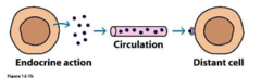
endocrine action
the hormone is distributed in blood and binds to distant target cells
if growth factor signaling is increased to control hematopoiesis, what happens?
there is increased production and decreased apoptosis
if growth factor signaling is decreased to control hematopoiesis, what happens?
there is decreased production and increased apoptosis
hematopoietic growth factors
glycoproteins - promote hematopoietic cell proliferation, maturation & survival
-produced in the marrow (paracrine and autocrine) and/or by cells in peripheral tissues (endocrine)
i.e. cytokines such as colony stimulating factors (CSFs), interleukins, and "-poietins"
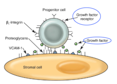
hematopoietic cells must have appropriate surface receptor to respond to a specific growth factor
what signals the bone marrow to produce new RBCS? **
Erythropoietin (EPO)
Erythropoietin (Epo) ***
-glycoprotein growth factor
-proliferation, differentiations, and survival of mature stem and progenitor cells
-main mechanism = inhibits apoptosis **
increased concentration of Epo -> decreased marrow transit time and promotes early release of "stress" reticulocytes (large forms)
produced in the kidney
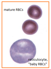
where is Epo synthesized?
mainly the kidney
-renal interstitial cells (endocrine action/signaling)
minimally, there is extrarenal production
-liver (esp. mammalian fetus)
-bone marrow macrophages and erythroid cells
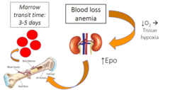
what is the marrow transit time for erythrocytes?
3-5 days
blood loss anemia can lead to decreased __________ which can result in increased _________ production
decreased oxygen (tissue hypoxia) and then increased Epo production in the kidneys to compensate
Why is this slide indicative or regenerative anemia in a dog? **
1) polychromasia (more "young" red blood cells, i.e. reticulocytes)
2) presence of nRBCs (nucleated red blood cells)
3) presence of H-J bodies (only has 1 piece of nuclear material)
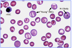
how can you differentiate H-J bodies from punctate reticulocytes?
H-J bodies will typically have 1 nuclear remnant which will stain purple; punctate will have multiple nuclear remnants which will not stain unless methylene blue
what can inhibit erythropoiesis?
1- insufficient/ lack of EPO -> results in more apoptosis (remember, EPO's main action is to inhibit apoptosis)
2- inflammatory cytokines at high concentrations
(contributes to anemia of inflammatory/chronic disease)
3- inhibitors themselves may induce apoptosis directly
what nutrients are required for ertyhropoiesis?
amino acids and essential fatty acids
metallic ions
-iron -> heme synthesis
-copper -> hephaestin, ceruloplasmin
-cobalt -> component of vitamin B12 (cobalamine)
vitamins
-vitamin B6 (pyridoxine) -> heme synthesis
-vitamin B12 -> folate metabolism
-tetrahydrofolic acid -> DNA, RNA, protein synthesis
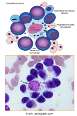
describe erythroid development
erythroid cells develop around macrophages
macrophages become nurse cells to support the development of erythrocytes
-source of early/intermediate acting growth factors (SCF (stem cell factor), IL-3 (interleukin 3) , GM-CSF (granulocytic-monocytic colony stimulating factor))
-erythropoietin (endocrine action/signaling)
-can be inhibited by inflammatory cytokines
-phagocytose expel nuclei and damaged/aged erythrocytes
-iron is recycled from the phagocytosed erythrocytes
what is the major growth factors for cells of the erythroid lineage? **
Erythropoietin (Epo)
where is Erythropoietin (Epo) produced and what effect do increased concentrations have on the erythroid lineage?
Epo is mainly produced in the kidney via renal interstitial cells (a minor amount is produced extra renally in the liver and bone marrow)
increased concentration of Epo decrease bone marrow transit time and promote early release of "stress" reticulocytes (larger form)
what is the marrow transit time for neutropoiesis?
6-9 days
neutropoiesis
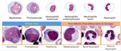
Granulocyte colony stimulating factor (G-CSF)
produced by fibroblasts, endothelial cells, T-lymphocytes, and mononuclear phagocytes (macrophages)
-produced in response to inflammatory cytokines
increase production of myeloid progenitors/precursors and decrease transit time
important for granulocyte development
granulocyte/monocyte colony stimulating factor (GM-CSF)
produced by fibroblasts, endothelial cells, T-lymphocytes, and mononuclear phagocytes (macrophages)
-produced in response to inflammatory cytokines
increase production of myeloid progenitors/precursors and decrease transit time
important for both granulocyte and monocyte development
how is granulopoiesis inhibited?
-mature neutrophils in marrow can release inhibitory substances
-neutrophils "clear" circulating G-CSF by binding to receptors (a negative feedback system)
-mature neutrophils in peripheral tissue remove stimulus for granulopoiesis
eosinophils in the bone marrow
-marrow transit ≤ 1 week
-marrow storage pool like neutrophils
-IL-3 and GM-CSF for early progenitors
*****
-IL-5 from TH2 lymphocytes for terminal maturation
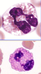
what specific interleukin is responsible for the terminal differentiation/maturation of eosinophils? **
IL-5 (interleukin-5).
basophils in the bone marrow
produced from a bi-potential basophil/mast cell progenitor
-similar growth factors for eosinophils (IL-3, GM-CSF, IL-5)
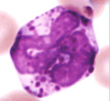
mast cells in the bone marrow
the mast cell progenitor MaP is released from the marrow to the blood
-Stem cell factor (SCF) is especially important for formation
terminal differentiation occurs in tissues
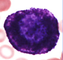
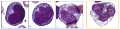
monocytopoiesis
production of monocytes
-growth factors IL-3, GM-CSF, M-CSF, and IL-34
may become macrophages or dendritic cells in tissues (influenced by amounts of various cytokines)
marrow transit time: 3 days
what is the marrow transit time for monocytes?
3 days
lymphocytes in the bone marrow
arise from common lymphoid progenitor (CLP)
B-lymphocytes
- discovered in Bursa of Fabricius - birds
- mature in Bone marrow - mammals
T-lymphocytes
-mature in the Thymus
Natural Killer (NK) cells
-develop in multiple tissue sites
what are G-CSF and GM-CSF?
Granulocyte colony stimulating factor (G-CSF) and granulocyte/monocyte colony stimulating factor (GM-CSF).
they are produced in response to inflammatory cytokines, and act as growth factors to increase production of myeloid progenitors/precursors and decrease transit time.
they are important for granulocyte/monocyte development
over what cell lineages do G-CSF and GM-CSF exert influence?
-basophils
-neutrophils
-eosinophils
-mast cells
-monocytes (GM-CSF only)