05a - Intro to Autonomic Pharmacology
 Call Kai
 Learn
Learn Practice Test
 Spaced Repetition
 Match
Match Flashcards
 Knowt Play
1/12

There's no tags or description
Looks like no tags are added yet.
Name | Mastery | Learn | Test | Matching | Spaced | Call with Kai | Chat |
|---|
No analytics yet
Send a link to your students to track their progress
13 Terms
autonomic nervous system (ANS)
controls involuntary functions including heart rate, blood pressure, pupil size, digestion, bronchial tone, gland secretion, sweating, urinary bladder infection, blood vessel tone
outflow comes from CNS, specifically the spinal cord and brainstem
CNS = brain + spinal cord
sympathetic and parasympathetic systems differ anatomically by where their outflow exits the CNS
sympathetic = thoracolumbar outflow
parasympathetic = craniosacral outflow
sympathetic and parasympathetic systems also differ by where their ganglia sit
sympathetic = near spinal cord and paravertebral chain
short preganglionic fiber; long postganglionic fiber
diffuse output pattern → chain allows signals to travel up/down the chain before synapsing
parasympathetic = near or inside target organ
long preganglionic fiber; short postganglionic fiber
targeted output pattern → specific and localized response
dual innervation → most organs are innervated by both sympathetic and parasympathetic fibers
sympathetic nervous system
“fight or flight” and “restore and repair” response
“fight or flight” → increased heart rate, increased blood pressure, pupil dilation, bronchodilation, blood flow redistribution, energy mobilization
“restore and repair” → maintain fluid balance, increase ADH, increase aldosterone, activate renin-angiotensin system, promote coagulation after injury, increase plasminogen activator inhibitor activity
outflow comes from thoracic and lumbar spinal cord
short preganglionic fiber; long postganglionic fiber
diffuse output pattern → chain allows signals to travel up/down chain before synapsing
parasympathetic nervous system
“rest and digest” response
“rest and digest” → digestion, sleep/rest, GI motility, glandular secretion, slower heart rate, urination, pupillary constriction
outflow comes from cranial and sacral regions
long preganglionic fiber; short postganglionic fiber
targeted output pattern → specific and localized response
afferent vs efferent neurons
afferent arrives at CNS; efferent exits CNS
efferent neurons → carry signals from CNS to effector organ
afferent neurons → carry sensory signals from periphery to CNS
autonomic neurotransmitters
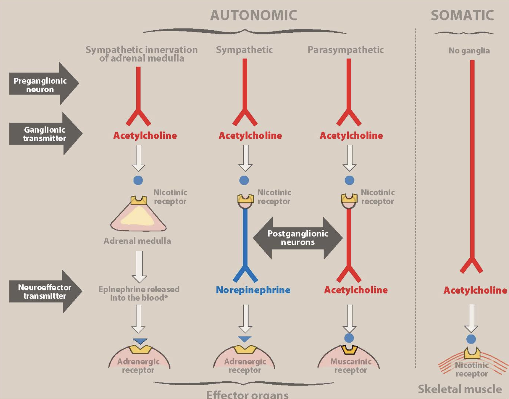
sympathetic innervation of adrenal medulla:
preganglionic neuron → acetylcholine → nicotinic receptor on chromaffin cells of adrenal medulla → epinephrine → adrenergic receptor of effector organs
epinephrine released in blood and acts as a hormone because it is distributed systemically
sympathetic innervation:
preganglionic neuron → acetylcholine → nicotinic receptor on postganglionic neurons (Nn) → norepinephrine → adrenergic receptor of effector organs (⍺/β)
parasympathetic innervation:
preganglionic neuron → acetylcholine → nicotinic receptor on postganglionic neurons (Nn) → acetylcholine → muscarinic receptor on effector organs
somatic innervation:
somatic motor neuron → acetylcholine → nicotinic receptor on skeletal muscle cells (Nm)
somatic nervous system
controls voluntary skeletal muscle movement
release acetylcholine and act on nicotinic muscle receptors (Nm)
innervates skeletal muscle
do not have ganglia
myelinated and produce fast responses
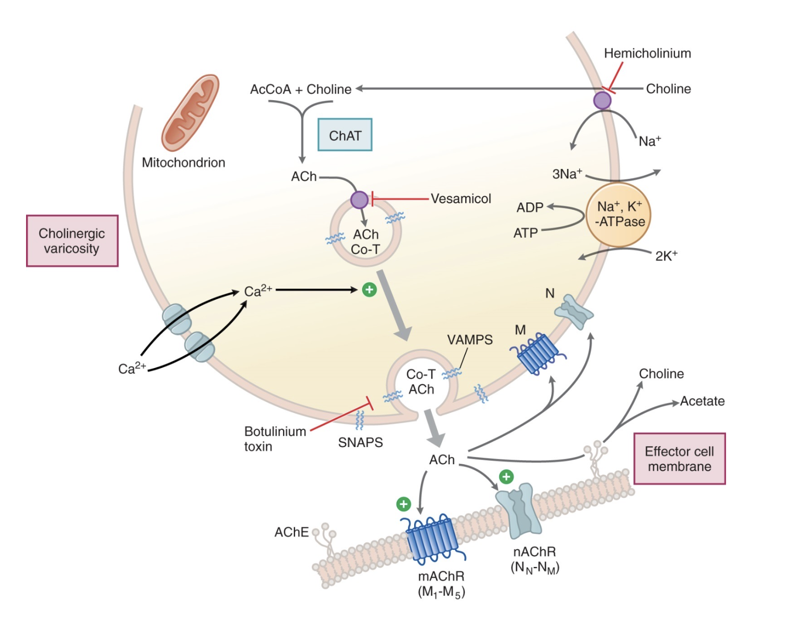
generalized cholinergic junction
choline uptake in nerve terminal via active transport
choline comes with acetyl-CoA, catalyzed by choline acetyltransferase (ChAT), to form acetylcholine
ACh is transported into vesicles
action potential reaches nerve terminal, and Ca2+ enters
vesicle fuses with membrane via vesicle attachment membrane proteins (VAMPS)
synaptosome-associated proteins (SNAPS) dock vesicle to membrane
ACh is released by exocytosis into synaptic cleft
ACh binds muscarinic (M1 - M5) or nicotinic receptors (Nn, Nm)
muscarinic receptors → GPCRs that activate phospholipase C to produce DAG and IP3, increasing intracellular release of Ca2+
nicotinic receptors → ligand-gated ion channel that stimulates flow of Na+ through channel
OR ACh is rapidly broken down by acetylcholinesterase on effector membrane, into choline and acetate
anticholinesterases inhibits AChE, causing stimulation of cholinergic receptors from increased ambient levels of ACh, due to decreased ACh breakdown
choline is recycled into the nerve terminal
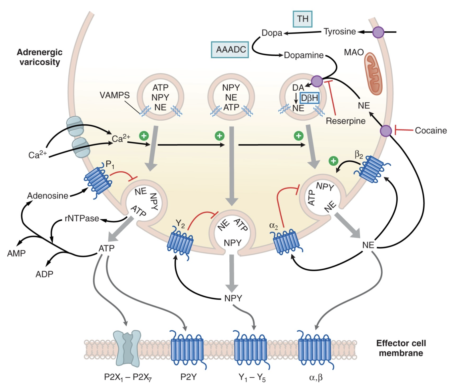
generalized noradrenergic junction
tyrosine enters nerve terminal
tyrosine hydroxylase converts tyrosine to DOPA
aromatic L-amino acid decarboxylase (AAADC) converts DOPA to dopamine
dopamine enters vesicle
dopamine-β-hydroxylase (DβH) converts dopamine to norepinephrine
reserpine → inhibits dopamine uptake into vesicle
NE released into nerve terminal with nerve stimulation
NE acts on ⍺ and β receptors on effector cell
OR NE can act on ⍺2 and β2 receptors on the nerve itself presynaptically
⍺2 → autoreceptor that inhibits further NE release
β2 → heteroreceptor that stimulates NE release
NE is a weak β2 agonist; epinephrine can also stimulate β2 receptor
OR NE can be taken back up by NET
80-90% of NE is reclaimed by adrenergic nerve terminal
cocaine → indirect adrenergic agent that inhibits NET to block NE uptake, allowing more NE to be taken up by ⍺ and β receptors
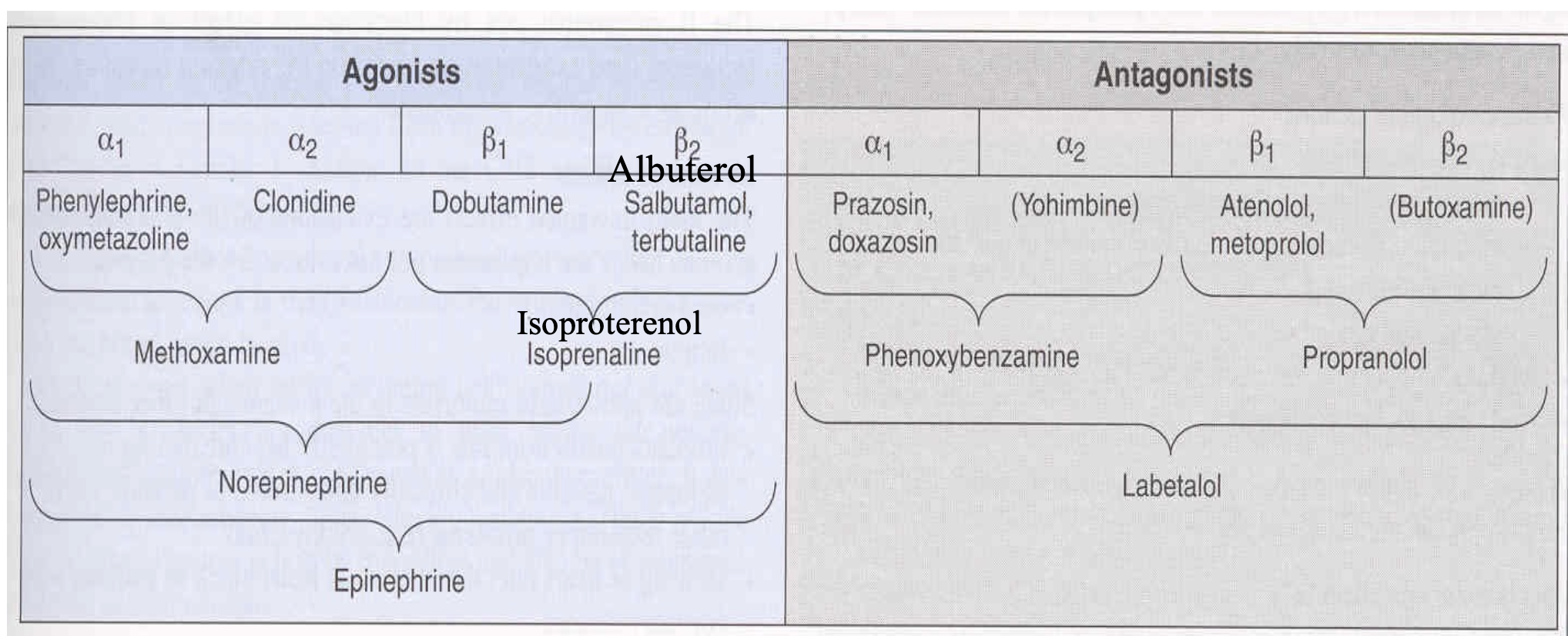
direct-acting adrenergic agonists
directly stimulate adrenergic receptors; responses may be potentiated by cocaine, reserpine, and guanethidine
selective:
⍺1 → phenylephrine
⍺2 → clonidine (presynaptic)
β1 → dobutamine
β2 → terbutaline
non-selective:
⍺1, ⍺2 → oxymetazoline
β1, β2 → isoproterenol
⍺1, ⍺2, β1 → norepinephrine
⍺1, ⍺2, β1, β2 → epinephrine
indirect-acting adrenergic agents
increase adrenergic signaling without directly activating receptors
releasing agents → amphetamine, tyramine
response abolished by prior treatment with reserpine or guanethidine
uptake inhibitor → cocaine (on NET)
monoamine oxidase (MAO) inhibitors → selegiline
responsible for breaking down NE in presynaptic nerve terminal
catechol-O-methyltransferase (COMT) inhibitors → entacapone
present in effector cells postsynaptically
mixed-acting adrenergic agonists
can stimulate adrenergic receptors directly, promote NE release, and affect uptake/metabolism pathways
⍺1, ⍺2, β1, β2 → ephedrine
response reduced with prior treatment with reserpine or guanethidine
adrenergic antagonists
⍺1 → prazosin, doxazosin
⍺2 → yohimbine
⍺1, ⍺2 → phenoxybenzamine
β1 → atenolol, metoprolol
β2 → butoxamine
β1, β2 → propranolol
⍺1, ⍺2, β1, β2 → labetalol
structures of the chambers of the eye
circular/sphincter muscle → muscarinic receptor
contraction causes miosis (pupil constriction)
radial/dilator muscle → ⍺1 adrenergic receptor
contraction causes mydriasis (pupil dilation)