gastrointestinal disorders
 Call Kai
 Learn
Learn Practice Test
 Spaced Repetition
 Match
Match Flashcards
 Knowt Play
1/56

There's no tags or description
Looks like no tags are added yet.
Name | Mastery | Learn | Test | Matching | Spaced | Call with Kai | Chat |
|---|
No analytics yet
Send a link to your students to track their progress
57 Terms
pain >2months
define chronic abdominal pain
green
what color is bilious vomiting
bilious vomiting
Bilious vomiting in a neonate or infant should be assumed to be intestinal obstruction requiring urgent surgical evaluation until proven otherwise.
Think of:
Malrotation with midgut volvulus (must not miss)
Duodenal atresia
Jejunal/ileal atresia
Meconium ileus
Hirschsprung disease with obstruction
Incarcerated hernia
Immediate management
NPO
NG tube for decompression
IV fluids ± electrolyte correction
Urgent pediatric surgery consult
Abdominal X-ray → if concern for malrotation, upper GI contrast series (gold standard)
Board pearl
Green (bilious) vomiting = obstruction distal to the ampulla of Vater until proven otherwise.
Projectile, non-bilious vomiting in a 2–8 week-old infant instead suggests hypertrophic pyloric stenosis, which is not the same emergency.
surgery for this type of vomiting until proven otherwise
-small bowel obstruction
-volvulus
as well as …..
Malrotation with midgut volvulus (must not miss)
Duodenal atresia
Jejunal/ileal atresia
Meconium ileus
Hirschsprung disease with obstruction
Incarcerated hernia
differential diagnoses of bilious vomiting
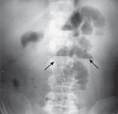
small bowel obstruction
Mnemonic: SBO = 3 S's
Step-ladder air-fluid levels
Swollen (dilated) small bowel (>3 cm)
Scarce distal colonic gas
-presents with sudden bilious vomiting, abdominal distention
NPO, NG tube for decompression
IV fluids
Emergent surgical consult
management of SBO
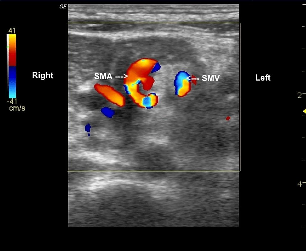
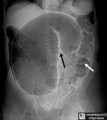
volvulus
Presentation: sudden onset bilious vomiting, abdominal distension, pain, rapid progression to shock
Evaluation:
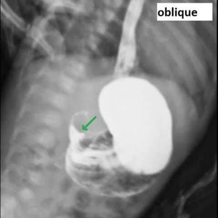
Upper GI series → corkscrew appearance
u/s: whorlpool
-acute gastroenteritis
-increased intracranial pressure
-GERD
-metabolic derangements
-peptic ulcer disease
-pyloric stenosis
differential diagnoses of nonbilious vomiting
gastric ulcer isease
pain worse with food. Weight loss due to not eating. MC H. pylori or NSAIDS overuse.
____
____________________
______________________________
duodenal ulcer disease
Zollinger-Ellison Syndrome (ZES) — High Yield
Definition:
A gastrin-secreting tumor (gastrinoma) → ↑ gastrin → ↑ gastric acid production → severe peptic ulcer disease.
Pathophysiology
Gastrinoma (usually pancreas/duodenum)
⬇
↑ Gastrin
⬇
↑ Parietal cell H⁺ secretion
⬇
Hyperacidity → recurrent ulcers + diarrhea
pain better with food. Weight gain. H. pylori. Zollinger Ellison syndrome.
Zollinger-Ellison Syndrome (ZES) — High Yield
Definition:
A gastrin-secreting tumor (gastrinoma) → ↑ gastrin → ↑ gastric acid production → severe peptic ulcer disease.
Pathophysiology
Gastrinoma (usually pancreas/duodenum)
⬇
↑ Gastrin
⬇
↑ Parietal cell H⁺ secretion
⬇
Hyperacidity → recurrent ulcers + diarrhea
Gastrin-secreting tumor associated with ulcers
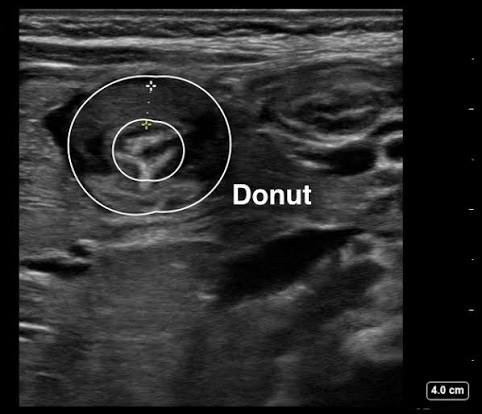
pyloric stenosis
Pt presents usually male,
2-8 weeks
was normal but now projectile vomiting.
Olive shaped mass, peristaltic waves.
noninfectious diarrhea
Clinical Presentation
Chronic (>2 weeks) or intermittent diarrhea
May be associated with weight loss, poor growth, or extraintestinal symptoms
Inflammatory bowel disease
Celiac disease
Lactose/fructose intolerance
Functional diarrhea (toddler's diarrhea)
Malabsorption syndromes (CF, pancreatic insufficiency)
ddx of noninfectious diarrhea
Functional constipation
Caused by holding feces in too long
-Has a normal transit time in the GI system, but the delay in getting stool out causes it to become hard (water gets absorbed back into body)
-most common cause of diarrhea
Hematemesis (bright red or coffee-ground emesis)
Melena if bleed is slower or more distal in upper GI tract
Signs of anemia, shock if severe
clinical presentation of upper GI bleed
Esophagitis, gastritis, PUD
Varices (portal hypertension)
Mallory-Weiss tear
Swallowed blood (epistaxis)
Ddx of upper GI bleed
Hematochezia (bright red blood per rectum)
± abdominal pain, diarrhea
presentation of lower GI bleed
Anal fissure (most common in children)
Polyps (juvenile polyp)
Meckel's diverticulum
IBD
Infectious colitis
Intussusception
Ddx of lower GI bleed
acetaminophen, anticonvulsants
main culprits of drug induced hepatitis
Trauma (most common in pediatrics)
Gallstones
Medications (valproate, azathioprine, steroids)
Infections (mumps)
Metabolic (hypertriglyceridemia, hypercalcemia)
Anatomic anomalies (pancreas divisum)
Ddx for acute panncreatitis causes
Pain that wakes the child from sleep
Progressive increase in severity/frequency
Localized pain away from umbilicus (especially RUQ or RLQ)
Associated systemic symptoms:
Unexplained fever
Weight loss, poor growth
Night sweats
Fatigue
GI bleeding (hematemesis, melena, hematochezia)
Persistent vomiting (especially bilious)
Chronic diarrhea
Family history of IBD, celiac disease, peptic ulcer
red flags of abdominal pain
downn syndrome
30% of cases of duodenal atresia are associated with
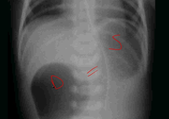
Duodenal atresia
What is the name for failed recanalization of the duodenum resulting in polyhydramnios, bile-containing vomitus, and a distended stomach?
double bubble sign
XR sign of duodenal atresia
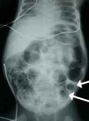
abdominal pain, rectal bleeding, hiighly associated with prematurity
clinincal features of necrotizing enterocollitis
Necrotizing Enterocolitis (NEC)
-pneumatosis intestinalis on XR
failure to pass meconium
initial presentation of Hirschsprung
Malrotation with midgut volvulus
presents similarly to NEC
-associated with VACTERL anomalies
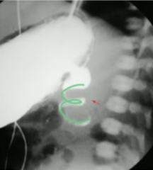
corkscrew sign
upper GI seriies finding of malrotation with midgut volvulus
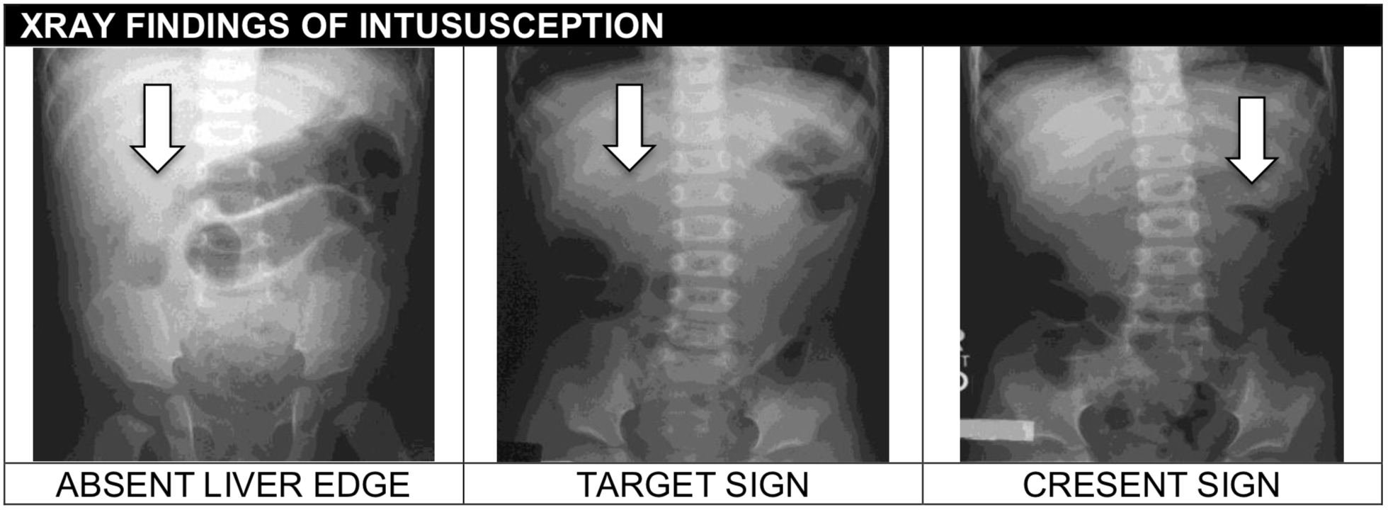
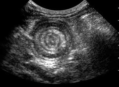
intussusception
-presents at 3-24 months
-currant jelly stool-intermittent, severe abdominal pain
-RUQ mass
target sign
Ultrasound finding of intussusception
first 24-48 hrs
when does duodenal atresia present
bilious vomiting
vomiting due to issue distal to the ampulla of vater
Chronic constipation, abdominal distension, poor growth
Explosive stool passage after digital rectal exam
how does hirschsprung present in infants/children
Rectal suction biopsy → absence of ganglion cells (aganglionosis)
gold standard to diagnose hirschsprung
Stabilize: Bowel decompression, IV fluids
Definitive: Surgical resection of aganglionic segment (pull-through procedure)
Monitor for enterocolitis (can be life-threatening)
management of hirschsprung
whirpool sign on US
US finding of malrotation
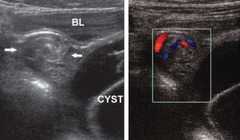
intussusception
Most common cause of intestinal obstruction in children 6 mo-3 yrs
Intermittent colicky abdominal pain,
currant jelly stools (blood + mucus),
vomiting
classic triad off intussusception
intussusception
Sausage-shaped abdominal mass (usually RUQ)
Child may draw knees to chest during pain episodes
Lethargy can be prominent
obstruction of the lumen of the appendix (fecalith, lymphoid hyperplasia, vegetables/seeds, parasites, or neoplasm)
most common cause of appendicitis
gliadin and gluten
celiac is an autoimmune intolerance of
food protein-induced enterocolitis syndrome (FPIES)
Adverse reaction to food protein MC cows milk, soy, or grains occurring 1-4 hours after exposure.
Pt: vomiting, lethargy, pallor, watery diarrhea sometimes can be bloody.
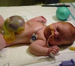
Gastroschisis
Extrusion of abdominal content through abd fold, usually right of umbilicus. Not covered by peritoneum. Diagnosed clinically
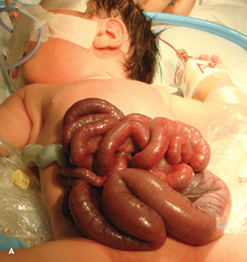
silo: push it back in slowly
management of gastroschisis
omphalocele
extrusion of abd content through abd fold, covered by peritonium, usually midline. Diagnosed clinically.
-frequent small volume diarrhea with urgency
-bloody diarrhea
frequent features of ulcerative colitis
-diffuse inflammation in mucosa or submucosa
-crypt architecture distortion
histopath of ulcerative colitis
Antineutrophil cytoplasmic antibodies (ANCAs)
serologic marker of UC
crohns disease
-discontinuous transmural asymmetric lesions
-mainly involving ileum and right sided colon
-cobblestone appearance
-deep fissures; transmural inflammation
-granulomatous inflammation
Anti-Saccharomyces cerevisiae antibodies (ASCA)
serologic marker of crohn's disease
meconium ileus
Failure to pass meconium. Associated with CF w/ + prenatal screen or pt who never had screen.
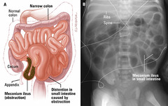
water enema
tx of meconium ileus
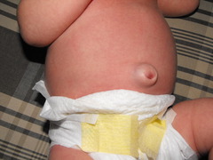
umbilical hernia
Herniation of abd content through umbilical opening from failure of umbilical ring to close. Diagnosed clinically.
Most require observation and close spontaneously by age 5.
Meckel's diverticulum
outpouching of distal ileum
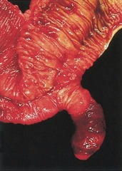
2% of the poppulation
M:F raio 2:1
2 ft from ileocecal valve
2 in in length
2% developing complications
2 types of heterotropic mucossa
define the rule of 2s of meckel's