IB Bio B1.1 Carbohydrates and Lipids / B3.3 Muscle and Motility (HL) / C3.2 Defence Against Disease
 Call Kai
 Learn
Learn Practice Test
 Spaced Repetition
 Match
Match Flashcards
 Knowt Play
1/86

Earn XP
Description and Tags
KILL ME
Name | Mastery | Learn | Test | Matching | Spaced | Call with Kai | Chat |
|---|
No analytics yet
Send a link to your students to track their progress
87 Terms
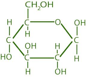
What monosaccharide is this?
Glucose

What disaccharide is this?
Maltose
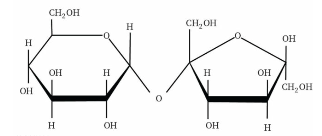
What diasaccharide structure is this?
Sucrose

What diasaccharide structure is this?
Lactose

What polysaccharide structure is this?
Starch

What polysaccharide structure is this?
Glycogen
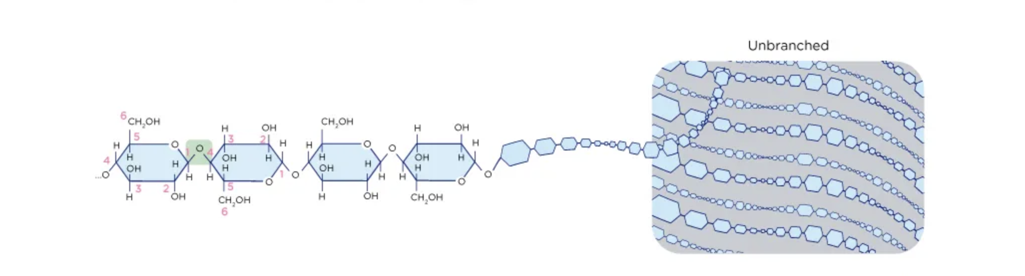
What polysaccharide structure is this?
Cellulose
What is the first step in blood coagulation?
The damaged cells of blood vessel walls release clotting factors
What is the second step in blood coagulation?
Clotting factors are released from activated platelets
What is the third step in blood coagulation?
Prothrombin is converted to thrombin
What is the fourth step in blood coagulation?
Fibrinogen is converted into the protein fibrin
What is the fifth step in blood coagulation?
Fibrin protein fibers form a network at the site of damage
What is the sixth step in blood coagulation?
The network of fibirin traps erythrocytes (RBCs) and forms a clot
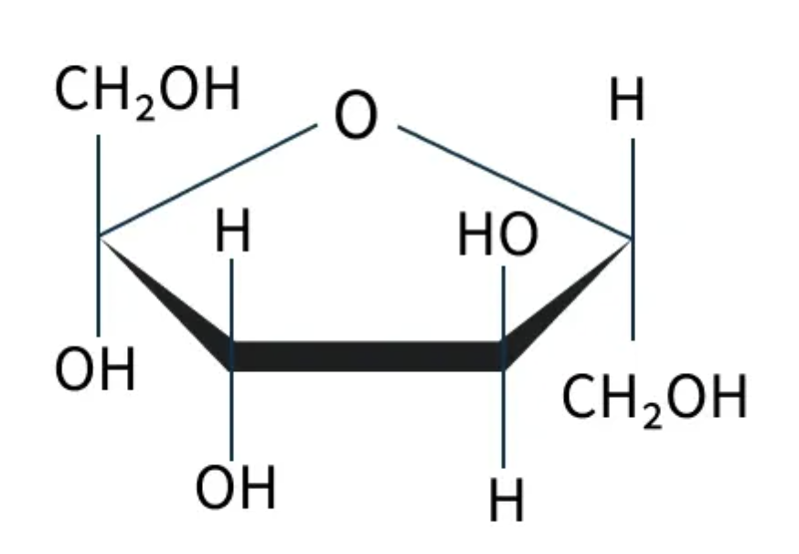
What monosaccharide is this?
Fructose

What polysaccharide is this? (type of starch)
Amylose

What polysaccharide is this? (type of starch)
Amylopectin
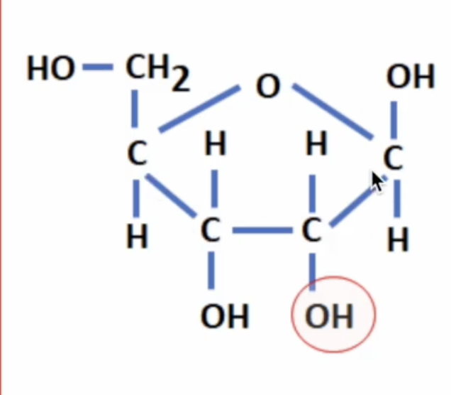
What pentose sugar is this?
Ribose (OH)

What pentose sugar is this?
Deoxyribose (H)

What lipid is this?
Phospholipid

What lipid is this?
Triglyceride

What fatty acid is this?
Saturated fatty acid

What fatty acid is this?
Unsaturated fatty acid

What fatty acid is this?
Monosaturated fatty acid

What fatty acid is this?
Polysaturated fatty acid
Condensation
The chemical process where monomers join together to form a polymer, releasing one H2O molecule per bond formed.
Hydrolysis
The chemical breakdown of a polymer into monomers by the addition of water.
Reactants required for hydrolysis
A polymer and water (H2O).
Saturated fats
Lipids that contain no C=C double bonds in their hydrocarbon chains.
Physical state of saturated fats at room temperature
Solid (e.g., butter), because their straight hydrocarbon chains pack tightly together.
Unsaturated fats
Lipids containing one or more C=C double bonds in their hydrocarbon chains.
Physical state of unsaturated fats at room temperature
Liquid (oils), because kinks in their hydrocarbon chains prevent tight packing.
Monounsaturated fat
An unsaturated lipid containing exactly one C=C double bond.
Polyunsaturated fat
An unsaturated lipid containing more than one C=C double bond.
Healthier type of dietary fat
Unsaturated fats (especially polyunsaturated fats), which are linked to lower LDL cholesterol and reduced heart disease risk.
Health impact of excess saturated fat consumption
Elevated blood cholesterol levels.
Structural components of a phospholipid
A hydrophilic (polar) phosphate head and two hydrophobic (non-polar) fatty acid tails.
Amphipathic
Describing a molecule that possesses both hydrophilic (water-loving) and hydrophobic (water-repelling) regions.
Reason phospholipids spontaneously form bilayers in water
Their amphipathic nature causes hydrophilic heads to face outward toward water while hydrophobic tails cluster inward away from water.
Initial event in blood clotting
Blood vessel damage occurs, triggering platelets to adhere and form a platelet plug.
Event following platelet plug formation
Release of clotting factors by activated platelets and damaged tissue.
Conversion of prothrombin to thrombin
An enzymatic step in blood clotting activated by clotting factors that requires Ca2+ ions.
Role of thrombin in blood clotting
Converts soluble fibrinogen into insoluble fibrin threads.
Role of fibrin in blood clotting
Forms a fibrous mesh network that traps red blood cells to produce a stable clot.
Sequential steps of blood clotting
Vessel damage & platelet plug → 2. Clotting factor release → 3. Prothrombin to thrombin (requires Ca2+) → 4. Fibrinogen to fibrin → 5. Fibrin mesh traps RBCs.
Selection pressure from antibiotic overuse
Antibiotics kill susceptible bacteria, allowing rare resistant mutants to survive, reproduce, and dominate.
Mechanism linking antibiotic overuse to increased resistance
Eliminating susceptible competitor bacteria frees up space and resources for resistant strains to proliferate.
Entity that evolves antibiotic resistance
The bacterial population evolves resistance, not the human host.
Non-specific (innate) immunity
A rapid immune response that acts generically against any pathogen without conferring immunological memory.
Examples of innate immune defenses
Physical barriers (skin, mucus), phagocytes, and inflammation.
Specific (adaptive) immunity
A targeted immune response against a specific antigen that develops slower initially but produces long-lasting immunological memory.
Primary cell types involved in adaptive immunity
Lymphocytes, including B cells and T cells.
Primary cell types involved in innate immunity
Phagocytes (macrophages, neutrophils, dendritic cells), natural killer cells, and mast cells.
Primary function of B lymphocytes
Synthesis and secretion of antibodies.
Key types of T lymphocytes in adaptive immunity
Helper T cells, cytotoxic (killer) T cells, and memory T cells.
Zoonosis
An infectious disease naturally transmissible from vertebrate animals to humans.
Examples of zoonotic diseases
Rabies, COVID-19, and vector-borne malaria (transmitted via mosquitoes).
Proportion of human infectious diseases that are zoonotic
Over 60%.
Movement (in biology)
Any change in position of an organism or its internal structures (e.g., heart beating or peristalsis).
Locomotion
Movement of an entire organism from one location to another (e.g., walking or swimming), requiring muscles and a skeleton.
Main components of a synovial joint
Bones, articular cartilage, synovial fluid, synovial membrane, joint capsule, and ligaments.
Function of articular cartilage
Reduces friction and absorbs shock between opposing bone surfaces.
Function of synovial fluid
Lubricates joint surfaces to allow smooth movement.
Function of the synovial membrane
Secretes synovial fluid into the joint cavity.
Function of the joint capsule
Encloses the joint cavity and holds the components of a synovial joint together.
Function of ligaments
Connect bone to bone, providing joint stability and preventing dislocation.
Structural classification of the hip joint
Ball-and-socket joint.
Structure forming the ball of the hip joint
The head of the femur.
Structure forming the socket of the hip joint
The acetabulum of the pelvis.
Storage site for Ca2+ ions in muscle fibers
The sarcoplasmic reticulum.
Effect of Ca2+ binding to troponin
Causes troponin to change shape and pull tropomyosin away from actin's myosin-binding sites.
Consequence of moving tropomyosin off actin's binding sites
Exposes binding sites, allowing myosin heads to bind actin and form cross-bridges.
Reason muscle contraction fails without Ca2+
Tropomyosin physically blocks myosin-binding sites on actin, preventing cross-bridge formation.
Composition of arthropod exoskeletons
Chitin combined with protein (and calcium carbonate in crustaceans).
Key functions of an arthropod exoskeleton
Protection, waterproofing (preventing desiccation), muscle attachment, and structural support.
Major growth constraint of an exoskeleton
It cannot expand and must be shed through moulting (ecdysis) to permit growth.
Vulnerability of arthropods during moulting
The old shell is shed and the new exoskeleton has not yet hardened, leaving the organism defenseless.
Sliding filament model
Mechanism of muscle contraction where myosin heads bind actin and pull filaments toward the sarcomere center, causing filaments to slide past each other.
Muscle cross-bridge
The temporary structural connection formed when a myosin head binds to actin.
Power stroke
The pivoting motion of a myosin head that pulls the actin filament toward the center of the sarcomere.
Role of ATP in the cross-bridge cycle
ATP binding causes myosin to detach from actin; ATP hydrolysis provides energy to re-cock the myosin head.
Cyclic steps of muscle contraction
- Myosin binds actin → 2. Power stroke pulls actin → 3. ATP binds causing detachment → 4. Myosin head re-cocks.
Overall sarcomere behavior during contraction
The sarcomere shortens.
Behavior of the I-band and H-zone during contraction
Both narrow (decrease in width).
Behavior of Z-discs during muscle contraction
Move closer together toward the center of the sarcomere.
Behavior of the A-band during muscle contraction
Remains unchanged in length.
Filament length changes during contraction
Neither actin nor myosin filaments change length; they only slide past each other.
Events during muscle relaxation
Ca2+ is actively pumped back into the sarcoplasmic reticulum using ATP, tropomyosin re-blocks actin binding sites, and the sarcomere lengthens.