NM cardiology Combined
1/1037
Earn XP
Description and Tags
literally....everything....
Name | Mastery | Learn | Test | Matching | Spaced |
|---|
No study sessions yet.
1038 Terms
What are the two methods of dynamic cardiac imaging we can perform in nuclear medicine?
first pass study
gated blood pool imaging (AKA MUGA, RVG, ERNA or RNA)
What are the indications for a First pass study?
eval of pts with:
LV dysfunction
Interventricular shunts
myocardial ischemia
MI
What are the advantages of first pass studies?
tracer activity is limited to 1 chamber at at time
background is decreased
rapidly completed
What are the disadvantages of first pass studies?
gamma cameras must be able to acquire data at 200,000 counts/sec or greater
speical multi-crystal cameras with high count rate capabilities are optimal but not widely available
increased count rate = increased sensitivity
What is the minimum dosage for a first pass study?
10 mCi
What types of imaging would we perform for a first pass study?
Gated images
What types of Tc tracers would we use for a first pass study?
sestamibi, tetrofosmin, pentetate, pertechnetate
What tracers should we NOT use for a first pass study? (hint: think about all Tc tracers, and cardio tracers)
MAA
SC (sulfur colloid)
Tl-201
When performing a first pass exam, the volume should be greater than ____ for a good bolus
1ml
How fast should a bolus be pushed when performing a LVEF first pass study? What should follow it?
over 2-3 sec
10ml flush
How fast should a bolus be pushed when performing a RVEF first pass study? What should follow it?
over 2-3 sec
10ml flush
When performing a first-pass study, what baseline test should be performed before the injection of the tracer? Why?
baseline ECG to assess rhythm
Through which veins are we injecting for a first pass study?
in Right AC (median Basilic vein) or jugular for direct path to superior vena cava
How can we tell if its a good bolus during a first pass study?
time activity curve over superior vena cava, calculate FWHM
What is the rate of data acquisition for a first pass study?
16-30 frames/sec or 1,200 frames in 60 sec
What matrix size would we use for a first pass study?
64×64
What type of collimator would we use when performing a first pass study?
high sensitivity
When imaging a patient during a first pass study, what can be done to ensure we positioned the patient properly?
1mCI dose can be injected to check positioning or perform a transmission scan
When positioning the patient under the camera for a first pass study, _____ should be positioned at the top of the FOV, and the ____ should be within the FOV
sternal notch (at top of FOV)
xifoid (within the FOV)
How would the camera be oriented when assessing the LVEF during a first pass study?
supine or upright
LAO view
How would the camera be oriented when assessing the RVEF during a first-pass study?
RAO
How would the camera be oriented when assessing both ventricles during a first pass study?
anterior
During a first pass study, describe the sequential visualization a good bolus would give
uSuperior vena cava
uRight atrium
uRight ventricle
uPulmonary artery to lungs
uPulmonary veins
uLeft atrium
uLeft ventricle
uAorta
When interpreting a first pass study, what are we looking for?
EF
left to right shunt
right to left shunt
When interpreting a first-pass study, if a patient has a left-to-right shunt, some of the oxygenated blood returning from the lungs will _____
will go through the shunt and circulate back to the lungs instead of the body
When interpreting a first-pass study, if a patient has a right-to-left shunt, some of the deoxygenated blood returning from the body will _____ instead of
go through the shunt and be sent out to the body
On a time activity curve, do the high points represent diastole or systole?
diastole
On a time activity curve, do the low points represent diastole or systole?
systole
How would a left to right shunt be interpreted on a first pass study?
Rapid recirculation of tracer to lungs once it enters the left side of the heart.
How would a right-to-left shunt be interpreted on a first pass study?
Appearance of bolus in left side of the heart and aorta before the appearance of lung activity
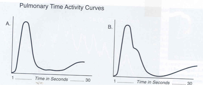
When interpreting a time activity curve for a Left to right shunt, A represents _______ and B represents _______
normal flow (1 peak)
left to right shunt (2 peeks)
In Gated blood pool imaging, how is data collected?
over many cardiac cycles using ECG gating
Gated equilibrium is another name for _______
Gated blood pool imaging
gated cardiac blood pool study is another name for a _______ procedure
Gated blood pool imaging
equilibrium radionuclide angiography (ERNA) is another name for a _______ procedure
Gated blood pool imaging
radionuclide ventriculography (RVG) is another name for a _______ procedure
Gated blood pool imaging
multiple gated acquisition study (MUGA) is another name for a _______ procedure
Gated blood pool imaging
What are the indications for Gated blood pool imaging?
Assessment of cardiac function in chemotherapy patients
Quantification of ejection fraction (LVEF, RVEF)
Detection or assessment of CAD
MI
Estimation of wall motion abnormalities
Eval of function of pts with valvular disease
follow-up of medical/surgical therapy
Why would we perform a Gated blood pool imaging instead of a full stress test?
Its own exam bc pts don’t need stress test, just ejection fraction
What is the patient prep for a gated blood pool imaging?
none
What is the most commonly used radiopharmaceutical for a.gated blood pool imaging?
Tc-99m labeled RBC’s
What are the 3 ways we can label RBC’s?
in vitro
modified
in vivo
When using the In Vivo labeling method, blood is labeled ________
inside the body
When using the In Vitro labeling method, blood is labeled ________
outside the body, using an Ultratag kit
The advantage(s) of using an In Vivo/In Vitro method of blood labeling is:
high efficiency (~95%)
absence of blood manipulation
The in Vivo method of blood labeling has a ____% labeling efficiency
60-90%
Describe the steps taken when labeling blood using the In Vivo method
Inject stannous ion
Wait 20-30 min
Inject 99mTc-Pertechnetate
Free 99mTc is secreted through gastric mucosa and kidneys
Not able to see bleed in stomach, small bowel, and/or the colon
What are the disadvantages of using the In Vivo method?
Labeling constancies variable (60-90%)
Free 99mTc is secreted through gastric mucosa and kidneys
Not able to see bleed in stomach, small bowel, and/or the colon
What is the advantage of using the in Vivo method?
Convenient and easy
What blood labeling methods use Stannous Pyrophosphate?
in Vivo
modified in vivo/in vitro
What is cold Stannous Pyrophosphate used for in blood labeling?
to pretreat RBC’s for labeling
What is the optimal dose of Stannous Pyrophosphate when labeling RBC’s?
0.5-1.0mg
What if too little of Stannous Pyrophosphate is used when labeling RBC’s?
dose of Tc will not properly label RBCs
What will happen if too much of Stannous Pyrophosphate is used when labeling RBC’s?
some of tin will circulate freely and tag to Tc outside of RBC’s
What wil happen to image quality if too much or too little of the Stannous Pyrophosphate is administered when labeling RBC’s?
results in increased background as a result of free Tc
Describe the steps taken when labeling blood using the In Vivo/In Vitro method
IV injection of stanous ion
Blood sample is collected into a syringe containing 99mTc-Pertechnetate and anticoagulant
Re-injected
Describe the steps taken when labeling blood using the In Vitro method:
1-3ml of blood withdrawn into syringe containing anticoagulant
let sit for 5 min
Add sodium hypochlorite and acid-citrate-dextrose (ACD)
To oxidize the extracellular stannous ion
Add 99mTc-Pertechnetate (diffuses into the RBC where it is reduced and trapped)
Wait 20 minutes (no longer than 60min) and inject
T/F: when using the In vitro method to label blood, once the tracer has been added, we can use a sample that has been sitting for more than 60min
false
T/F: When using the In vitro method to label blood, we can inject imedietly after adding the Tc-99m Pertechnetate
false
T/F: When using the In vitro method to label blood, we have to wait at least 60 minutes after the Tc-99m Pertechnetate has been added before injecting.
false
T/F: When using the In vitro method to label blood, once the Tc-99m Pertechnetate has been added, we have to wait at least 20 minutes, but no more than 60 min before injecting
true
What is the advantage of using an In Vitro method for labeling blood?
high labeling efficiency, superior image quality
Describe the procedure for a gated equilibrium study.
In Vivo, In Vitro, or modified In Vivo/In Vitro labeling of RBC’s w/ Tc-99m
3 gated planar images obtained
Anterior
LAO
Lft Lateraral
What views are obtained when performing a gated equilibrium study?
planars:
Anterior
LAO
Lft Lateraral
T/F: When performing a MUGA (multiple gated acquisition study), an ECG rhythm strip should be obtained and reviewed before injection
True
Why should a ECG rhythm strip be obtained and reviewed before injection during a MUGA procedure?
Rapid atrial fibrillation or frequent PVCs (premature ventricular contractions) are contraindications to study
One cardiac cycle can be divided into intervals of ___
8 (16, 24. or 32) frames
What triggers gating in cardiac images?
R wave
During a gated study, 24 frames per cardiac cycle are obtained. If a patient’s heart rate is 65 bpm, the length of time per frame is:
a. 38 msec
b. 3.8 msec
c. 41 msec
d.4.1 msec
b. 38 msec
In a gated study, if the patient’s heart rate is 65 bpm, and 24 frames per cardiac cycle was obtained, How do you calculate the length of time per frame in msec?
60 sec/min divided by 65 beats/min = 0.92 sec/beat
divide by 24 frames = 0.038 sec X 1,000 = 38 msec
Which view provides the best separation between the L. and R. ventricles during a gated study?
LAO
How should a technologist reposition the camera if trying to separate the atria from the ventricles in a gated study?
apply a 5-10 degree caudal tilt
From the best LAO view, the anterior and lateral views are obtained ± ____ degrees.
45 degrees
how would the camera be positioned during a gated study if trying to visualize the Right ventricle?
20 degrees anterior (5 degrees of caudal tilt)
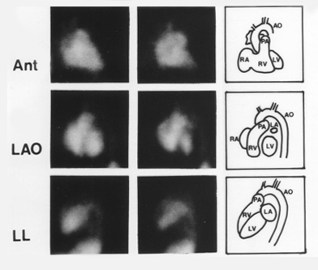
Based on this image, which is systole and which is diastole?
1st row diastole, 2nd row systole
During a gated study, in what views would we potentially apply a caudal tilt?
only for LAO or RAO
When performing a gated study, can we use a caudal tilt for anterior or lateral views?
no
If we apply a caudal tilt to the camera, we are tilting the camera towards the _______
Pelvis (caudus)
How does a caudal tilt affect images?
used to elongate ventricles
When performing a gated blood pool imaging, are we acquiring rest or stress images?
rest
During a gated equilibrium study, what energy window are we using?
20% window centered at 140keV
What type of collimator are we using during a gated equilibrium study?
LEHR
What is the R-R acceptance window in a gated equilibrium study?
10-15%
What matrix size are we using for a gated equilibrium study?
64×64×16
When taking images for a gated equilibrium study, we have to acquire a minimum of ________ counts per frame, or about ___ to ___ minutes per view
250,000 counts
~5-10 min
T/F: If the heart rate is irregular, imaging during a gated equilibrium study will take longer
True
How is a gated equilibrium study data reconstruted?
added together to form a representative cardiac cycle
simulates the heart beating
Qualitative anaysis of a gated equilibrium study can be used to assess:
cardiac chamber size
overall biventricuar function
regional wall motion
extra cardiac abnormalities such as aneurysms
What does Akinesis mean?
absence of wall motion
What does Hypokinesis mean?
decreased wall motion
What is Dyskinesis?
outward bulge during systole
What is stroke volume?
volume of blood ejected by either ventricle during systole
What is the cardiac output?
volume of blood that heart pumps per minute
CO = SV at heart rate
What is the ejection fraction?
% of blood ejected from ventricles during each contraction
What does qualitative analysis of a gated equilibrium study used to analyze?
LVEF
How are images processed for quantitative analysis during a gated equilibrium study?
ROI’s drawn on LV at diastole and systole
Background ROI selected about 3-6 o’clock from LV
ROI can be manually drawn or generated by computer
In images taken at end diastole, the ventricle is _____, and the counts obtained would be _______.
relaxing
highest
In images taken at end systole, the ventricle is _____, and the counts obtained would be _______.
contracted
lowest
How do you calculate Ejection fraction?
[(net diastolic counts - net systolic counts) / net diastolic counts] X 100
Calculate the Ejection fraction if ED (end diastolic counts) are 90,400 and ES (end systolic counts) are 40,000
(90,000 - 40,000) / 90,400 = 0.557
0.557 X 100 = 55.7%