Genetics - Chromosomes Mitosis (Week 1)
1/13
There's no tags or description
Looks like no tags are added yet.
Name | Mastery | Learn | Test | Matching | Spaced | Call with Kai |
|---|
No analytics yet
Send a link to your students to track their progress
14 Terms
Review the similarities and differences between prokaryotic and eukaryotic cells.
Prokaryotic: Single cell, doesn’t have nucleus, seen in bacteria and plant, dont have a nuclear envelope or membrane organelles, chloroplasts
Eukaryotic: Has a nucleus (containing genetic material, and its a membrane bound cell), Nucleous (with a nucleoulus organizer region or NOR), and other membrane-bound cells, Cytoplasm, Mitochondria
have centriole: cytoplasmic organelle composed of nine groups of microtublues; function in the gneration of cilia and flagella and serve as foci for the spindles in cell division (located at the centrosome) help control the spindles in mitosis/meiosis
Differentiate between nonhomologous chromosomes, homologous chromosomes, and sister chromatids.
Homologous chromosomes: Pair of chromosomes that are nearly identical to each other—> same length, type, gene, but DIFFERENT ALLELE
Nonhomologous chromosomes: not same type of chromosomes or length (different)
Sister Chromatids: Pair of chromosomes that are duplicates of each other—> same length, type, gene, and alleles
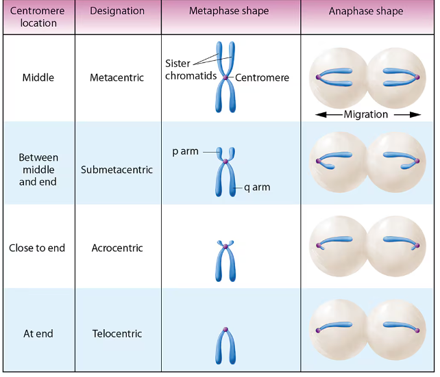
Determine if a chromosome is telocentric, acrocentric, submetacentric or metacentric.
Telocentric: Not seen in humans, does NOT have p arms or telomere
Arocentric: small p arms, large q arms, repeatable sequences
Submetacentric: most of our human genes are in this form
Metacentric: centromere is in the middle
If given a location on a chromosome (such as 2p23), identify it on a chromosome map.
2 is the Chromosome itself
p is which arm its located
23 is what region/sub region is at
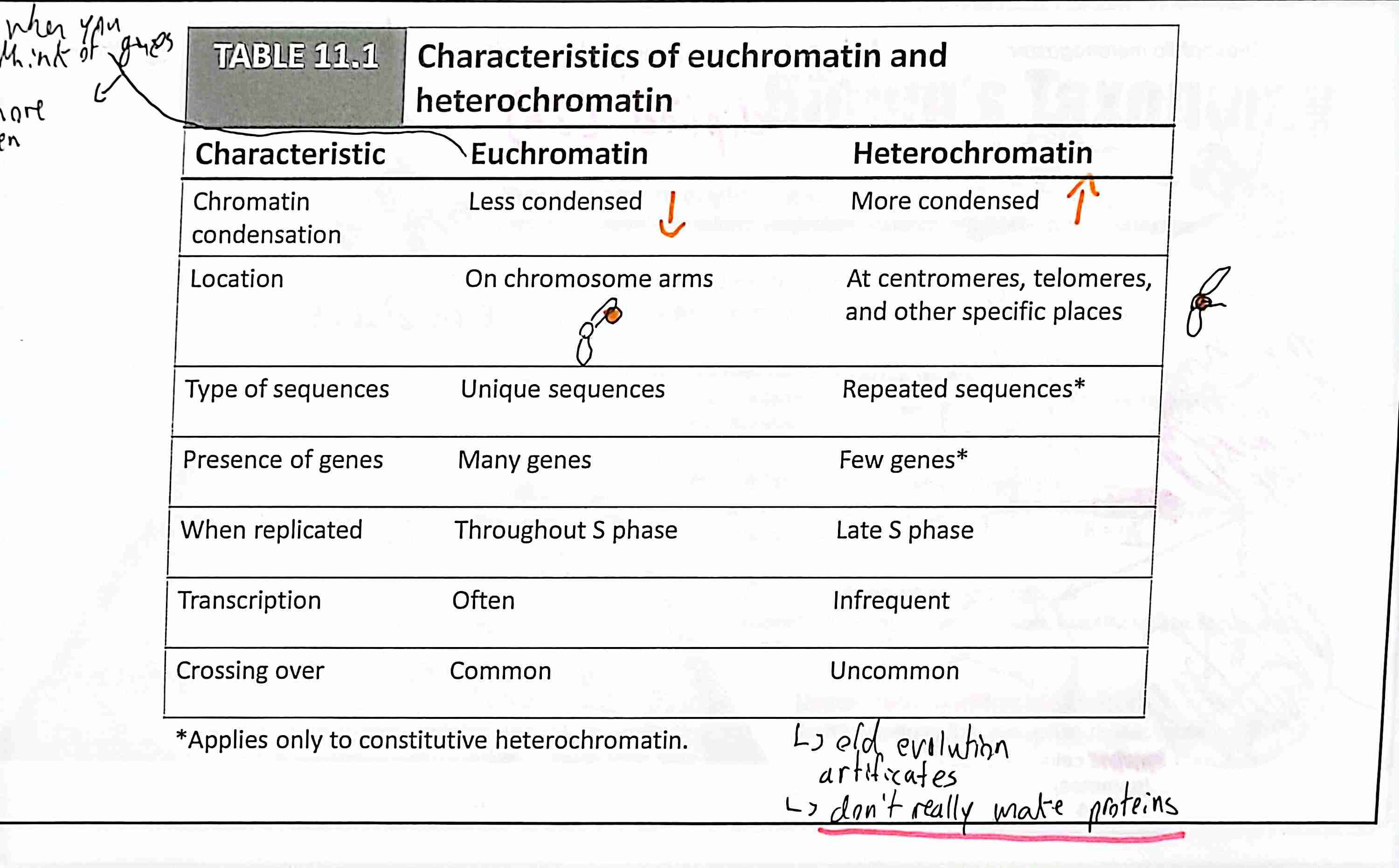
Differentiate between heterochromatin and euchromatin
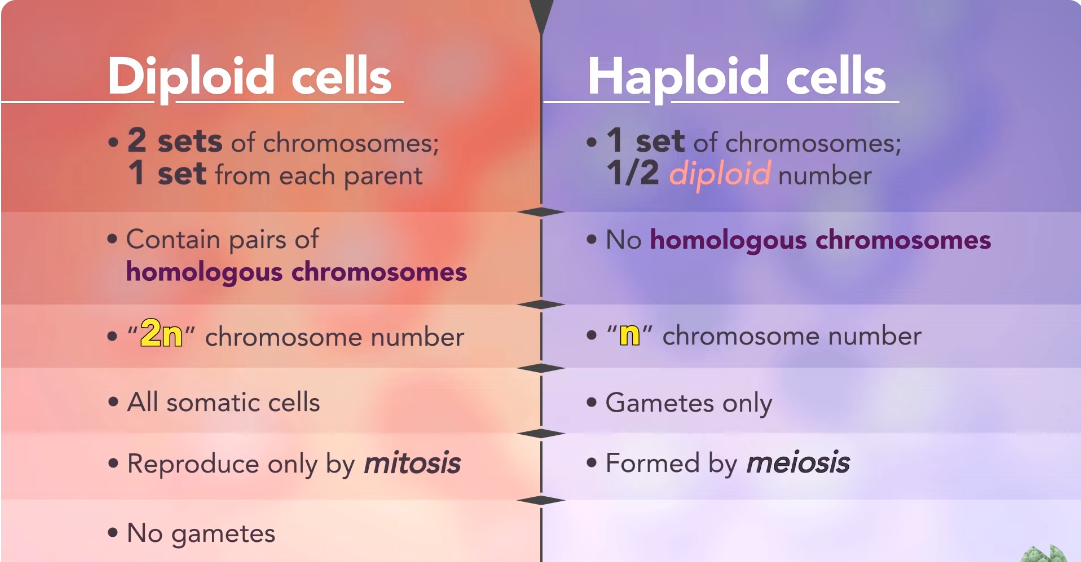
Differentiate between the diploid number and the haploid number.
Diploid (2n): The completed pair of chromosomes that are received from both parents (23 from mom and 23 from dad)
Haploid (n): The number of homologous chromosome pairs characteristic of an organism or species —> equal to ½ of a diploid (just 1 chromosome)
they are the gametes (i.e. sperm and egg)
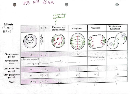
Describe the events that characterize each stage of mitosis, including the location and structure of chromosomes, centrosomes, mitotic spindle, nuclear membrane, and cell membrane.
Interphase: initial stage of cell cycle (interval between divisions)—> the replication of the DNA of each chromosome
Prophase: just outside of the nuclear envelope (centrosomes) 1. the migration of two pairs of centrioles to opposite ends of the cell 2. nuclear envelop breaks down and disappears + nucleolus disintegrates. 3. diffuse chromatin begin to condense into visible chromosomes which are sister chromids held together by cohesin.
prometaphase:
Stage of cell division during which the spindle fibers are assembled and attached to the centromeres of chromosomes, which migrate to the opposite side of the cell
metaphase: Stage of cell division in which condensed chromosomes lie in a central plane between the two poles of the cell and during which the chromosomes become attached to the spindle fibers —metaphase plate: the midline region of the cell perpendicular to the spindle fibers axis
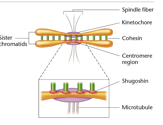
1. lines up in the metaphase plate and sister chromids are attached to the spindle fibers by the protein kinetochore. 2. cohesin is degraded by separase (except the centromere region protected by shugoshin) 3 each centromere is aligned at the metaphase plate with the chromosomes arms extending outwards in a random array
anaphase: 1. sister chromatids disjoin from each other by disjunction: (1) shugoshin must be degraded, reversing its protective role; (2) the cohesin complex holding the centromere region of each sister chromosome is then cleaved by separase; and (3) sister chromatids of each chromosome are pulled toward the opposite poles of the cell 2. the migrating chromatids ar now called daughter chromosomes 3. Movement of daughter chromosomes to the opposite poles of the cell is dependent on the kinetechore–spindle fiber attachment. (helped by molecular motors: specialized proteins that facilitate the movement of cellular components by hydrolysis of ATP shorting the microtubles )
stage of cell division in which chromosomes begin moving to the opposite poles of the cell (shortest stage)
telophase: 1. two complete sets of chromosomes are present at each pole. 2. Cytokensis: two new cells being produced from 3. cell membrane poduces cell furrow that constrics the cell into two cells 4. chromosomes begin to uncoil and be diffuse chromitds again. —> enters into interphase again
Stage of cell division in which the daughter chromosomes have reached the opposite pole of the cell and reverse the stage characteristic of prophase, reforming the nuclear envelopes and uncoiling the chromosomes. It ends during cytokenesis, which divides the cytoplasm and splits the parental cell into two daughter cells
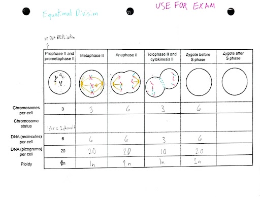
Predict the outcomes for mitosis for a cell, in particular the amount of DNA, number of chromatids, ploidy, and/or number of chromosomes.
Explain the proteins (cohesion, separase, shugoshin) involved in segregation during mitosis, and what happens when the process goes wrong
cohesion: A protein complex that holds sister chromatids together during mitosis and meiosis and facilitates attachments of spindle fibers to kinetochores (formed during S phase)
kinetochore are the protein that the microtubule attaches too (forms opposite of the centromere
Separase: an enzymes that degrades cohesion proteins
Shugoshin: protein that protects cohesin from being degraded by separase at the centromeric regions
What mutations occur: potentially leads to errors during chromosomes migration, altering the diploid content of daughter cells —> and damaged cells would divid incontrollable
Summarize the events that characterize each stage of meiosis, including the location and structure of chromosomes, centrosomes, meiotic spindle, nuclear membrane, and cell membrane. —> 5 steps of prophase I.
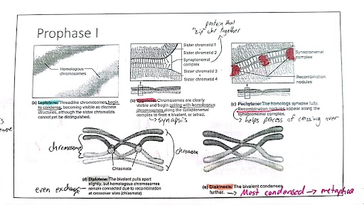
Prophase 1
Leptotene: Thread like chromosomes begin to condense becoming visible as discrete structures, although the sister chromatids can’t be distinguished
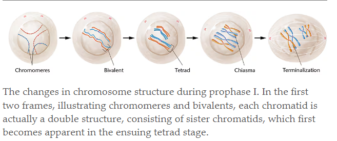
Zygotene: Chrom. are clearly visible and begin pairing with other homologous chrom. along the synaptonemal complex to form bivalents, or tetrads —> SYNAPSIS
Pachytene: The homolgs synapse fully. Recombination’s nodules appear along the synaptonemal complex —> helps with crossing over
Diplotene: The bivalent pulls apart slightly, but homolog chrom. remain connected due to recombination at crossover sites (chiasmata)
Diakinesis: The bivalent condense further.
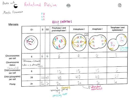
Metaphase 1: chromosomes have maximally shortened and thickened—>
location and structure of chromosomes: in the middle of cell in the metaplate (look like X)
centrosomes: still attached to the chr and sister chromatids
meiotic spindle: enlongated and connected to the kinochores on the centromeres and pulling the chr to the other side of the cell
nuclear membrane: gone
cell membrane: more oval/enlogated because of the centriols
Anaphase I:
location and structure of chromosomes: migrating to the other side of cell, (they still look like X)
centrosomes: still attached to 2 sister chromatids
meiotic spindle: actively pulling the homolog pair way and shortening themselves to do so
nuclear membrane: gone
cell membrane: still elongated
Telophase I:
location and structure of chromosomes: 23 in one cell and 23 in the other (still look like X)
centrosomes: still attached to their sister chromatids
meiotic spindle,: detach from the chro
nuclear membrane: begins to reform
cell membrane: cytokinesis occurs and the cleavage furrow pinches the two new cells off of each other
Prophase II:
location and structure of chromosomes, inside the nucleus (which is breaking) (still look like X)
centrosomes: still attach to sis chromatids
meiotic spindle: beginning to form and extend outwards
nuclear membrane: beginning to fragment
cell membrane.: nothing happening
Metaphase II:
location and structure of chromosomes: begin to line up at the metaplate (still look like X)
centrosomes: attached to sis chromatids but also to the microtubules by the kinochore protein
meiotic spindle: extended out and attaching to the centromeres
nuclear membrane: completely fragmented and gone
cell membrane: slightly elongated
Anaphase II:
location and structure of chromosomes: new chromosomes from the chromatids of the sisters are moving to the opposite side of the cell (now they look like I)
centrosomes: fragmented and now attached to each new chromosomes after separation
meiotic spindle: shortening and pulling the chr to the other side of the cell
nuclear membrane: still gone
cell membrane: elongated
Telophase II
location and structure of chromosomes: inside the new nucleus in the new daughter cell, (look like I)
centrosomes: attached to the chro (looks like chromatids)
meiotic spindle: detached the centromeres and are outside of the nucleus in the new cell
nuclear membrane: reforms around the new chromosomes
cell membrane: goes through cytokinesis and cleavage furrow pinches the 4 new daughter cells off
Make sure to explain why homologous chromosomes must pair during meiosis, and the importance of crossing over to this process and increasing genetic variation
maternal and paternal Homolog chrom. must pair up with each other in order for there to be the production of a new, unique gamete cell. These cell pair up during synapsis and exchange information through the process of crossing over; where nonsister chromatids swap places. This creates the genetic variation
Predict the outcomes for meiosis for a cell, in particular the amount of DNA, number of chromatids, ploidy, and/or number of chromosomes.
Explain the molecular processes (cohesion, separase, shugoshin, patronus) involved in segregation during meiosis, and what happens when the process goes wrong.
cohesion: ring-like multi-protein complex that holds the homologous pairs together
separase: a protease, Rec8 phosporylation is REWUIRED for cleavage of cohesion by the enzyme
shugoshin: protein that protects cohesin cleavage by separase at centromeric location during anaphase 1
patronus: protein that protects cohesin cleavage separase at centromeric locations during interkinesis
if it goes wrong, there might be an addition or one less chromosomes in a cell which can cause genetic disorder/muations
Compare and contrast the detailed processes of mitosis and meiosis.
Compare: Involved in making new cells, both start with Diploid cells (2n), both go through PMAT, Prophase 1 both condense the chromosomes, metaphase met up in the middle at the metaplate (in metaphase for mit and metaphase 2 in mei), anaphase (mit and mei 2) pull chromosomes part to opposite polls
Contrast:
Mitosis: Body (somatic) cell, the genetic info in the daughter cells is identical to the original, goes through PMAT once, metaphase- single file line, anaphase- pull the sister chromatids away. telo +cyto= idnetical diploid cells
Meiosis: Gamete (sex)cells, Prophase 1 involves crossing over genetic infor for variation, goes through PMAT twice, homologous pair up with eachother during Synapsis, resulting in Tetrads, metaphase 1- paired up line w./ homo pair. Anaphase 1- pulling the pair oh homologous chromosomes away, telo 1 and cyto= another reduction is required to happen. once meiosis 2 finishes = non-idnetical + gametes (haploid)