Week 2: Dysrhythmias: Life Threatening Rhythms
 Call Kai
 Learn
Learn Practice Test
 Spaced Repetition
 Match
Match Flashcards
 Knowt Play
1/34

There's no tags or description
Looks like no tags are added yet.
Name | Mastery | Learn | Test | Matching | Spaced | Call with Kai | Chat |
|---|
No analytics yet
Send a link to your students to track their progress
35 Terms
Premature Ventricular Contractions (PVCs) description
premature =
rhythm
P wave
PR
QRS
T wave
• Premature (early)
• Underlying rhythm usually regular, irregular with PVC
• P waves not present with PVC
• PR – none with the PVC
• QRS wide – usually 0.12 seconds or greater
• T wave is usually large

ECG PQRST
PR interval and segment
QRS complex
ST
QT

Premature Ventricular Contractions (PVCs)
associated with what? (4)
what can happen to a normal heart (1) vs diseased heart (3)
assess for what (1)
treatment (4)
• Associated with stimulants, electrolyte imbalances, hypoxia, heart disease
• Not harmful with normal heart. In diseased heart - CO reduction, angina, and HF can result.
• Assess apical-radial pulse deficit
Treatment:
• Correct the cause
• Antidysrhythmic – B-blockers, amiodarone, lidocaine
A patient has a diagnosis of acute myocardial infarction, and his cardiac rhythm is sinus bradycardia with 6 to 8 premature ventricular contractions (PVCs) per minute. The pattern that the nurse recognizes as the most characteristic of PVCs is
a. An irregular rhythm.
b. An inverted T wave.
c. A wide, distorted QRS complex
d. An increasingly long PR interval.
C
Ventricular Tachycardia (VT, V Tach)
causes (5)
Types (4)
why is it life threatening
Causes: heart disease, electrolyte imbalances, stimulants, drugs, CNS disorder
Types:
Monomorphic (look the same)
polymorphic (different appearance)
sustained
non-sustained
Considered life-threatening because of decreased CO and the possibility of deterioration to ventricular fibrillation

Ventricular Tachycardia
intervention (2)
what does sustained VT cause? (1) which can cause (4)
Stable vs unstable Pt
interventions for stable Pts (5)
interventions for unstable Pts (5)
Find precipitating cause(s) and treat
• e.g. hypoxia, electrolyte disturbance
Sustained VT causes severe decrease in CO
• Hypotension, pulmonary edema, decreased cerebral blood flow, cardiopulmonary arrest
Stable (patient has a pulse)
• Airway, oxygen, monitor, synchronized cardioversion if serious S/S; if no serious S/S and QRS > 0.12 follow ACLS algorithm
Unstable (pulseless) - Advanced Cardiovascular Life Support (ACLS) algorithm
• CPR, Defibrillation, IV, CPR, Epinephrine….. repeat
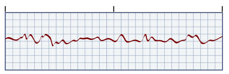
Ventricular Fibrillation
associated with (4)
Pt characteristics (3)
if not treated what will happen (1)
treatment (5)
• Associated with MI, ischemia, disease states, procedures
• Unresponsive, pulseless, and apneic
• If not treated rapidly, death will result
• Treat with immediate CPR and ACLS (same as for pulseless VT)
• Defibrillation
• Drug therapy (epinephrine, vasopressin)
Epinephrine
indications (5)
considered after what in symptomatic bradycardia (1)
drug class (1)
actions (5)
dosage (1)
Indications: cardiac arrest (pulseless VT, VF, asystole, considered after atropine in symptomatic bradycardia)
• Drug class- alpha & beta adrenergic agonist
• Actions – increases SVR, increased HR, enhanced myocardial contraction, coronary vasodilation, enhanced brain perfusion
• Dosage – 1 mg (1:10,000 solution
Pulseless Electrical Activity
define (3)
prognosis (1)
treatment (4)
• Electrical activity can be observed on the ECG, but no mechanical activity of the heart is evident, and the patient has no pulse (e.g. idioventricular rhythm (IVR), ventricular escape rhythms, post-defibrillation IVR)
• Prognosis is poor unless underlying cause quickly identified and treated
• Treatment – CPR, IV/IO access, Epinephrine, find and treat cause
causes of PEA - 6 Hs, 5 Ts
don’t need to memorize

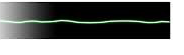
Asystole
describe
Pt characteristic (3)
must assess (1)
usually a result of (3)
treatment: (4)
prognosis (1)
• Represents total absence of ventricular electrical activity
• No ventricular contraction
• Patient unresponsive, pulseless, apneic
• Must assess in more than one lead
• Usually result of advanced cardiac disease, severe
conduction disturbance, or end-stage HF
• Treat with immediate CPR and ACLS measures
• Epinephrine
• Intubation
• Poor prognosis
Sudden Cardiac Death (SCD)
define
results from (2)
• Death from a cardiac cause
• Majority of SCDs result from ventricular dysrhythmias
• Ventricular tachycardia
• Ventricular fibrillation
DEFIBRILLATION
treatment of choice for what (2)
what is it not for (2)
Most effective when completed within ____ of dysrhythmia onset
action (2)
monophasic vs biphasic defibrillators
• Treatment of choice for VF and pulseless VT. NOT for asystole nor PEA.
• Most effective when completed within 2 minutes of dysrhythmia onset
• Passage of DC electrical shock through the heart to depolarize myocardial cells
• Allows SA node to resume pacemaker role (120-200 joules)
• Monophasic defibrillators deliver energy in one direction (360 joules)
• Biphasic defibrillators deliver energy in two directions
• Use lower energies
• Fewer post-shock ECG dysrhythmia
which type of defilation is preferred
Biphasic
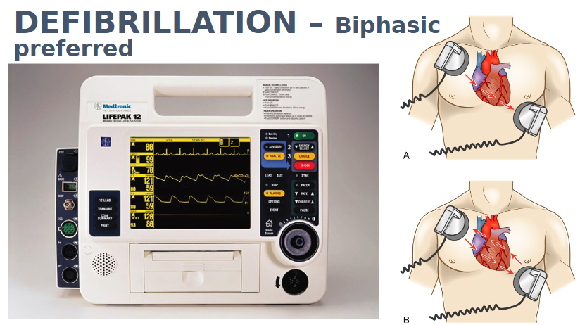
DEFIBRILLATION process (8)
1. Start CPR while obtaining and setting up defibrillator
2. Turn on and select energy
3. Make sure sync button is turned off
4. Apply gel pads
5. Charge
6. Position paddles firmly on chest
7. Ensure “All clear”!!!!!
8. Deliver charge
Synchronized Cardioversion (DCCV)
used for (3)
action
similar to defibrillation except ___
if pt is stable ___ prior
initial ____ energy
_____ joules (biphasic)
____ joules for afib
what to do if pt becomes pulseless
Used for STABLE Tachyarrhythmias -
A-fib, A-flutter; VT with pulse
• Synchronized circuit delivers a countershock on the R wave of the QRS complex of the ECG
• Procedure similar to defibrillation except sync button turned ON
• If patient stable, sedate prior
• Initial lower energy
• 50-100 joules (biphasic)
• 120 joules for afib
• If patient becomes pulseless, turn
sync button off and defibrillate
A patient in the coronary care unit develops ventricular fibrillation. The first action the nurse should take is to
a. Prepare for synchronized cardioversion.
b. Perform defibrillation
c. Initiate CPR.
d. Administer IV antidysrhythmic drugs per protocol.
B
NCLEX key terms
p wave =
QRS wave =
lack of QRS complexes =
wide bizarre QRS complexes =
chaotic or unorganized =
chaotic rhythm with no p wave =
chaotic rhythm without QRS complexes

The monitor of a patient in the coronary care unit abruptly
shows a chaotic rhythm without QRS complexes. The
first action the nurse should take is to
a. Prepare for synchronized cardioversion.
b. Perform defibrillation
c. Initiate CPR.
d. Administer IV antidysrhythmic drugs per protocol
B
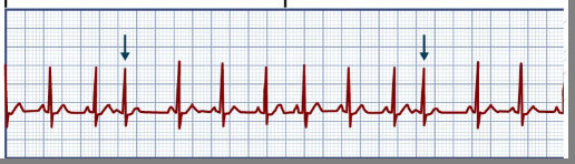
Premature atrial contraction
define
p wave (3)
QRS (1)
could be caused by (10) (5 big ones)
Contraction originating from ectopic focus in atrium in location other than SA node
Travels across atria by abnormal pathway, creating distorted P wave
P wave could be flattened, notched, or
hidden in T wave
QRS normal but comes early
Could be caused by…
Stress
anxiety
Caffeine
Tobacco
Alcohol
Fatigue
Hypoxia
Electrolyte imbalance
Disease states e.g. HF
Digitalis toxicity
Premature Atrial Contraction (PACs)
manifestations (feels like…) (3)
treatment (5)
Manifestations (Feels Like…)
Palpitations
Heart “skips a beat”
Unknown, unaware
Treatment – treat cause & slow it down
Withhold sources of stimulation
β-blockers
Monitor for more serious dysrhythmias
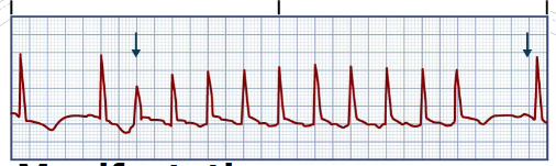
Paroxysmal Supraventricular Tachycardia (PSVT)
manifestations (7) what is the key? (1) (HR)
what to think about
reentrant phenomenon
paroxysmal refers to ____
associated with (6)
Manifestations
HR is 150–250 beats/minute
HR > 180 leads to decreased cardiac
output and stroke volume
Recognizing unstable tachycardia is KEY
Hypotension, Dyspnea, Angina, Altered cognition, Acute heart failure
Is the tachycardia causing the S/S or are S/S producing the tachycardia
reentrant phenomenon:
PSVT triggers a run of repeated premature beats
Paroxysmal refers to an abrupt onset and termination
Associated with
overexertion, stress, deep inspiration, stimulants, disease, digitalis toxicity
Paroxysmal Supraventricular Tachycardia (PSVT)
treatment (5)
if symptoms persist (1)
Treatment – slow it down
Vagal stimulation (Valsalva maneuver)
IV β-blockers
Calcium channel blockers
IV adenosine (slows heart)
If symptoms persist – synchronized cardioversion

Atrial fibrillation
define (HR)
controlled when
uncontrolled when
3 major chacteristics
assessing pulse with a fib where?
Atria – fibrillating 350-600/min (inadequate filling or contraction)
Ventricular response
Controlled when HR 60-100
Uncontrolled when HR > 100
3 major characteristics
1. No P waves
2. Normal QRS configuration
3. Fibrillatory waves (F waves)
Assessing pulse with A-fib - apical
Atrial fibrillation (Afib)
most common ____
define
prevalence increases with __
occurs in pts with (6)
high risk of ____ is ___
Paroxysmal (comes & goes) or persistent
Most common dysrhythmia
Prevalence increases with age
Usually occurs in patients with underlying heart disease e.g. MI, HTN, valve disease
Other – digitalis toxicity, COPD
High risk of clots and stroke if HR >100
Atrial fibrillation (Afib)
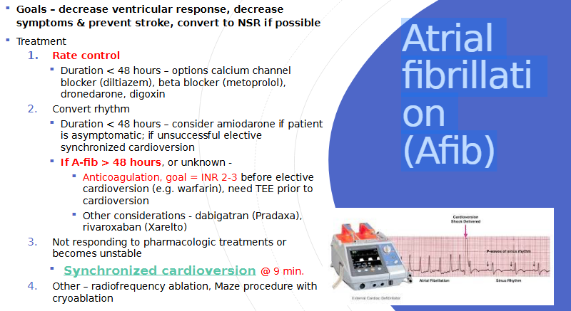
goals (3)
treatment:
rate control: duration and options (4)
convert rhythm: duration and options (2)
if a-fib for > 48 hours or unknown (3) other considerations (2)
if not responding to tx or becomes unstable (1) other (2)
- dabigatran (Pradaxa), rivaroxaban (Xarelt) = blood thinners used to prevent and treat dangerous blood clots
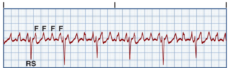
Atrial Flutter
rate?
P wave
PR
Atrial oscillations
must calculate what (2)
associated with (1)
s/s result from ___ which causes __
increased risk of ___
Atrial Rate generally – 250-350
Atria so rapidly that the AV node can’t respond to every impulse. The ventricular rate depends on the number of atrial impulses that pass through the AV node (will see ventricular response)
No P waves. No PR-interval to measure
Atrial oscillations – flutter waves (f waves), saw-toothed or picket fence appearance
must calculate atrial and ventricular rates
Typically associated with disease
Symptoms result from high ventricular rate and
loss of atrial “kick” → decreased CO → heart
failure
Increases risk of strok