5 - Basic Information for Bacteria
 Call Kai
 Learn
Learn Practice Test
 Spaced Repetition
 Match
Match Flashcards
 Knowt Play
1/112

There's no tags or description
Looks like no tags are added yet.
Name | Mastery | Learn | Test | Matching | Spaced | Call with Kai | Chat |
|---|
No analytics yet
Send a link to your students to track their progress
113 Terms
d. Crystal Violet
[Gram Stain]
Primary stain used in Gram staining.
a. Safranin
b. Iodine
c. Alcohol
d. Crystal Violet
c. Mordant/fixer
[Gram Stain]
Role of iodine in Gram staining.
a. Primary stain
b. Decolorizing agent
c. Mordant/fixer
d. Secondary stain
d. Decolorizing agent
[Gram Stain]
Role of alcohol in Gram staining.
a. Primary stain
b. Mordant/fixer
c. Secondary stain
d. Decolorizing agent
c. Safranin
[Gram Stain]
Secondary stain/counter stain used in Gram staining.
a. Crystal Violet
b. Iodine
c. Safranin
d. Alcohol
d. Violet
[Gram Stain]
Color of Gram-positive organisms after Crystal Violet step.
a. Red
b. Colorless
c. Pink
d. Violet
c. Violet
[Gram Stain]
Color of Gram-negative organisms after Crystal Violet step.
a. Red
b. Colorless
c. Violet
d. Pink
d. Violet
[Gram Stain]
Color of Gram-positive organisms after Iodine step.
a. Red
b. Colorless
c. Pink
d. Violet
d. Violet
[Gram Stain]
Color of Gram-negative organisms after Iodine step.
a. Red
b. Colorless
c. Pink
d. Violet
c. Violet
[Gram Stain]
Color of Gram-positive organisms after Alcohol decolorizing step.
a. Colorless
b. Red
c. Violet
d. Pink
d. Colorless
[Gram Stain]
Color of Gram-negative organisms after Alcohol decolorizing step.
a. Violet
b. Red
c. Pink
d. Colorless
d. Violet
[Cell Wall]
Final color of Gram-positive organisms after Safranin counterstain.
a. Red
b. Colorless
c. Pink
d. Violet
d. Red
[Cell Wall]
Final color of Gram-negative organisms after Safranin counterstain.
a. Violet
b. Colorless
c. Pink
d. Red
d. Violet
[Cell Wall]
Final color of Gram-positive organisms in Gram staining.
a. Red
b. Colorless
c. Pink
d. Violet
d. Teichoic acid
[Cell Wall]
Antigenic determinant of Gram-positive cell wall.
a. Lipid A
b. Porin
c. LPS
d. Teichoic acid
c. Thick layer of peptidoglycan
[Cell Wall]
Type of peptidoglycan layer present in Gram-positive cell wall.
a. Thin layer of peptidoglycan
b. No peptidoglycan
c. Thick layer of peptidoglycan
d. Single layer of peptidoglycan
c. Amino acids and disaccharide
[Cell Wall]
Components that make up the peptidoglycan layer in Gram-positive cell wall.
a. Lipid A and polysaccharide
b. Teichoic acid and porin
c. Amino acids and disaccharide
d. LPS and outer membrane
d. Transpeptidase
[Cell Wall]
Enzyme responsible for cross-linking peptidoglycan in Gram-positive cell wall.
a. Lysozyme
b. Beta-lactamase
c. Penicillinase
d. Transpeptidase
c. Beta-lactam
[Cell Wall]
Antibiotic class that inhibits transpeptidase in Gram-positive cell wall synthesis.
a. Aminoglycosides
b. Macrolides
c. Beta-lactam
d. Fluoroquinolones
b. Inhibit transpeptidase
[Cell Wall]
Effect of beta-lactam antibiotics on transpeptidase.
a. Activate transpeptidase
b. Inhibit transpeptidase
c. Replace transpeptidase
d. Duplicate transpeptidase
c. Cross-linked via transpeptidase
[Cell Wall]
Method by which peptidoglycan components are connected in Gram-positive cell wall.
a. Hydrogen bonding
b. Ionic bonding
c. Cross-linked via transpeptidase
d. Covalent bonding via lysozyme
d. Red
[Cell Wall]
Final color of Gram-negative organisms in Gram staining.
a. Violet
b. Pink
c. Colorless
d. Red
c. Endotoxin
[Cell Wall]
Property automatically present in all Gram-negative organisms.
a. Teichoic acid
b. Thick peptidoglycan
c. Endotoxin
d. Transpeptidase
c. Porins
[Cell Wall]
Structure in Gram-negative cell wall that acts as passageway of substances.
a. Teichoic acid
b. Lipid A
c. Porins
d. LPS
b. Somatic O antigen
[Cell Wall]
Outer membrane component of Gram-negative organisms that contributes to somatic O antigen.
a. Lipid A
b. Somatic O antigen
c. Core polysaccharide
d. Porin
c. Core polysaccharide
[Cell Wall]
Outer membrane component of Gram-negative organisms that contributes to core polysaccharide.
a. Somatic O antigen
b. Lipid A
c. Core polysaccharide
d. Porin
d. Lipid A
[Cell Wall]
Outer membrane component of Gram-negative organisms that is the endotoxin and part of cell wall.
a. Somatic O antigen
b. Core polysaccharide
c. Porin
d. Lipid A
c. LPS
[Cell Wall]
Another term for the lipopolysaccharide of Gram-negative outer membrane.
a. Teichoic acid
b. Peptidoglycan
c. LPS
d. Endotoxin only
d. Lysozymes
[Cell Wall]
Content of the periplasmic space in Gram-negative cell wall.
a. Teichoic acid
b. Lipid A
c. Transpeptidase
d. Lysozymes
d. 1 layer
[Cell Wall]
BEQ: Thickness of peptidoglycan layer in Gram-negative cell wall.
a. Thick multilayer
b. No peptidoglycan
c. 2 layers
d. 1 layer
c. 1 layer
[Cell Wall]
BEQ: Number of peptidoglycan layers in Gram-negative cell wall.
a. 3 layers
b. 2 layers
c. 1 layer
d. No layers
c. Cocci
[Morphology]
Bacterial morphology described as "round."
a. Bacilli
b. Spirilla
c. Cocci
d. Pleomorphic
d. Staphylococcus
[Morphology]
Round-shaped bacterium arranged in clusters.
a. Streptococcus
b. Spirilla
c. Coccobacilli
d. Staphylococcus
c. Streptococcus
[Morphology]
Round-shaped bacterium arranged in chains.
a. Staphylococcus
b. Coccobacilli
c. Streptococcus
d. Spirilla
b. Bacilli
[Morphology]
Bacterial morphology described as "rod."
a. Cocci
b. Bacilli
c. Spirilla
d. Pleomorphic
d. Coccobacilli
[Morphology]
-shaped bacterium with a rounded appearance between cocci and bacilli.
a. Spirilla
b. Streptococcus
c. Staphylococcus
d. Coccobacilli
d. Spirilla
[Morphology]
Bacterial morphology described as curved.
a. Cocci
b. Bacilli
c. Pleomorphic
d. Spirilla
c. Pleomorphic
[Morphology]
Bacterial morphology described as varied morphology.
a. Spirilla
b. Bacilli
c. Pleomorphic
d. Cocci
c. Flagella
[Virulence Factors]
Virulence factor used for forward locomotion in bacteria.
a. Pili
b. Capsule
c. Flagella
d. Fimbriae
b. Chemotaxis
[Virulence Factors]
Movement of bacteria toward a chemical gradient using flagella.
a. Phototaxis
b. Chemotaxis
c. Geotaxis
d. Thermotaxis
c. H antigen
[Virulence Factors]
Antigen associated with bacterial flagella.
a. O antigen
b. K antigen
c. H antigen
d. Vi antigen
d. Atrichous
[Virulence Factors]
Flagella type with no flagella.
a. Monotrichous
b. Lophotrichous
c. Peritrichous
d. Atrichous
c. Shigella dysenteriae
[Virulence Factors]
BEQ: Example of an atrichous bacterium that is non-motile.
a. Vibrio cholerae
b. Helicobacter pylori
c. Shigella dysenteriae
d. Campylobacter jejuni
No flagella
Non motile
[Virulence Factors]
Characteristic of Shigella dysenteriae as an atrichous bacterium.
a. Comma shaped
b. Gull wing shaped
c. No flagella, non motile
d. Flagella on both ends
c. Monotrichous
[Virulence Factors]
Flagella type with one flagella.
a. Atrichous
b. Lophotrichous
c. Monotrichous
d. Amphitrichous
d. Vibrio cholerae
[Virulence Factors]
BEQ: Example of a monotrichous bacterium that is comma shaped.
a. Shigella dysenteriae
b. Campylobacter jejuni
c. Helicobacter pylori
d. Vibrio cholerae
c. Lophotrichous
[Virulence Factors]
Flagella type with more than one flagella.
a. Atrichous
b. Monotrichous
c. Lophotrichous
d. Amphitrichous
c. Helicobacter pylori
[Virulence Factors]
BEQ: Example of a lophotrichous bacterium that is the number one cause of peptic ulcer disease.
a. Vibrio cholerae
b. Shigella dysenteriae
c. Helicobacter pylori
d. Campylobacter jejuni
d. Helicobacter pylori
[Virulence Factors]
BEQ: Number one cause of peptic ulcer disease.
a. Vibrio cholerae
b. NSAIDs
c. Campylobacter jejuni
d. Helicobacter pylori
c. NSAIDs
[Virulence Factors]
BEQ: Second most common cause of peptic ulcer disease.
a. Helicobacter pylori
b. Campylobacter jejuni
c. NSAIDs
d. Shigella dysenteriae
d. Amphitrichous
[Virulence Factors]
Flagella type with flagella on both ends.
a. Atrichous
b. Monotrichous
c. Peritrichous
d. Amphitrichous
c. Campylobacter jejuni
[Virulence Factors]
Example of an amphitrichous bacterium described as gull wing shaped.
a. Vibrio cholerae
b. Shigella dysenteriae
c. Campylobacter jejuni
d. Helicobacter pylori
d. Peritrichous
[Virulence Factors]
Flagella type with flagella all over the bacterial body.
a. Atrichous
b. Monotrichous
c. Amphitrichous
d. Peritrichous
b. E. coli
c. Shigella dy
[Virulence Factors]
Example of a peritrichous bacterium.
a. Vibrio cholerae
b. E. coli
c. Shigella dysenteriae
d. Campylobacter jejuni
d. Pili
[Virulence Factors]
Virulence factor that is longer and few in number compared to fimbriae.
a. Flagella
b. Fimbriae
c. Capsule
d. Pili
c. Fimbriae
[Virulence Factors]
Virulence factor that is shorter and many in number compared to pili.
a. Flagella
b. Pili
c. Fimbriae
d. Capsule
c. Sex pili
[Virulence Factors]
Type of pili used for conjugation.
a. Adhesion pili
b. P fimbriae
c. Sex pili
d. Common pili
d. Adhesion pili
[Virulence Factors]
Type of pili used for attachment.
a. Sex pili
b. P fimbriae
c. Common pili
d. Adhesion pili
c. P fimbriae
[Virulence Factors]
Type of fimbriae seen in E. coli that attaches to the urinary tract.
a. Sex pili
b. Adhesion pili
c. P fimbriae
d. Common fimbriae
d. E. coli
[Virulence Factors]
Bacterium that uses P fimbriae to attach to the urinary tract.
a. Shigella dysenteriae
b. Vibrio cholerae
c. Campylobacter jejuni
d. E. coli
c. Attach to the urinary tract
[Virulence Factors]
Function of P fimbriae in E. coli.
a. Conjugation
b. Forward locomotion
c. Attach to the urinary tract
d. Chemotaxis
b. Pili are few; fimbriae are many
[Virulence Factors]
Characteristic that differentiates pili from fimbriae in terms of quantity.
a. Pili are many; fimbriae are few
b. Pili are few; fimbriae are many
c. Both are equal in number
d. Pili are absent; fimbriae are many
c. Polysaccharide
[Virulence Factors]
Material that all capsules are made of.
a. Lipopolysaccharide
b. Peptidoglycan
c. Polysaccharide
d. Teichoic acid
d. Bacillus anthracis
[Virulence Factors]
All capsule are made of “polysaccharide” except for ______ which are made of D-glutamic acid
a. E. coli
b. Neisseria spp.
c. Klebsiella pneumoniae
d. Bacillus anthracis
b. D-glutamic acid
[Virulence Factors]
Material that makes up the capsule of Bacillus anthracis instead of polysaccharide.
a. Lipid A
b. D-glutamic acid
c. Teichoic acid
d. Peptidoglycan
c. Firmly attached and organized
[Virulence Factors]
Characteristic that differentiates capsule from slime layer.
a. Made of polysaccharide
b. Loosely attached and disorganized
c. Firmly attached and organized
d. Made of peptidoglycan
d. K antigen
[Virulence Factors]
Antigen associated with bacterial capsule.
a. H antigen
b. O antigen
c. Vi antigen
d. K antigen
b. Prevent phagocytosis
[Virulence Factors]
Role of capsule in bacterial virulence.
a. Forward locomotion
b. Prevent phagocytosis
c. Conjugation
d. Attachment to urinary tract
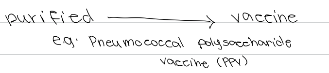
b. Pneumococcal polysaccharide vaccine (PPV)
[Virulence Factors]
Capsule purified to produce a vaccine against encapsulated organisms.
a. Pneumococcal conjugate vaccine
b. Pneumococcal polysaccharide vaccine (PPV)
c. BCG vaccine
d. Meningococcal conjugate vaccine
c. Quellung reaction
[Virulence Factors]
Test used to detect bacterial capsule through antibody binding causing capsular swelling.
a. Gram staining
b. India ink staining
c. Quellung reaction
d. Nigrosin staining
c. Streptococcus pneumoniae
[Virulence Factors]
Organism detected by Quellung reaction and treated with PPV.
a. Haemophilus influenzae
b. Klebsiella pneumoniae
c. Streptococcus pneumoniae
d. Neisseria spp.
c. Pneumococcal polysaccharide vaccine (PPV)
[Virulence Factors]
Streptococcus pneumoniae is treated with:
a. BCG vaccine
b. Meningococcal vaccine
c. Pneumococcal polysaccharide vaccine (PPV)
d. MMR vaccine
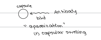
b. Opsonization and capsular swelling
[Virulence Factors]
Mechanism of Quellung reaction involving antibody binding to capsule.
a. Capsular dissolution
b. Opsonization and capsular swelling
c. Agglutination
d. Complement activation
d. E. coli
[Virulence Factors]
Encapsulated organism that appears green in methylene blue staining.
a. Haemophilus influenzae
b. Klebsiella pneumoniae
c. Neisseria spp.
d. E. coli
Haemophilus influenzae
Klebsiella pneumoniae
Streptococcus pneumoniae
Neisseria spp.
E. coli
📌Mnemonic: "HKSNE"
[Virulence Factors]
Encapsulated organism include ______
a. Post-splenectomy
[Virulence Factors]
Removal of spleen.
a. Post-splenectomy
b. Post-nephrectomy
c. Post-hepatectomy
d. Post-thyroidectomy
b. Encapsulated bacteria
[Virulence Factors]
Type of bacteria that post-splenectomy patients are prone to.
a. Gram-negative bacteria
b. Encapsulated bacteria
c. Acid-fast bacteria
d. Anaerobic bacteria
c. Post-splenectomy
[Virulence Factors]
Condition that makes a patient prone to encapsulated bacteria.
a. Malnutrition
b. Diabetes mellitus
c. Post-splenectomy
d. Immunocompromised state
d. Vaccine
[Virulence Factors]
Remedy for post-splenectomy patients prone to encapsulated bacteria.
a. Antibiotic prophylaxis
b. Immunoglobulin therapy
c. Corticosteroids
d. Vaccine
a. Spores
[Virulence Factors]
_____- are considered highly resistant due to presence of Calcium dipicolinate
a. Spores
b. Capsule
c. Flagella
d. Pili
Bacillus spp.
Clostridium spp.
[Virulence Factors]
Spore-forming bacterium [2]
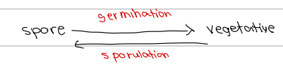
b. Germination
[Virulence Factors]
Process by which spore converts to vegetative form.
a. Sporulation
b. Germination
c. Conjugation
d. Transformation
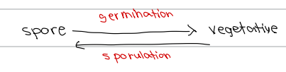
c. Sporulation
[Virulence Factors]
Process by which vegetative form converts back to spore.
a. Germination
b. Conjugation
c. Sporulation
d. Transformation
d. Calcium dipicolinate
[Virulence Factors]
Component responsible for high resistance of spores to physical and chemical agents.
a. Teichoic acid
b. D-glutamic acid
c. Peptidoglycan
d. Calcium dipicolinate
d. Gram-positive and Gram-negative
[Toxin]
Source of exotoxin.
a. Gram-negative only
b. Fungi only
c. Gram-positive only
d. Gram-positive and Gram-negative
d. High
[Toxin]
Toxicity of exotoxin.
a. Low
b. Moderate
c. Variable
d. High
b. High
[Toxin]
Potency of exotoxin.
a. Low
b. High
c. Moderate
d. Variable
d. Heat-labile
[Toxin]
Heat characteristic of exotoxin.
a. Heat-stable
b. Heat-neutral
c. Heat-resistant
d. Heat-labile
c. A region
[Toxin]
Region of exotoxin responsible for activity.
a. B region
b. C region
c. A region
d. D region
c. B region
[Toxin]
Region of exotoxin responsible for binding.
a. A region
b. C region
c. B region
d. D region
d. IgM and IgG
[Toxin]
Defense against exotoxin.
a. IgA and IgE
b. IgD and IgA
c. IgE and IgD
d. IgM and IgG
d. Formaldehyde
[Toxin]
Significance of exotoxin as source of toxoid through inactivation via:
a. Heat treatment
b. Alcohol
c. Radiation
d. Formaldehyde
c. Artificially acquired active immunity
[Toxin]
Type of artificially acquired immunity produced from exotoxin-derived toxoid.
a. Natural active immunity
b. Natural passive immunity
c. Artificially acquired active immunity
d. Artificially acquired passive immunity
d. Diphtheria toxoid
[Toxin]
Example of a toxoid vaccine derived from exotoxin.
a. BCG
b. PPV
c. MMR
d. Diphtheria toxoid
b. Tetanus toxoid
[Toxin]
Example of a toxoid vaccine alongside Diphtheria toxoid.
a. BCG
b. Tetanus toxoid
c. MMR
d. PPV
c. Gram-negative bacteria
[Toxin]
Primary source of endotoxin.
a. Gram-positive bacteria
b. Fungi
c. Gram-negative bacteria
d. Protozoa
d. Listeria spp.
[Toxin]
Gram-positive bacterium that is an exception possessing endotoxin.
a. Staphylococcus aureus
b. Bacillus anthracis
c. Streptococcus pneumoniae
d. Listeria spp.
d. Low
[Toxin]
Toxicity of endotoxin.
a. High
b. Moderate
c. Variable
d. Low
b. Low
[Toxin]
Potency of endotoxin.
a. High
b. Low
c. Moderate
d. Variable
c. Heat-stable
[Toxin]
Heat characteristic of endotoxin.
a. Heat-labile
b. Heat-neutral
c. Heat-stable
d. Heat-resistant
d. Septic shock = decreased BP
[Toxin]
Clinical significance of endotoxin.
a. Source of toxoid
b. Artificially acquired active immunity
c. IgM and IgG defense
d. Septic shock = decreased BP