Case 5: Get Along Little Buddy
 Call Kai
 Learn
Learn Practice Test
 Spaced Repetition
 Match
Match Flashcards
 Knowt Play
1/33

There's no tags or description
Looks like no tags are added yet.
Name | Mastery | Learn | Test | Matching | Spaced | Call with Kai | Chat |
|---|
No analytics yet
Send a link to your students to track their progress
34 Terms
True = common leads to left atrial enlargement
In our canine patients, mitral valve disease is the most common acquired heart disease.
TRUE OR FALSE
False
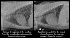
The only pathologic process that causes the trachea to be elevated from its normal position (diverging from the spine) is cardiomegaly.
TRUE OR FALSE
True
Patient positioning is critical when evaluating the cardiac silhouette.
TRUE OR FALSE
Size and shape
What two Roentgen signs are important when evaluating the abnormal image?
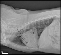
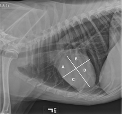
A. Right atrium
B. Left atrium
C. Right ventricles
D. Left ventricles
Identify the labeled structures
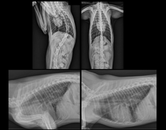
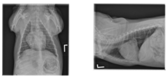
Canine Left lateral and VD thoracic projections are available for review.
Describe the study (views and anatomy)
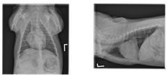
Patient positioning is adequate for interpretation.
Study technique is adequate for interpretation.
note: there is good superimposition of the rib heads
Describe patient positioning and technique
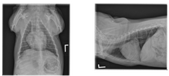
The cardiac silhouette is enlarged, being both too tall and too wide. There is marked left atrial enlargement which is causing left mainstem bronchus compression. The trachea is elevated. The pulmonary vessels are not enlarged and the pulmonary parenchyma is unremarkable. The pleural space is unremarkable. No abnormalities are identified within the extra-thoracic structures.
Describe your findings using roentgen signs
Left atrial enlargement and left mainstem bronchus compression with no radiographic evidence of pulmonary venous congestion or pulmonary edema. These findings are most consistent with mitral valve disease.
Describe your conclusion: Give your radiographic diagnosis and list possible differential diagnoses for all your findings
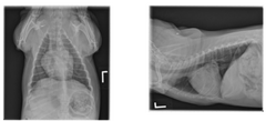
Echocardiography could be considered to further evaluate cardiac structure and function.
Describe Next Steps: Are there any additional imaging suggestions that would help clarify your findings? Are there critical next steps to take from a stabilization/management/treatment plan standpoint?
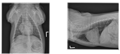
Middle
What part of the mediastinum is the cardiac silhouette located in?
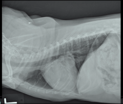
Trachea is elevated dorsally
This is a left lateral of a dog. Heart is too tall and too wide which you can tell WHY? Because of deviation of what other structure?
Soft tissue muscle of the heart and the fluid within the heart are the same opacity = cannot delineate margins
Why is it hard to interpret the cardiac silhouette of an animal on radiographs?
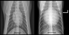
Left atrium = can tell from "cowboy sign" which is when the trachea comes down and the primary/principal bronchus curves out
What structure sits here in the VD view? How can you tell it is enlarged?
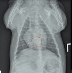
Number, size, opacity, margin, shape, position
List the roentgen signs
The one WITH the arrow - NORMAL
note: the one without the arrow is a patient with congestive heart failure
Which of these images is normal, the one with the arrow pointing to it OR the one without the arrow?
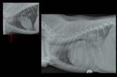
Caudal margin of the cardiac silhouette is not visible because of increased opacity in the caudodorsal lung fields, which is pulmonary edema
How can you tell the image on the right has congestive heart failure?
note: the left image is normal
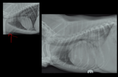
It is not likely = no pulmonary edema
Does this dog have congestive heart failure? How can you tell?
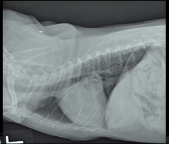
Systolic murmur
What kind of murmur will you hear with mitral valve disease at the 5th IC space?
Between the left atrium and left ventricle
Where is the mitral valve located?
Between the left ventricle and out to the circulatory system (aka body)
Where is the aortic valve located?
True
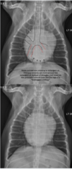
Left atrial enlargement often results in the "bow-legged cowboy" sign.
TRUE OR FALSE
xx
for reference on cowboy sign
b. Right Ventricle
What is structure A?
a. Main Pulmonary Artery
b. Right Ventricle
c. Caudal Lobar Arteries
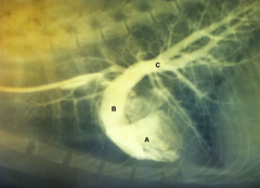
1. A) Left Atrium B) Left Ventricle
Identify the structures labeled A and B:
1. A) Left Atrium B) Left Ventricle
2. A) Left Ventricle B) Left Atrium
3. A) Ascending Aorta B) Left Ventricle
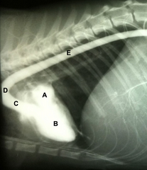
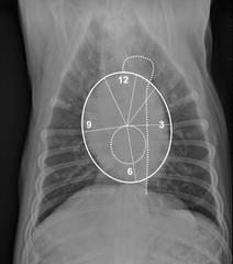
LINE: aorta
CIRCLE: left atrium
What are the DOTTED LINE and DOTTED CIRCLE representing, respectively?
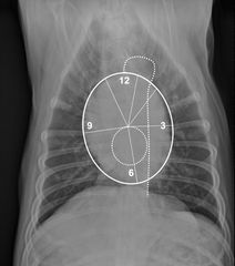
Aortic arch
What structure would be located at 12 o'clock?
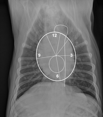
Main pulmonary artery
What structure would be located at 1-2 o'clock?
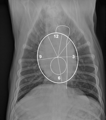
Left auricle
What structure would be located at 3 o'clock?
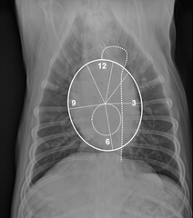
Left ventricle
What structure would be located at 3-6 o'clock?
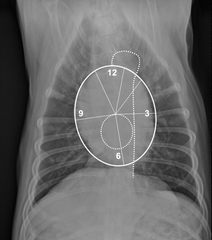
Right ventricle
What structure would be located at 6-9 o'clock?
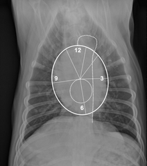
Right atrium
What structure would be located at 9-11 o'clock?
True
note: This is because the mitral valve becomes "leaky" and allows some blood to move in the wrong direction, back into the left atrium. The left atrium responds to this increase in volume by dilating
The main reason the left atrium gets big with mitral valve disease is because of left atrial volume overload.
TRUE OR FALSE
A. Cranial Lobar Artery
B. Cranial Lobar Vein
C. Enlarged Left Atrium
D. Caudal Vena Cava
What are the structures located at A, B, C, and D respectively?