PSYC 367 Midterm 1: Learning Objectives
1/37
There's no tags or description
Looks like no tags are added yet.
Name | Mastery | Learn | Test | Matching | Spaced |
|---|
No study sessions yet.
38 Terms
3 cues that listeners use to judge the distance of sound sources
relative intensity of sound: making our own assumptions
spectral composition of sounds: when sound sources are far away, higher frequencies decay first so sound sounds muddy
relative amounts of direct vs. reverberant energy: more reverberations = farther sound
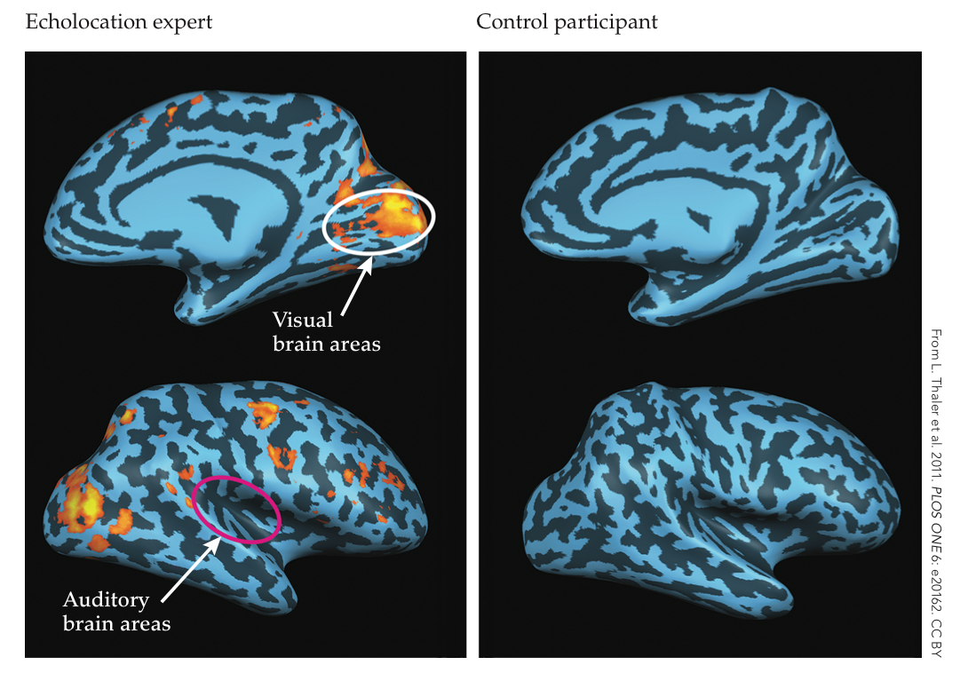
Describe the fMRI evidence for the use of echolocation by visually impaired listeners
found that visual areas of the brain are still activated when using hearing to identify where stimuli is the environment

Describe 2 important design components of modern electronic
hearing aids
(1) amplify sound signals while also (2) compressing intensity differences - keep highest intensities at comfortable listening level
Define hidden hearing loss and state the hypothesized
mechanism for it
exposure to moderately high levels of noise changing ability to use sound even when ability to detect sound remains normal - may explain difficulties we have in noisy situations even when audiograms are normal
Discuss the relationship between the active process that sharpens and amplifies the frequency response of the basilar membrane, otoacoustic emissions, and electromotility of outer hair cells
active process is intricately linked to the electromotility of outer hair cells (the cochlear amplifier)which enhances sound detection and frequency discrimination
electromotility generates otoacoustic emissions: measurable sounds produced by the cochlea
How is characteristic frequency related to place code for sound frequency
characteristic frequency: frequency to which an auditory nerve fibre is most sensitive (specific place on basilar membrane)
tontopic organization (high frequency sounds at base and low frequency sounds at apex of cochlea)
place code is based on tontopic organization
how is two-tone suppression related to place code for sound frequency?
response of nerve fibre to its characteristic frequency reduced when a second frequency is played at the same time - reduction in cochlear amplifier activity in response, leading to reduced basilar membrane response
place code: basilar membrane response to one frequency is inhibited by adjacent frequencies so the clarity and specificity of place code for a specific sound is improved
how is rate saturation related to place code for sound frequency?
auditory nerve fibre firing rate maxes out and no longer increases after a certain intensity level
place code: compensates by this potential limitation with different types of intensity coding - low-threshold (respond to soft sounds and quickly saturate with high) and high-threshold fibres (less sensitive to soft sounds, respond to high intensities)
how is phase locking related to temporal code?
temporal coding relies on when neurons fire in relation to sound wave to gather information about sound’s frequency (instead of relying on where along basilar membrane like place coding)
how is the volley principle related to temporal code?
crucial for temporal coding at frequencies above phase-locking limits of individual neurons (4000-5000 Hz) so timing of sound wave cycles can still be tracked
Discuss the coding of sound intensity information in the inner ear
primarily occurs through activity of auditory nerve fibres connected to hair cells in cochlea - key mechanisms:
firing rate of nerve fibres: low rate for low intensity sounds, high rate for higher sounds (potential rate saturation)
recruitment of additional nerve fibres as sound level increases
inner hair cells (signal to the brain) vs. outer hair cells (amplifying low intensity sounds
tonotopic organization
high-spontaneous vs. low-spontaneous fibres for encoding a range of intensities
List, in order, the 4 main stations (and any subdivisions) where synapses occur along the auditory pathways between the inner ear and the cortex. State whether the neurons are monaural or binaural in each station
Cochlear nucleus (brainstem) - first point of synapse for auditory nerve fibers, dorsal and ventral with monaural neurons (input from only one ear, ipsilateral)
superior olivary complex (pons) - first sight where both ears are synapse sights, medial (low-frequency) and lateral (high-frequency) with binaural neurons
inferior colliculus (midbrain) - receives both direct and indirect input from cochlear nuclei and SOC, no subdivisions, binaural neurons
medial geniculate body (thalamus) - final relay station before auditory cortex, ventral (primary relay) and dorsal/medial (processing complex sounds), binaural input
Describe the connections and function of the olivocochlear bundle
efferent fibres that originate in superior olivary complex and project to cochlea
2 divisions: medial (projects to outer hair cells [contralateral] and modulate activity of outer hair cells), lateral (projects to inner hair cells [ipsilateral], modulate activity of outer hair cells)
functions: protection from acoustic injury, improve signal-to-noise ratio, feedback control of cochlear amplification, bilateral sound processing, auditory attention
Identify the 3 main auditory cortical areas on a diagram of the brain, and describe their sub-regions and tonotopic organization
Primary auditory cortex (A1): superior temporal gyrus, regions organized based on sound frequency such that low frequencies are on one end and high are on the other
Secondary auditory cortex (A2): surrounds A1 (referred to as belt area) - belt (receives input from A1) and parabelt (region adjacent to belt), tontopic organization less defined than A1
Tertiary auditory cortex: surrounds belt and parabelt, extends into sulcus - includes regions responsible for abstract processing, less tonotopic organization
Describe the projections and function of the auditory “what” and
“where” pathways through the parabelt region and on to other
cortical areas
what pathway (ventral): identifying sounds (pitch, timbre, complex sounds) - originates in anterior parabelt region and projects to temporal lobe and prefrontal cortex, passing through anterior superior temporal gyrus and temporal sulcus
where pathway (dorsal): localizing source of sounds - originates in posterior posterior parabelt and projects to parietal lobe and prefrontal cortex, passing through temporal gyrus and parietal lobule
Explain how to obtain an audibility curve with air-conducted or bone-conducted sound.
air-conduction: tests the entire ear by delivering sound through headphones - audiometer generates pure tones at specific frequencies (Hz), threshold is identified for each frequency (dBs), low threshold = more sensitive, high = hearing damage
bone-conduction: tests cochlea (inner ear) specifically via vibrations through skull - bone conduction transducer placed on mastoid bone to vibrate it, same process as air-conduction (curve typically shows better hearing sensitivity than air-conduction)
if both air and bone conduction threshold are elevated, it points to sensorineural hearing loss
Describe a loudness matching experiment to obtain an equal loudness contour, and describe specific points on a set of contours in terms of their relative loudness
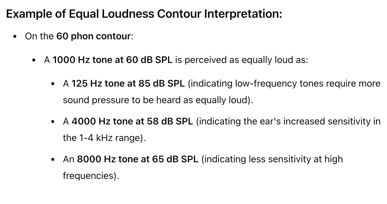
represent perceived loudness of sounds across different frequencies through equal loudness contours, relationship between sound pressure level and perceived loudness of tones
Procedure: show baseline reference tone then presented with tones of various frequencies (Hz) - for each frequency the listener adjusts the intensity until it is as loud as reference, these levels are plotted on graph where each contour represents tones that are perceived to be equally loud
show how sensitivity of human hearing varies
most sensitive to frequencies between 1000 Hz and 4000 Hz
phon = unit of perceived loudness lelel, each phon contour represents tones at various frequencies that sound equally loud to certain Hz

Distinguish between phons and sones
both are units of loudness
phons: unit of loudness level that corresponds to decibel level of a 1000 Hz tone, measured through loudness matching experiments - relative scale of perceived loudness, adjusts for human sensitivity
soones: unit of perceived loudness magnitude (linear relationship with perceived loudness, 1 sone = 1000 Hz at 40 phons (twice as loud = 2 sones), logarithmic and based on perceived doubling or halving loudness
Explain why loudness is not equivalent to intensity (dB)
loudness is the subjective perception of sound, while intensity (dB) is a physical measurement of sound
Discuss loudness discrimination and pitch discrimination in the
context of Weber’s law.
weber’s law allows us to understand how sensitive we are to changes in sound intensity - at low intensities, people can detect small changes in loudness, but as sound intensity increases, JND also increases
weber’s law allows use to understand our ability to discriminate between pitches and mid frequencies, for low frequencies we are sensitive, but as frequency increases, JND becomes larger
Describe a critical bandwidth masking experiment, explain why the pure-tone masking effect is asymmetrical, and discuss what masking experiments tell us about pitch perception.
investigates how frequency content and bandwidth of sound affects our ability to detect a target (pure) tone in presence of masking noise - determine frequency range where masker most effectively interferes (critical bandwidth)
asymmetrical: lower-frequency sound tend to mask higher-frequency sounds more effectively (upward spread of masking), due to high-frequency at base of basilar membrane and low frequencies at apex (low-frequency vibrations spread upward)
show that frequency spectrum is divided into critical bands - width shows how well we can distinguish between different pitches, why we can differentiate between two similar frequencies
auditory system is highly selective, which is our ability to perceive complex sounds
supports place theory of pitch perception - perceived pitch is related to location of max excitation on basilar membrane, regions of cochlea respond to different frequencies
State Ohm’s acoustical law, and discuss how the ear might act as a Fourier analyzer.
the perceived timbre (quality) of a complex sound is determined by the amplitudes of its individual harmonic components, but not by relative phases (ear is sensitive to frequencies and amplitudes but not to how these harmonics are aligned in time)
implies that ear acts like a Fourier analyzer - breaking down complex sounds into sine wave components
tontopic organization of basilar membrane sorts sounds into individual frequencies and hair cell coding by auditory nerve fibres
phase locking preseves the temporal structure of hearing
Distinguish between conductive and sensorineural hearing loss in terms of the location of the damage, the ability to hear air-conducted and bone-conducted sound, and current treatment options.
location
conductive: problem with outer or middle ear that prevents sound from reaching inner ear (blockages, infections, otosclerosis, perforation of eardrum, fluid in middle ear, ossicle damage)
sensorineural: damage to inner ear (cochlea) or auditory nerve paths (damage to hair cells, noise exposure, genetics, acoustic trauma, infections, tumours)
ability to hear
conductive: air conducted is significantly reduced, bone conducted is less affected since sound is directly transmitted to inner ear
sensorineural: air-conducted and bone-conducted both significantly reduced because damage affects ability to convert sound to signals
treatment
conductive: earwax removal, antibiotics, surgery, hearing aids, surgical reconstruction
sensorineural: hearing aids, cochlear implants, auditory brainstem implants, assistive listening devices
Define otitis media, otosclerosis, retrocochlear dysfunction,
presbycusis; state the type of hearing loss each usually produces
otitis media: inflammation/infection of the middle ear - impedes transmission of sound from eardrum to ossicles, reducing sound conduction
otosclerosis: abnormal bone growth in middle ear (particularly stapes) preventing proper vibration - diminished sound transmission since stapes is transmitting vibrations properly
retrocochlear dysfunction: damage/disorders that occur beyond cochlea (auditory nerve) - sensorineural hearing loss, impairs ability to process sound signals (unilateral)
presbycusis: age-related degeneration of inner ear/auditory pathways - sensorineural hearing loss, high-frequency sounds being most affected (bilateral)
Draw the pathway for ipsilateral and contralateral acoustic reflexes, and describe how these reflexes are affected by inner ear damage vs. retrocochlear dysfunction
ipsilateral
sound detection from outer ear to cochlea
transduction of hair cells into electrical signals in cochlea
signals travel from auditory nerve to cochlear nucleus on the same side of ear
signal is sent to superior olivary complex on the same side
SOC processes and sends signals to facial nerve on same side
facial nerve innervates stapedius muscle on same side
stapedius muscle contracts, dampening movement of stapes to reduce sound transmission
contralateral
sound detection from outer ear to cochlea
transduction of hair cells into electrical signals in cochlea
signals travel from auditory nerve to cochlear nucleus on the same side of ear
signal is sent to SOC on opposite side via interneurons that cross the midline
SOC processes and sends signal to contralateral facial nerve
facial nerve innervates stapedius muscle on opposite side
stapedius muscle on contralateral side contracts
inner ear damage may lead to elevated reflex thresholds, reduced reflex magnitude, possible absence of reflex (reduces ability of hair cells, weaker signals sent via auditory nerve)
retrocochlear dysfunction may lead to absent reflexes or asymmetrical reflex patterns (impairs transmission of signals to brain stem)
Describe two types of binaural information that humans can use to locate the position from which a sound comes at a certain elevation. Describe the brainstem physiology for processing this information
Interaural time difference: difference in time it takes for a sound to reach each ear which allows estimation of the sound’s direction - most effective for localizing low-frequency sounds
processed in medial superior olive, receive input from both ear
Interaural level differences: difference in sound intensity reaching each ear, processing in lateral superior olive and MNTB - effective for high-frequency sounds
head shadow effect occurs when sound come from one side, causes sound to be less intense at opposite ear
Describe the brainstem physiology for processing interaural level differences
lateral superior olive: detecting/processing ILDs (excitatory input from ipsilateral ear and inhibitory from contralateral - relayed through medial nucleus of trapezoid body), also tontopically organized
medial nucleus of trapezoid body: inhibitory output to LSO, excitatory input from contralateral cochlear nucleus
superior olivary complex: receives relays from LSO and integrates them, detects ILDs (high frequency sounds)
Describe how head movements and pinna shape allow us to localize sounds with ambiguous intensity and time differences
head movements: dynamic change in binaural cues - turning/tilting the head changes spatial relationship between ears and sound source and alters ITD and ILD, continuous updating of cues, eliminates ambiguities
pinna shape: aids localization especially for sounds from different elevations, provides monaural spectral cues, spectral filtering from unique shape of pinna, front-back sounds filtered differently
Name the 3 major planes and 3 major axes of the human body and
use them to describe the 3 directions of rotation, 3 directions of
translation and 2 directions of tilt (when upright)
3 major planes
sagittal plane: divides body into left and right halves (nodding head)
frontal (coronal) plane: divides body into front and back halves (shaking head)
transverse (horizontal) plane: divides body into upper and lower halves (rotating head)
3 major axes and 3 directions of rotation
roll: rotation around x-axis (left-right) - nodding head
pitch: rotation around y-axis (up-down) - turning the head
yaw: rotation around z-axis (front-back) - tilting head
3 directions of translation
forward-backward (x-axis) - moving forward/backward in space
left-right (y-axis) - sliding from left to right
up-down (z-axis) - jumping or squatting
2 directions of tilt (when upright)
pitch tilt - forward or backward (leaning forward/backward)
roll tilt - left or right (lean sideways)
List the 5 organs of the vestibular system in each inner ear. Describe the sensory receptors in each organ, and state the effective physical stimulus for each
utricle (vestibule of inner ear): hair cells embedded in macula (connected to single kinocilium and underneath otoconia), detects linear acceleration and head tilts in horizontal plane
saccule (in vestibule): hair cells embedded in macula (connected to single kinocilium and underneath otoconia), detects linear acceleration and head tilts in vertical plane
anterior semicircular canal (vertical, perpendicular to horizontal canal): hair cells in crista and embedded in cupula, which are housed by the ampulla, detects rotational acceleration around sagittal place (nodding) - endolymph fluid lags and causes cupula to bend when head rotates
posterior semicircular canal (vertical, at angle to anterior and horizontal canals): ampulla with crista and cupula, detects rotational acceleration around frontal plane (tilting head to shoulders)
horizontal semicircular canal (horizontal): ampulla with crista and cupula, detects rotational acceleration around transverse place (shaking head)
Describe transduction and the coding of direction and amplitude in the semicircular canals and in the otolith organs
semicircular canals (rotational movement)
endolymph fluid in canals moves in response to head rotation
coding of direction: inertia of endolymph causes it lag and push against cupula, causing it to bend - either depolarizes or hyperpolarizes the hair cells depending on direction (toward kinocilium = depolarization, away = hyperpolarization)
coding of amplitude: greater rotation = greater deflection of cupula → greater deflection = more stereocilia bend (larger change in firing rate of hair cells and larger intensity)
otolith organs (linear movement and tilt)
utricle detects horizontal movements, saccule vertical
otoconia are sensitive to changes in gravity and inertia
direction: otoconia shift due to force of gravity causing stereocilia to bend (toward kinocilium = depolarization, away = hyperpolarization), different orientations of hair cell allows detection of movement in multiple directions
amplitude: stronger tilts causes greater shift of otoconia = greater change in firing rate, duration of tilt is also important
Use vestibular system dynamics to explain: alcohol-induced head spinning, the oculogyral illusion, and the oculogravic illusion.
alcohol spins: alcohol, which has a lower density, enters the endolymph and alters the fluid’s normal dynamics
endolymph begins to move from the alcohol which causes abnormal movement of the cupula due to imbalance between them
oculogyral illusion: after rapid head rotation - semicircular canals detect motion through movement of endolymph because of its lag, so it continues to move after stopping
oculogravic illusion: sudden change in linear acceleration (takeoff in airplane) causing false sense of til, otolith organs sense movement of otoconia in response to changes in motion - a rapid forward acceleration creates a force that otolith organs interpret as a tilt backward even though head remains upright
Describe how to measure discrimination thresholds with: (i) the method of constant stimuli, (ii) the method of limits, and (iii) the method of adjustment
method of constant stimuli: proportion of "yes” responses to stimulus presence plotted as a function of stimulus intensity - JND = stimulus intensity that participant can identify 50% of the time
method of limits: calculated as average intensity at which participant switches response from no difference to difference - responses from ascending and descending trials are averaged
method of adjustment: participant adjusts intensity of comparison until it matches standard stimulus - threshold = average setting across trials where participant adjusted stimulus to match
Discuss the advantages and disadvantages of each of the 3 classical psychophysical methods
method of constant stimuli:
advantages: accurate/detailed picture of participant sensitivity to range of stimuli intensity, randomization prevents anticipation bias
disadvantages: time-consuming, requires many trials, inefficient
method of limits
advantages: require fewer trials, more efficient
disadvantages: anticipation biases, less precise
method of adjustment
advantages: quick/intuitive for participants, requires fewer trials
disadvantages: less reliable (participant bias), prone inconsistent results
Describe how to measure detection thresholds with the staircase method, and discuss the advantages and disadvantages of this method
stimulus intensity is increased or decreased based on whether/when participant detects the stimulus - goal is to converge on detection threshold by stepping toward it
if participant detects stimulus, intensity of next stimulus is decreased by a small amount
if participant does not detect stimulus, intensity of next stimulus is increased by a small amount
advantages: efficiency, accuracy and precision, adaptability
disadvantages: susceptibility to response bias, learning and fatigue effects
Distinguish between “yes-no” and 2-alternative forced-choice paradigms, and explain the advantages offered by the latter paradigm.
yes-no paradigm: participant responds with either yes or no if they detect a stimulus
2-alternative choice” participant is given two choices, one containing the signal and one without - must choose what stimuli is different, forced to make a choice
reduces response bias, more accurate threshold measurement, controls guessing
Give a description and an equation for Weber’s law
relationship between intensity of a stimulus and the smallest detectable difference - JND is a constant proportion of the original stimulus intensity (depends on relative difference between stimulus intensities)
as stimulus increases, difference required to notice a change increases
equation: ΔI / I =k
Give a description and an equation for Fechner’s law. Explain how Fechner’s law makes use of Weber’s law.
Fechner proposed that subjective sensation of a stimulus increases logarithmically with objective intensity of the stimulus - each additional unit of perceived sensation requries a larger increase in stimulus intensity