Part 8: Different types of scanners
1/32
There's no tags or description
Looks like no tags are added yet.
Name | Mastery | Learn | Test | Matching | Spaced | Call with Kai |
|---|
No study sessions yet.
33 Terms
The CT scanner in tx planning.
What does it do?
Year?
Which body region was initially the only one that could be scanned?
Images 360 cross-section of the body
1972
head region
Typically, the patient must be in their treatment position for CT contour. What is an exception to this rule?
give examples.
If a limb has to be abducted out of the tx field because the CT bore is too small to extend the limb.
e.g. breast tx
e.g. hodgkin’s mantel field
What is the shape of the table of a typical CT unit? What adjustments do we need.
Concave
A table attachment to make the table flat
CT is useful in 2 aspects of tx planning. List them.
providing quantitative data for tissue heterogeneity
Translation: providing large amounts of data about different densities of tissue
Delineation of target volume and surrounding normal structures (localization)
In CT the image scale must be accurate in which dimension?
X and Y dimensions
Spiral CT pros
Produces images faster = ↓ patient movement artifacts
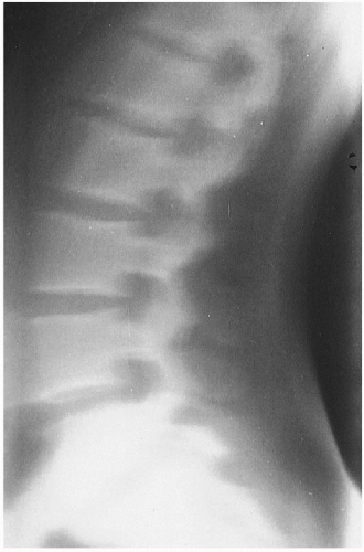
MRI
aka
Year
MR
1980s
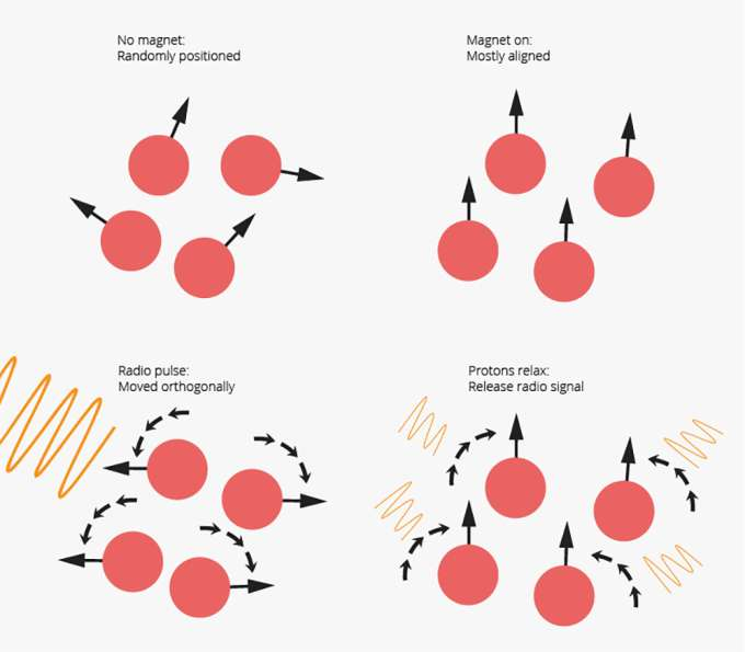
How does MRI work?
Turns body’s atoms into radio transmitters that come together to form an image
Planes in which the following machines form images
CT
MRI
Axial (transverse)
transverse, sagittal, coronal and obliques
Advantages of MRI vs CT
Wider variety of views (i.e. planes)
no ionizing radiation
higher contrast
better imaging of soft tissues
the ability to record small images that are close together as separate images
resolution
Disadvantages of MRI compared to CT
lower resolution
Not as good for bones and calcifications
longer scan times
smaller magnet hole (bore)
fewer contrast agents (used to be none)
provides cross-sectional info of internal structures in relation to contour. These images are generally not nearly as good as CT or MR (or even IVP).
Transverse Tomography
Tomographic image quality
not as good as CT or MRI
Ultrasound
Image quality
Poor (compared to MRI or CT)
Ultrasound Advantages
no ionizing radiation
lower cost
Ultrasound uses in RTT
obtaining chest wall depth
Localization of:
Breast
Abdomen
Retroperitoneum(behind
Scar Boosts
Chest wall depth?
Why would be want to know it?
Form skin surface to lungs
varies from person to person
For a mastectomy patient to avoid treating lungs
Ultrasound frequencies used
1-20 MHz
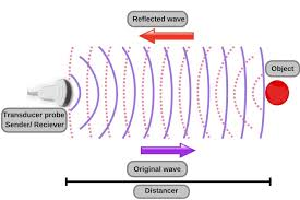
how does ultrasound work.
How are the images produced?
How does it differentiate between tissues?
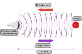
Produces images by transmission/ reflection (usually echo reflection)
Reflections (echos) are caused by variations in acoustic impedance (bc some materials block more sound waves than others)
impedance mismatch
↑ difference in Z# between 2 tissues = ↑ impedance mismatch = ↑ contrast (better image)
The highest mismatches occur at interfaces (junction) of:
air/tissue
tissue/bone
chest wall/ lung
The physics effect responsible for the production of ultrasound images
Piezoelectric Effect
In a nutshell, what is the piezoelectric effect
A transducer converts electrical energy into ultrasound and vice-versa
PET stands for
Positron Emission Tomography
How do PET scans work?
Patient receives a radionuclide that resembles a substance found in the body (e.g. sugar)
Positrons (anti-matter: e+) collide with nearby electrons to produce annihilation rxn (matter/anit-matter rxb)
Gamma rays produced by annihilation rxn are emitted from the patient’s body
The gamma rays produce the images seen in scans
Common Radionuclides
fuck.cn.org
Flourine-18 (HCC)
Carbon-11
Nitrogen-13
Oxygen-15
Rubidium-82
Gallium-68
What can a PET scan image?
Structure AND function
Mental function and psychiatric abnormalities
(e.g. brain: taken up in areas that consume sugar)
What produces the radionuclides used for PET scans?
Cyclotrons
PET/CT fusion
aka
What does it do?
image fusion
forms images with the “best of both words” Structure AND function seen together = more dynamic view
Nuclear Medicine Scanning
What does SPECT stand for
Single-photon emission computed tomography
BEV stand for
Beam’s Eye View
What is BEV?
A tx display in which tx plan is viewed from the POV of the radiation source
Images reconstructed from CT data that represent the patient’s anatomy and defined volumes from the POV of the tx beam