Anemias + Granulocyte/Lymphocyte Disorders
 Call Kai
 Learn
Learn Practice Test
 Spaced Repetition
 Match
Match Flashcards
 Knowt Play
1/42

Earn XP
Description and Tags
Covers anemias, inclusions, and disorders of granulocytes and lymphocytes
Name | Mastery | Learn | Test | Matching | Spaced | Call with Kai | Chat |
|---|
No analytics yet
Send a link to your students to track their progress
43 Terms
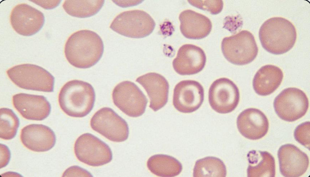
What abnormal RBC shape is shown? What anemia is this MOST associated with?
Codocyte (“Target Cell”); Thalassemia
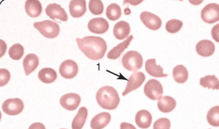
What abnormal RBC shape is indicated?
Dacrocyte (“Teardrop Cell”)
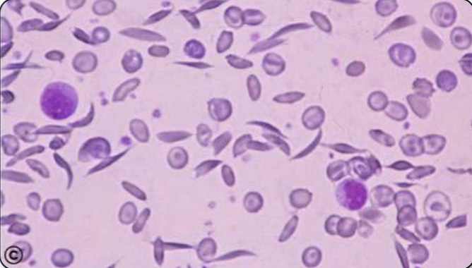
What is the predominant abnormal RBC shape shown? What type of hemoglobin does it have? Why does it form this shape?
Drepanocyte (“Sickle cell”); HbS; low oxygen conditions cause polymerization of HbS
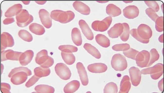
What abnormal RBC shape is shown?
Elliptocyte (“Pencil/Cigar cell”)
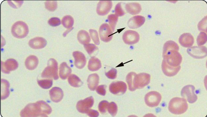
What abnormal RBC shape is shown?
Blister cell
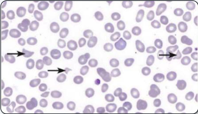
What abnormal RBC shape is indicated?
Keratocyte (“Helmet cell”) (also technically a schistocyte)
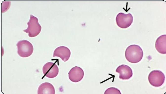
What abnormal RBC shape is indicated? Why is part of the cell missing?
Degmacyte (“Bite cell”); cellular inclusions pitted out of cell in the spleen
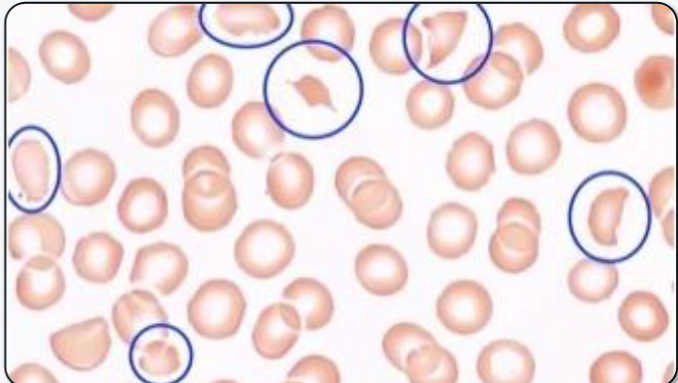
What abnormal RBC shape is shown?
Schistocyte (“RBC fragment”)
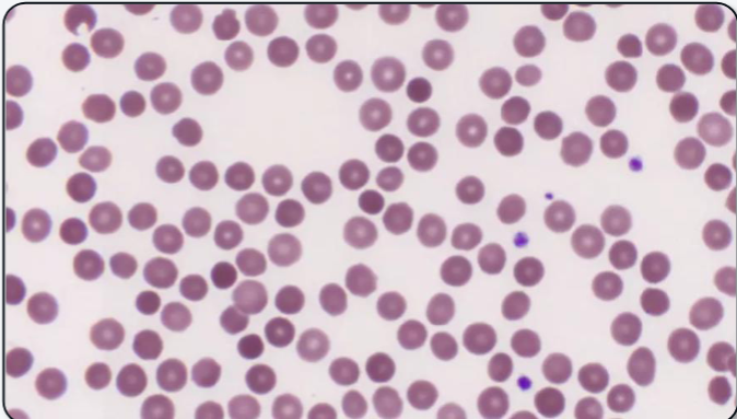
What abnormal RBC shape is shown? What anemias (2) is it most commonly associated with?
Spherocyte; Hereditary spherocytosis AND Autoimmune Hemolytic Anemia (AIHA)
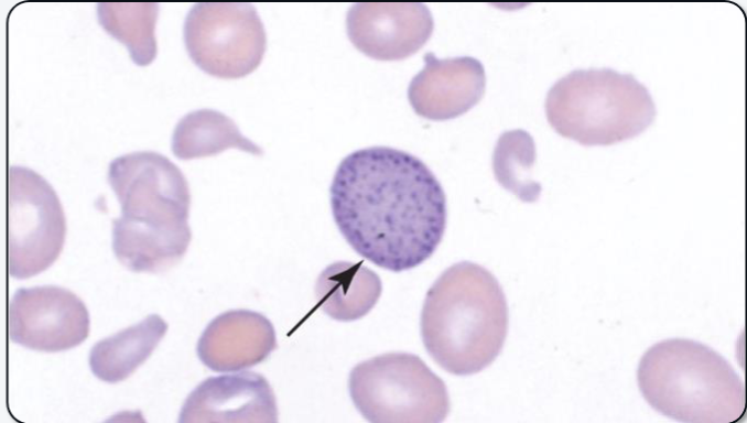
What cellular inclusion is shown? What is it composed of? What anemia is it most associated with?
Basophilic stippling; ribosomal RNA (rRNA); Thalassemia
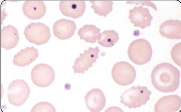
What cellular inclusion is shown? What is it composed of? What anemia is it most associated with?
Howell-Jolly bodies; DNA (nuclear remnant); Megaloblastic anemia
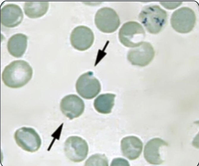
What cellular inclusion is shown? What is it composed of? What anemia is it most associated with?
Heinz bodies; denatured hemoglobin; G6PD deficiency
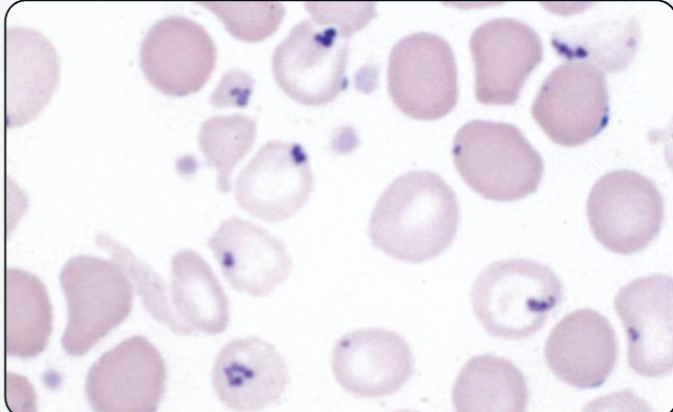
What cellular inclusion is shown? What is it composed of? What anemia is it most associated with?
Pappenheimer bodies; iron-bound proteins (iron aggregates); sideroblastic anemia
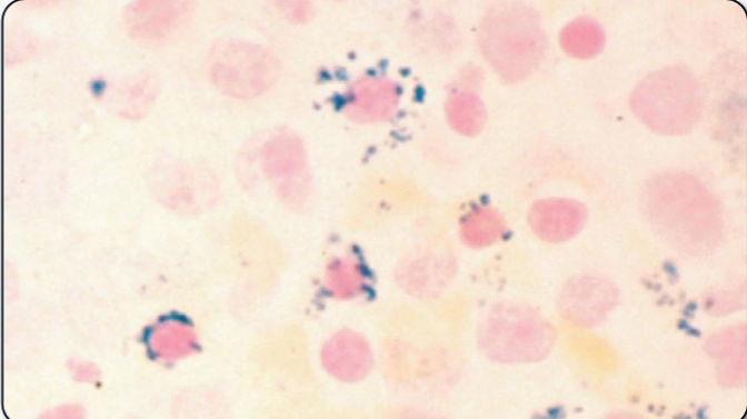
What is the name for these iron-containing RBC precursors?
Sideroblasts
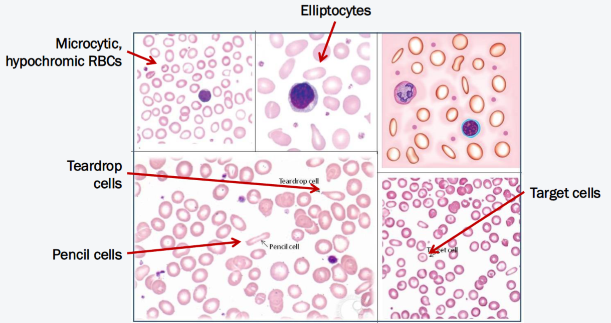
What anemia do these peripheral blood smears indicate?
Iron Deficiency Anemia (IDA)
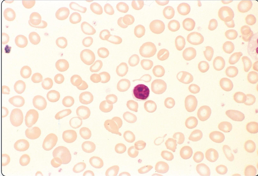
What anemia does this peripheral blood smear indicate? What are the key distinguishing features?
Iron Deficiency Anemia (IDA); microcytic, hypochromic cells / elliptocytes / dacrocytes
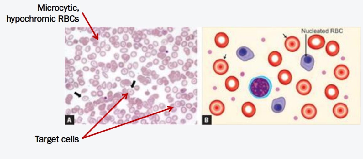
What anemia do these peripheral blood smears indicate?
Thalassemia
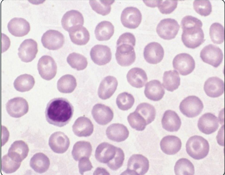
What anemia does this peripheral blood smear indicate? What are the key distinguishing features?
Thalassemia; microcytic cells / codocytes
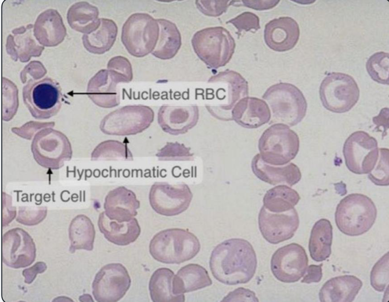
What anemia does this peripheral blood smear indicate? What is another name for nucleated red blood cells? Why are they in the peripheral blood here?
Thalassemia; Orthochromatic erythroblasts; Hemolysis causes bone marrow stress and premature release of maturing RBCs to circulation
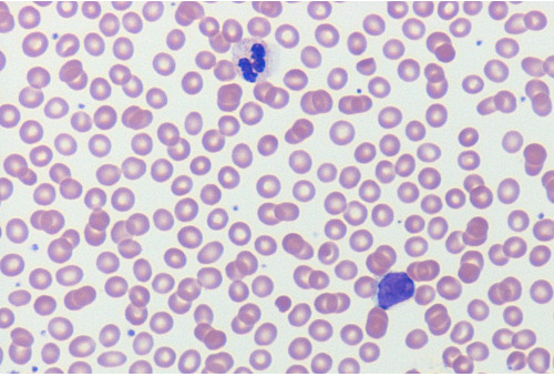
What anemia does this peripheral blood smear indicate?
Anemia of Chronic Disease (I know it’s heard to tell without a normal lymphocyte, but these RBCs are normocytic, normochromic)
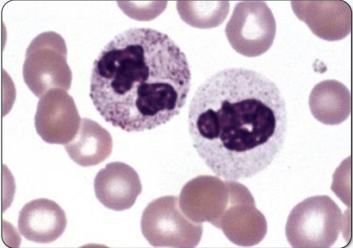
What neutrophilic change is present here?
Toxic granulation (primary granules overexpressed in mature PMN)
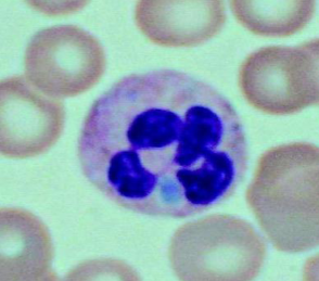
What neutrophilic change is present here? What is the blue dot made of?
Döhle bodies; rough endoplasmic reticulum remnants
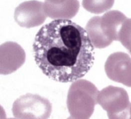
What neutrophilic change is present here?
Cytoplasmic vacuoles
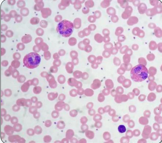
What is the name for this granulocytic condition? What are the most common causes 1) worldwide and 2) in the US?
Hypereosinophilia; 1) parasitic infection, 2) allergies
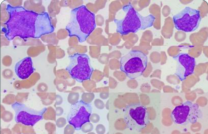
What type of cell is shown?
Reactive lymphocyte (atypical)
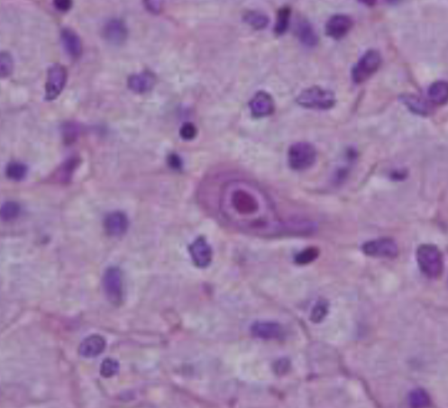
What lymphocytotic pathogen causes this cell shape?
Cytomegalovirus (CMV)
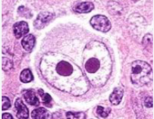
What is this cellular inclusion called? What pathogen causes it?
“Owl’s eye”; CMV
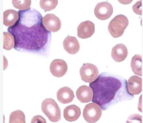
What are the large cells called? What nickname is given to them? What disease and pathogen is most associated with these cells?
Reactive lymphocytes; “Ballerina skirt cells”; Infectious mononucleosis / Epstein-Barr Virus (EBV)
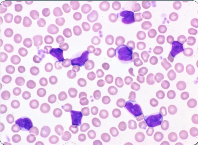
What lymphocytotic pathogen/disease causes this immune response? What is the cause of this absolute lymphocytosis?
Bordetella pertussis (whooping cough); pertussis toxin blocks lymphocyte migration from circulation to tissue
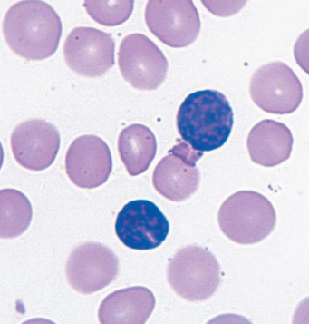
What lymphocytotic pathogen causes is displayed in this smear? What characteristic of the lymphocytes shows this?
Bordetella pertussis; clefted nucleus / “soccer-ball” chromatin
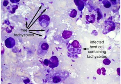
What pathogen/disease is portrayed here? What is the most common source?
Toxoplasma gondii (toxoplasmosis); cat feces
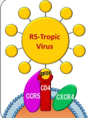
What type of cell does this HIV target?
Macrophages
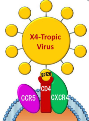
What type of cell does this HIV target?
CD4+ Helper T-cells
What test most easily distinguishes an EBV from a B. pertussis infection? What does this test for the presence of?
Monospot (EBV is +, Pertussis is -); heterophilic antibodies
What are the two deficiencies that cause megaloblastic anemia? Which one causes neurological effects?
Vitamin B12 and Folate; B12 deficiency causes demyelination of neurons
What hepatic hormone is associated with anemia of chronic disease? What does it do?
Hepcidin; prevents release of iron from gut epithelial cells and splenic macrophages → causes iron sequestration
What defect results in sideroblastic anemia?
Mutation/defect in heme synthesis enzymes (like ALA synthase)
What is the main cause of normocytic anemia?
Anemia of Chronic Disease (early disease)
What is the real name of “Bubble boy” disease? What cell is it characterized by the lack of?
Severe combined immunodeficiency syndrome (SCID); T-cells
What are the main causes of immediate neutrophilia? Is it a true increase in neutrophil count?
Exercise/stress/drugs; No, aka. pseudoneutrophilia (neutrophil shift from marginating zone to circulation)
What is the main cause of acute neutrophilia? Where do the neutrophils come from? What is seen in the peripheral blood?
Bacterial infection/toxin; non-mitotic pool of bone marrow; Left Shift (bands and metamyelocytes)
What is the main cause of chronic neutrophilia? Where do the neutrophils come from? What is seen in the peripheral blood?
Persistent bacterial infection; mitotic pool of bone marrow; profound Left Shift (bands, metamyelocytes, and maybe even myelocytes)
What are the 3 main mechanisms of neutropenia (general)?
1) Decreased bone marrow production or release, 2) increased neutrophil utilization or autoimmune destruction, 3) Pseudo-neutropenia (shift to marginating zone/lab artifacts)