homeostasis and excretion
 Call Kai
 Learn
Learn Practice Test
 Spaced Repetition
 Match
Match Flashcards
 Knowt Play
1/23

There's no tags or description
Looks like no tags are added yet.
Name | Mastery | Learn | Test | Matching | Spaced | Call with Kai | Chat |
|---|
No analytics yet
Send a link to your students to track their progress
24 Terms
|
the ability to maintain a constant internal environment despite environmnetal changes
Describe the maintenance of a constant internal body temperature in humans.
(describe endotherms and ectotherms and how they regulate their temps)
Endotherms
An Animal that can regulate its own body temperature
E.g Birds and Mammals
Body temperature tends to stay the same regardless of the environment
ectotherms
An Animal that cannot regulate its own body temperature
E.g Reptiles and Fish. Body temperature changes with the temperature of
the environment
How do endotherms regulate their temperature? Metabolic Reactions
How do ectotherms regulate their temperature? Through behaviour e.g lying in the sun
Advantages and disadvantages of endotherms
adv:
Enzymes can work efficiently no matter what the outside temperature
Enzymes can work efficiently no matter what the outside temperature metabolism keeps going, even when it is cold.
High stamina: Fast metabolism allows quick bursts of energy and muscle recovery.
Diverse habitats: Species can live successfully across polar, temperate, and changing climates
disadv:
Energy to keep warm needs to come from somewhere (they can not produce their own energy) -
endotherms need to eat a lot more than ectotherms during food shortages
High energy cost: Constant heat production burns through many calories.
advtanges and disadvantages of ectotherms:
adv:
-Do not waste calories generating internal heat.
-Require significantly less food than endotherms.
-Survive long periods without eating during shortages.
Channel more food energy into growth and reproduction.
disadv:
Rely entirely on external temperatures to function.
Limited Activity Windows due to climate.
Vulnerability to Predators when body temperatures are low.
Restricted from permanently colonising extremely cold polar regions.
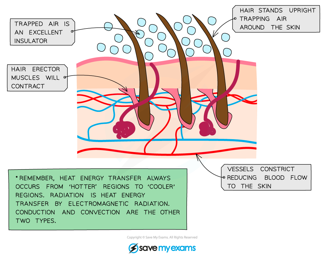
know the functions of specific body parts in thermoregulation also labeling the skin
-skin blood vessels and hypothalamus

know the functions of specific body parts in thermoregulation (hypothalamus)
hypothalamus: acts as a body thermostat, it signals the body to:
-conserve heat or to generate more heat.
too COLD:
Conserving heat:
erector muscles in our skin contracts, causing the hairs on our skin to stand and it traps a thick layer of warm air next to skin to warm up (insulate)
vasoconstriction- arterioles constrict in size so small amount of blood flow travels within, this stops blood from losing heat energy to air.
generating heat:
shivering- muscles contract and release quickly. the metabolic rates increase which generates heat and the blood that flows thru muscles is warmed by the heat and then distributed thoughout the body
too HOT:
-hypothalamus tells the body to lose heat
lose heat:
the erector muscles in our skin relax so the hairs on body lies flat, causing no warm air trapped.
vasodilation- arterioles dilate (increase in size) so lots of blood flow through so blood loses heat to air.
sweat- sweat glands secrete sweat and water evaporates taking heat from skin and cooling body
explain the concept of negative feedback
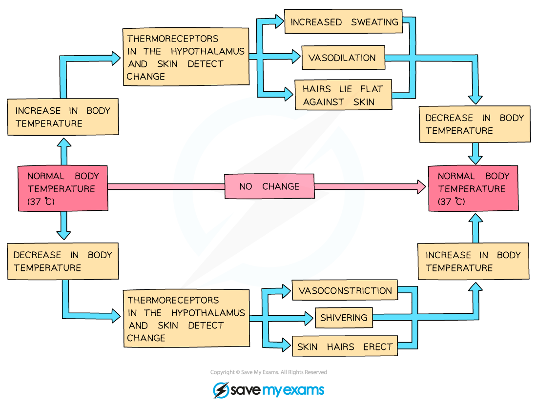
When one parameter changes in a particular direction, this is sensed and measures are put in place to change it back towards the norm
-aims to put ur internal temp at 37 degrees
thermoreuglation negative feedback loop
STIMULUS / IMBALANCE
Body temperature rises above or falls below ~37°C.
↓
SENSOR / RECEPTOR
Thermoreceptors in the skin and brain detect the change.
↓
CONTROL CENTRE
The hypothalamus processes data and activates a response.
↓
(IF TOO HOT)
Vasodilation (blood vessels widen)
Sweating (evaporative cooling)
Flattened body hairs
↓
(IF TOO COLD)Vasoconstriction (blood vessels narrow)
Shivering (muscles generate heat)
Goosebumps (traps air)
↓
HOMEOSTASIS
Body temperature returns to normal.
Negative feedback turns off the responses.
what are the 4 main components of the thermoregulation feedback loop?
stimulus, sensors (thermoreceptors), control centre (hypothalamus) , effectors
2 functions of the fatty tissue beneath the skin
The fatty tissue beneath the skin (subcutaneous fat)
=thermal insulation to help regulate body temperature by preventing heat loss,
=shock absorber or cushion to protect underlying muscles and bones from physical impact
vasodilation and vasoconstriction full depth
vasocontricition
Smooth Muscle Contraction in the arteriole walls causes the arterioles to contract.
Shunt Vessels Dilate: The smooth muscle in the shunt vessels relaxes (opens), allowing blood to take the shortcut deeper under the skin.
Reduced Blood Flow: Less blood flows through the capillaries located close to the skin surface.
the aim is that Less heat is lost from the blood to the external environment by radiation, keeping the core body warm
vasodilation
the smooth muscles relax in the ateriole walls causing the arterioles (small arteries) near the surface of the skin to dilate
More blood flows through the capillaries located close to the skin surface.
Heat is lost from the blood to the external environment by radiation and convection, cooling the body down
Identify and locate on a diagram of the skin; hairs, hair erector muscles, sweat glands, receptors, sensory neurones, blood vessels and fatty tissue.
(Glycaemic control)
Describe the control blood glucose concentration by the liver and the roles of insulin and glucagon from the pancreas (islets of langerhan)
If blood glucose concentration rises:
Sensor: Pancreas detects high glucose levels.
Hormone: Pancreas secretes insulin into the blood.
Target: Insulin travels to the liver and muscles.
Effect 1: Causes liver and muscle cells to absorb glucose from the blood.
Effect 2: Stimulates the conversion of glucose into glycogen for storage.
Outcome: Blood glucose concentration decreases back to the set point.
If blood glucose concentration falls:
Sensor: Pancreas detects low glucose levels.
Hormone: Pancreas secretes glucagon into the blood.
Target: Glucagon travels to the liver only.
Effect: Stimulates the breakdown of stored glycogen into glucose.
Outcome: Glucose is released into the blood, and concentration increases back to normal.
3. Critical Vocabulary Check (Zero Marks if Confused)
Examiners actively penalise students who mix up these three words. Memorise these exact definitions:
Glucagon: The hormone secreted by the pancreas.
Glycogen: The insoluble carbohydrate stored in the liver.
Glucose: The soluble sugar carried in the blood plasma.
Shared symptoms of Type 1 and type 2 diabetes
Frequent urination: The kidneys work overtime to filter excess sugar from your blood.
Extreme thirst: Frequent bathroom trips cause dehydration, making you feel constantly thirsty.
Constant hunger: Your body cannot move sugar into cells, leaving them starved for energy.
Extreme fatigue: Cells lack the fuel they need, making you feel exhausted.
Blurry vision: High blood sugar pulls fluid from your eye lenses, making it hard to focus.
Slow healing: High glucose levels impair blood flow and natural healing processes.
Symptoms of only type 1 diabetes
Rapid weight loss: Your body rapidly burns fat and muscle for energy instead of sugar.
Mood shifts: Sudden irritability and mood swings are common, especially in children.
Bedwetting: Children who previously stayed dry at night may start wetting the bed.
Fruity breath: A warning sign of dangerous chemical buildup (ketones) in the blood.
Symptoms of only type 2 diabetes
No symptoms at all: Many people live with it for years without noticing any signs.
Numbness or tingling: Nerve damage causes a pins-and-needles feeling in hands or feet.
Darkened skin: Dark, velvety patches (acanthosis nigricans) appear on the neck or armpits.
Frequent infections: Persistent yeast infections or bladder infections happen more often.
Explain the process of what our body does to excess proteins
How the body destroys the excess protein:
The liver destroys them by breaking them down into:
The Nitrogen Part (The toxic waste)
The Carbon Part (The leftover fuel)
Where do the two halves go?
Half 1: The Nitrogen Part turns into Pee
As we talked about, this is the dangerous part. The liver instantly turns this nitrogen into ammonia, quickly converts it into safe urea, and sends it to your kidneys so you can pee it out.
Half 2: The Carbon Part turns into Energy or Fat
This is the "excess protein" leftover that you are asking about! This part is not toxic at all. It is made of carbon, hydrogen, and oxygen—which is the exact same stuff that sugar and fat are made of.
The liver recycles this leftover carbon part based on what your body needs at that exact moment:
The remaining carbon part is turned into glycogen for storage or broken down in respiration to provide energy.
If your body already has enough energy: The liver converts this leftover carbon piece into fat and stores it around your body for later use.
Summary: The Ultimate Destination
Nitrogen half = Leaves your body completely through your urine.
Carbon half = Stays in your body as active energy or gets stored as body fat.
(excretion)
Describe the role of the liver in the deamination of amino acids to form urea.
body cannot store excess amino acids, so they travel to the liver, which is the exclusive site for deamination. During this process, liver enzymes remove the nitrogen-containing amino group from the amino acid molecule, while the remaining carbon-containing part is recycled into carbohydrate (glycogen for storage) or used in respiration for energy.
The detached amino group is instantly converted into ammonia, which is highly toxic to cells and cannot be allowed to accumulate. To protect the body, the liver immediately converts this toxic ammonia into urea, a much less toxic compound that safely dissolves into the blood plasma. This blood travels to the kidneys, where the urea is filtered out and ultimately excreted from the body in urine.
Exam Summary Checklist
body Cannot store excess amino acids.
excess proteins so the amino acids float to the liver for deamination to occur
Deamination = removal of the nitrogen-containing part of amino acids.
liver converts the excess amino acids into two parts: Carbon part which is converted to glycogen or used in respiration. nitrogen part into toxic ammonia immediately
Ammonia converted into less toxic urea. by adding carbon dioxide
urea is less toxic than ammonia and is highly soluable in water. it is then carried by blood plasma to kidneys excreted in urine.
State that carbon dioxide (lungs) and urea, excess water and salts (kidney) are excreted to limit the toxicity of carbon dioxide and urea
Carbon dioxide is excreted by the lungs, while urea, excess water, and excess salts are eliminated by the kidneys.
This excretion process limits the accumulation of toxins, as high carbon dioxide levels lower blood pH to damage cells, and excess urea poisons tissues while disrupting nervous system functions
Urea buildup → Causes uremia which Causes oxidative stress in tissues and Alters neurotransmitters and causes brain swelling.
carbon dixoide buildup → Causes hypercapnia which Forms carbonic acid and Drops blood pH (respiratory acidosis) Denatures enzymes.
Kidneys: Identify the ureters, bladder and urethra and outline the structure of the kidney.

Outline the structure and function of the kidney tubule including the role of the glomerulus and Bowman’s capsule in the ultrafiltration of blood and tubule in the selective reabsorption of all glucose, most of the water and some salts leading to the concentration of urea in the urine formed.
1. Ultrafiltration: The Glomerulus and Bowman’s Capsule
Ultrafiltration is the non-selective filtration of blood under high pressure, occurring at the junction between the circulatory system and the kidney tubule.
High Hydrostatic Pressure: Blood enters the glomerulus (a tight knot of capillaries) via the wide afferent arteriole and exits through the narrower efferent arteriole. This structural bottleneck creates an intense, high-pressure zone within the glomerular capillaries.
The Filtration Barrier: The high pressure forces fluid out of the blood plasma and across a three-layered sieve: the capillary endothelium (which has tiny pores), a basement membrane, and the podocyte cells of the Bowman’s capsule (which feature interlocking foot-like slits).
The Glomerular Filtrate: Small molecules are forced out of the bloodstream and into the hollow lumen of Bowman’s capsule.
What is filtered out: Water, glucose, salts (ions), amino acids, and urea.
What stays behind: Large blood proteins and blood cells, which are too big to pass through the filtration barrier.
2. Selective Reabsorption: The Role of the Tubule
As the glomerular filtrate leaves Bowman's capsule and travels down the kidney tubule, the body reclaims essential nutrients while leaving waste behind. This highly regulated, energy-demanding process is called selective reabsorption.
All Glucose is Reclaimed: In the Proximal Convoluted Tubule (PCT), 100% of the filtered glucose is actively reabsorbed back into the surrounding blood capillaries. The walls of the PCT are lined with microvilli to maximize surface area and packed with mitochondria to provide the energy (ATP) needed for active transport proteins.
Most Water is Reclaimed: Water is reabsorbed throughout the tubule—predominantly in the PCT, the descending limb of the Loop of Henle, and the collecting duct. This occurs passively via osmosis, driven by the movement of salts out of the tubule and into the surrounding kidney tissue.
Some Salts are Reclaimed: Critical ions like sodium and chloride are actively and passively reabsorbed in varying amounts along the PCT and the ascending limb of the Loop of Henle. The kidney precisely balances how much salt is kept or excreted to maintain homeostatic blood pressure and volume.
3. Concentration of Urea
Because the kidney tubule is structurally impermeable to urea across most of its length, urea cannot be easily reabsorbed like glucose, water, or salts.
As the filtrate flows downward, the massive reabsorption of all glucose, most water, and some salts drastically reduces the total volume of fluid inside the tubule.
Because the volume of water shrinks while the total amount of urea remains constant, the concentration of urea increases significantly.
By the time the remaining fluid reaches the collecting duct, it has transformed into concentrated urine, packed with a high percentage of waste urea, excess salts, and a minimal amount of water.
Describe the use of dialysis in kidney machines in terms of salt balance, the maintenance of glucose concentration and the removal of urea.
Removal of Urea
Who needs it: Patients suffering from kidney failure. Their natural nephrons no longer work.
When it is used: When a patient's kidneys can no longer filter blood on their own, making dialysis a critical life-support treatment that must be performed for several hours, multiple times a week, until they can receive a kidney transplant.
The Problem: Without a filtration mechanism, metabolic waste products build up to lethal levels, and the body's water and ion balances collapse entirely.
Urea is a toxic waste product that must be completely removed from the blood.
Dialysate Composition: The dialysis fluid contains no urea.
Mechanism: This creates a steep concentration gradient between the patient’s blood and the fluid. Urea moves out of the blood and into the dialysis fluid by diffusion across a partially permeable membrane.
Exam Tip: State that the fluid is constantly refreshed to maintain this steep concentration gradient so diffusion does not stop.
2. Maintenance of Glucose Concentration
Syllabus Fact: Glucose is needed by the body cells for respiration and must not be lost.
Dialysate Composition: The fluid contains a glucose concentration equal to a normal, healthy blood glucose level.
Mechanism: Because the concentrations are equal on both sides of the membrane, there is no concentration gradient. Therefore, there is no net movement of glucose out of the blood.
3. Salt Balance (Mineral Ions)
Syllabus Fact: The body must maintain an optimum level of salts to prevent tissue damage.
Dialysate Composition: The fluid contains a salt/ion concentration equal to the ideal homeostatic blood plasma level.
Mechanism:
If the patient has excess salts in their blood, a gradient exists, and the extra salts diffuse out into the fluid.
If the patient's salt levels are normal, no gradient exists, resulting in no net movement. This restores and maintains the optimum salt balance.
Substance | Concentration in Fluid compared to Blood | Process Involved | What Happens to the Blood? |
|---|---|---|---|
Urea | Lower/ None | Diffusion down gradient | Completely removed |
Glucose | Equal concentration | No gradient | Maintained (No net loss) |
Salts | Equal to ideal level | Diffusion (only if excess) | Balanced (Excess removed) |
The Big Picture: Why Dialysis is Needed for patients with kidney failure
Urea is highly soluble and travels freely in the blood plasma. However, if it builds up (a toxic condition called uraemia), it poisons body tissues, disrupts cell metabolism, causes severe vomiting, confusion, and will ultimately lead to coma and death.
How does the dialysis machine handle it?
The Setup: The dialysis fluid (dialysate) is formulated to contain zero urea
The Process: This creates an incredibly steep concentration gradient between the patient’s urea-loaded blood and the clean fluid.
The Movement: Because of this gradient, urea rapidly moves out of the blood, across the partially permeable membrane, and into the dialysis fluid by diffusion.
The Impact: The machine continuously flushes away used fluid and pumps in fresh, urea-free fluid (countercurrent flow). This keeps the gradient steep, completely stripping the toxin from the blood so the patient doesn’t suffer from systemic poisoning.
2. Glucose
What is it?
Glucose is a simple sugar carried in the blood plasma. It is not a waste product; it is the primary fuel your cells require to perform aerobic respiration and create energy (ATP).
Why must it be maintained?
Your body needs a highly regulated, constant concentration of glucose in the blood to function. If glucose levels drop too low during dialysis, your brain and muscle cells will be starved of energy, leading to hypoglycemia, fainting, cellular damage, and shock.
How does the dialysis machine handle it?
The Setup: The dialysis fluid is mixed to have a glucose concentration exactly equal to a normal, healthy blood glucose level.
The Process: Because the concentration of glucose is identical on both sides of the partially permeable membrane, there is no concentration gradient.
The Movement: Glucose molecules still bump across the membrane, but they do so at equal rates in both directions. This means there is no net movement (net diffusion) of glucose.
The Impact: The patient's blood glucose levels remain perfectly stable throughout the hours of treatment. The body's vital energy supply is fully preserved, preventing any negative metabolic impacts.
3. Salt Balance (Mineral Ions)
What is it?
These are essential dissolved mineral ions in your blood plasma, such as sodium (\(\text{Na}^{+}\)), potassium (\(\text{K}^{+}\)), and chloride (\(\text{Cl}^{-}\)).
Why must it be balanced?
Salts dictate the water potential of your blood and tissues.
If salts are too high: Water will be drawn out of your body cells by osmosis, causing cells to shrivel and dehydrate. High sodium also causes dangerous high blood pressure.
If salts are too low: Water will rush into your cells by osmosis, causing them to swell and burst (lyse). High potassium levels specifically can disrupt electrical signals in the heart, causing fatal cardiac arrest.
How does the dialysis machine handle it?
The Setup: The dialysate fluid is pre-formulated with the ideal, normal physiological concentration of salts.
The Process: This sets up a corrective concentration gradient that automatically adjusts to the patient's immediate blood chemistry.
The Movement via Diffusion:
If the blood has an excess of salts: The concentration in the blood is higher than the fluid. The extra ions diffuse down their gradient across the membrane into the dialysate.
If the blood is deficient in salts: The concentration in the fluid is higher than the blood. The missing ions diffuse out of the fluid and into the blood.
If the blood is perfectly balanced: The concentrations are equal, resulting in no net movement.
The Impact: This dynamic exchange corrects dangerous electrolyte imbalances perfectly. It restores optimum water potential to the blood, protecting cells from osmotic damage and keeping blood pressure stable.
Summary Checklist for IGCSE Exams
To secure full marks on an IGCSE question about dialysis, you must state:
Blood and dialysis fluid flow in opposite directions across a partially permeable membrane.
Urea is a toxic waste that diffuses out because the fluid contains no urea (steep gradient).
Glucose is a vital nutrient that experiences no net movement because the fluid has an equal concentration to healthy blood.
Salts are balanced because excess ions diffuse out into fluid that mimics ideal physiological levels, maintaining correct homeostatic water potential.
Discuss the advantages and disadvantages of kidney transplants compared to dialysis.
Advantages of Kidney Transplants (Compared to Dialysis)
Long-Term Cure: A transplant offers a long-term solution to kidney failure, whereas dialysis is only a temporary, short-term maintenance therapy.
No Long Hospital Sessions: Patients no longer need to spend 4–5 hours connected to a dialysis machine 3 to 4 times every week.
Freedom to Travel: Patients are not restricted to staying near a dialysis clinic and can travel freely without scheduling treatments.
Fewer Dietary Restrictions: Because a real kidney works continuously 24/7, the patient does not have to severely limit their salt, fluid, or protein intake like they do on dialysis.
Higher Quality of Life: Patients generally feel much less fatigued, experience fewer dietary disruptions, and can return to full-time work or study easily.
Cheaper in the Long Run: Although the initial surgery is very expensive, it costs the healthcare system and patient much less over several years than continuous, lifelong dialysis treatments.
Disadvantages of Kidney Transplants (Compared to Dialysis)
Risk of Organ Rejection: The patient's immune system may recognize the foreign donor kidney as non-self and attack it.
Lifelong Immunosuppressants: To prevent rejection, patients must take drugs that suppress their immune system for the rest of their lives.
Increased Infection Risk: Because immunosuppressants lower the body’s natural defenses, the patient becomes highly vulnerable to catching other infectious diseases, such as bacterial or viral illnesses.
Surgical Risks: A transplant requires a major, invasive operation under general anaesthetic, which carries risks of internal bleeding, blood clots, and surgical infections.
Severe Shortage of Donors: There are long waiting lists for a kidney. A patient might wait years on dialysis for a suitable donor match (either from a living relative or a deceased organ donor).
Limited Lifespan of the Organ: A transplanted kidney does not last forever; it typically functions for 10 to 15 years before the patient requires a new transplant or must return to dialysis.
Summary Comparison Table for Quick Revision
Feature | Kidney Dialysis | Kidney Transplant |
|---|---|---|
Availability | Available immediately to anyone | Very long waiting lists for a matching donor |
Lifestyle | High disruption (hours in hospital weekly) | Normal lifestyle after recovery |
Dietary Freedom | Very strict control over salt and water intake | Little to no restrictions |
Medication Needed | None specific to organ function | Lifelong immunosuppressant drugs |
Main Danger | Blood clots and infections from machine lines | Organ rejection and high vulnerability to illness |