Mechanoreception & Electroreception
1/7
There's no tags or description
Looks like no tags are added yet.
Name | Mastery | Learn | Test | Matching | Spaced |
|---|
No study sessions yet.
8 Terms
Define mechanoreceptor and know the 2 types of mechanoreceptor proteins and their characteristics
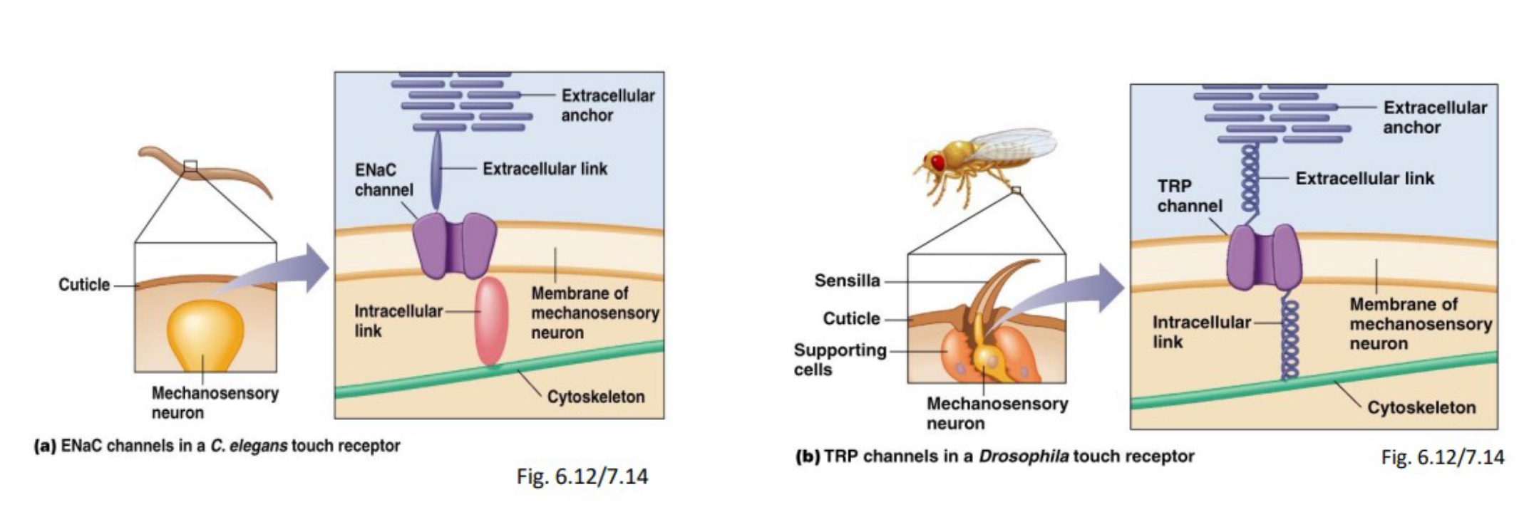
Mechanoreceptors: sensory receptors that transduce mechanical stimuli into electrical signals (required for touch, hearing, balance, and vertebrate blood pressure)
2 known types of mechanoreceptor proteins:
ENaCs (epithelial sodium channels)
TRP (transient receptor potential) channels
Mechanoreceptor signal transduction
Mechanical forces cause displacement of extracellular anchoring proteins and produces a conformational change in ion channels
Absence of intracellular cascade
Understand that there are many types of mechanoreceptors (vertebrates and invertebrates)
Types of mechanoreceptors
Touch and pressure
Baroreceptors (vertebrates)
Tactile receptors (vertebrates and invertebrates)
Proprioceptors (vertebrates and invertebrates)
Equilibrium and hearing
Statocysts (invertebrates)
Various hearing organs (invertebrates)
Hair cells (vertebrates)
Neuromasts (bony fish)
Describe the mechanoreceptors involved in touch and pressure in vertebrates and invertebrates (baroreceptors, tactile receptors, proprioceptors)
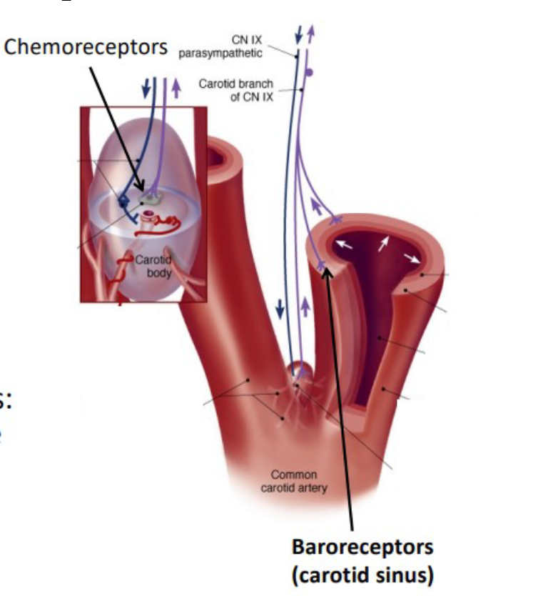
Baroreceptors
Vertebrates
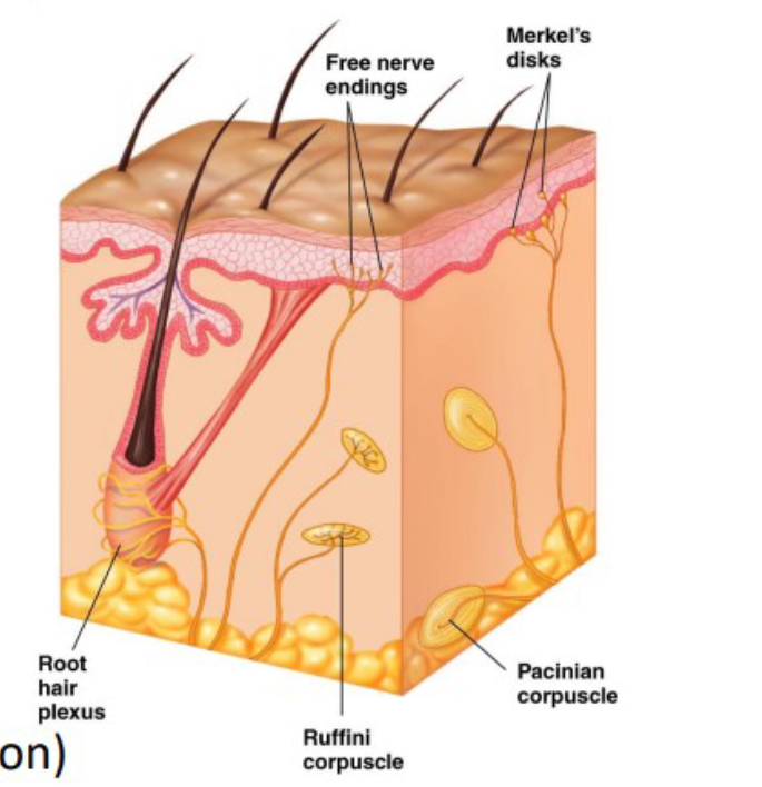
Tactile receptors
Vertebrates
Free nerve endings
Merkel’s Disks
Ruffini Corpuscle
Root hair plexus
Pacinian Corpuscle
Invertebrates
Trichoid sensilla
Campaniform sensilla
Proprioceptors
Vertebrates
Muscle spindles
Golgi tendon organs
Joint capsule receptors
Invertebrates
Chordonal organ
Describe baroreceptors
Baroreceptors (vertebrates)
Detect pressure changes (stretch-sensitive) in walls of blood vessels, parts of the heart, digestive, reproductive, and urinary tracts
E.g. Carotid sinus baroreceptors: monitor blood pressure to the brain
Describe tactile receptors
Tactile Receptors
(vertebrates)
Detect touch, pressure, vibration on the body
Free nerve endings
Sensory neurons with dendrites interspersed among epidermal cells
Receptor proteins on the dendrites
Merkel’s Disks
Enlarged epidermal cell (Merkel cells) associated with free nerve endings
Small receptive field (fine discrimination)
Tonic receptor cells
detect light touch and pressure on skin surface
Slowly adapting
Ruffini Corpuscle
dendrite endings with elongated capsule
located in connective tissue of skin and with connective tissue of joints and limbs
sensitive to stretching of skin and mvt of joints
help detect position of body in space (proprioceptor)
Root hair plexus
Nerve endings wrap around base of hair follicles (responds when hair is displaced)
Phasic receptor
Detect changes in movement across body surface
Pacinian Corpuscle
Sensory dendrite surrounded by lamellae
Located in skin (deep), muscles, joints, and internal organs
Phasic receptors, detect changes in pressure
Large receptive field, poor discrimination, especially sensitive to vibration
Phasic receptor cells
Detect movement or pressure on skin surface
Trichoid sensilla
Hair-like projection of cuticle, bends in response to touch or vibration
Accessory structure transfer mvt of sensilla to dendrite of bipolar sensory neuron
Open stretch-sensitive TRP ion channels
Very sensitive: detect small changes in air movements
Campaniform sensilla
Similar to trichoid sensilla except lack hair shaft
Dome-shape projection of cuticle
Found in clusters, especially near joints of limbs
Detect cuticle deformation as insect moves
Allows coordinated movements
Describe proprioceptors
Proprioceptors (vertebrates)
encode information about body position; essential for motor coordination
Tonic receptors: NO adaptation constant message to CNS
Muscle spindles
surface of skeletal muscles
consist of intrafusal fibers: modified muscle fibers enclosed in connective tissue capsule
monitor muscle length (AP firing rate related to spindle stretch)
Golgi tendon organs
at junction between skeletal muscle and tendon
sense tendon tension
Joint capsule receptors
located in capsules that enclose the joints
many types similar to Pacinian corpuscles, golgi tendon organs, free nerve endings, etc.
Detect pressure, tension and mvt in joint
Chordotonal organ
detects bending of the cuticle
grouping of functional units called scolopidia, consisting of a bipolar sensory neuron and scolopale cells (complex accessory cells) that surround sensory neuron dendrites
Describe the structure involved in equilibrium and hearing in vertebrates
vertebrate hair cells
Equilibrium
Mammalian inner ear
Vestibular apparatus
3 semicircular canals
2 vestibular sacs (utricle and saccule)
Hearing
Mammalian outer ear
Mammalian middle ear
Mammalian inner ear
Vertebrate hair cells
distributed in different sensory organs serving diverse functions
Epithelial sensory receptor cells
Synapse with afferent neuron
Transduce mechanical stimulus into electrical signal
1 long kinocilium (absent in mammals)
Many stereocilia in tight bundle:
contain few actin filament at base (flexible)
Linked by small fibers: tip links
connected to ion channels via elastic proteins (springs)
pull open channel or push it closed
Equilibrium:
mammalian inner ear
vestibular apparatus
3 semicircular canals
each arranged in a separate plane perpendicular to each other
acceleration of fluid in a particular canal is dependent on the plane of movement
Ampulla:
at rest: hair cells partially depolarized
with movement of head: fluid in canals moves and causes stereocilia to pivot
displacement in one direction depolarizes
displacement in other direction hyperpolarizes
2 vestibular sacks (utricle and saccule)
contain otoliths (mineralized particles) which are suspended in gelatinous matrix above membrane called macula
physical movements induce displacement of otoliths, pulls on macula, causing deformation of hair cells
Macula contains large number of hair cells (>100 000)
utricle macula: oriented horizontally
detects motion in the horizontal plane
saccule macula: oriented vertically
detects motion in the vertical plane
Hearing
Mammalian outer ear
outer ear:
pinna and auditory canal
funnel and concentrate sound wave
Mammalian middle ear
Middle ear amplifies sound, allowing sensitive detection of sound
Tympanic membrane
Larger surface area than oval window
Vibrations from sound
Malleus, incus, and stapes
Hinge-like connections between malleus, incus, stapes act as levers, amplifying vibrations
Transfer vibrations initiated at tympanic membrane across the middle ear space to oval window
Mammalian inner ear
Mammalian inner ear receives and transfers vibrations to hair cells in the cochlea, where they are transduced
Cochlea
2 fluid filled compartments
Outer: vestibular and tympanic ducts (perilymph)
Inner: cochlear duct (endolymph, high [K+])
cochlear duct is separated by basilar membrane and houses tectorial membrane
Organ of Corti
Space between basilar and tectorial membranes, contains hair cells
Hair cells are embedded in basilar membrane, and stereocilia contact the tectorial membrane’
Vibrations transmitted from stapes to oval window
Causes waves of pressure differences in fluid of vestibular duct (perilymph)
Waves travel to round window
Cause vibrations of basilar membrane
Hair cells embedded in basilar membrane move relative to stationary tectorial membrane
Electroreception in fish
Electroreception
Electroreceptors - Modified hair cells similar to neuromasts
Passive electrolocation
Detect fields (flow, animal muscle, or nerve activity)
“Electroreceptive” - detect fields only
Active electrolocation
Detect and produce fields (electric organ produces a field, electroreceptors detect perturbations in the field)
“Electrogenic” detect and produce fields