Case 12: A Little Hitch in My Giddy-Up
 Call Kai
 Learn
Learn Practice Test
 Spaced Repetition
 Match
Match Flashcards
 Knowt Play
1/31

There's no tags or description
Looks like no tags are added yet.
Name | Mastery | Learn | Test | Matching | Spaced | Call with Kai | Chat |
|---|
No analytics yet
Send a link to your students to track their progress
32 Terms
True
If you see an appointment on the schedule that says, "Puppy with a lameness issue," the top 2 things you are thinking about as a cause for that puppy's lameness are trauma or a congenital/developmental issue.
TRUE OR FALSE
b. Humerus + Radius + Ulna
The elbow joint is made up of the articulations of which 3 bones?
a. Humerus + Radius + Olecranon Process
b. Humerus + Radius + Ulna
c. Scapula + Humerus + Radius
d. Humerus + Radius + Scapula
a. proximal radial epiphysis which will become radial head
What is A?
a. proximal radial epiphysis which will become radial head
b. separate center of ossification for medial epicondyle of the humerus
c. separate center of ossification of olecranon process
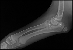
b. separate center of ossification for medial epicondyle of the humerus
What is B?
a. proximal radial epiphysis which will become radial head
b. separate center of ossification for medial epicondyle of the humerus
c. separate center of ossification of olecranon process
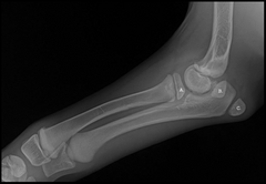
c. separate center of ossification of olecranon process
What is C?
a. proximal radial epiphysis which will become radial head
b. separate center of ossification for medial epicondyle of the humerus
c. separate center of ossification of olecranon process
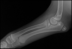
cranial caudal view
What is the radiographic view of A?
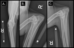
Flexed lateral
What is the radiographic view of B?
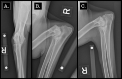
Lateral view
What is the radiographic view of C?
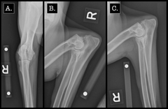
Olecranon Process
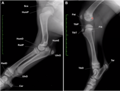
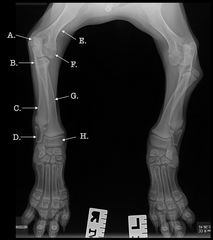
What is A?
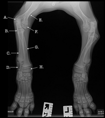
Proximal radial epiphysis
What is B?
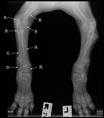
Ulna
What is C?
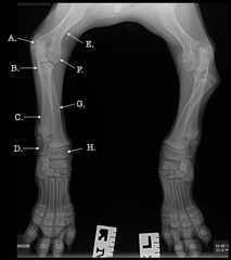
Distal ulnar epiphysis
What is D?
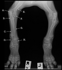
Humerus
What is E?
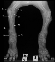
Medial humeral condyle
What is F?
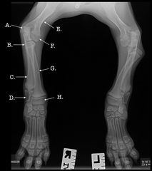
Radius
What is G?
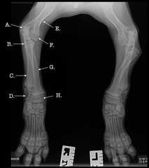
Distal Radial epiphysis
What is H?
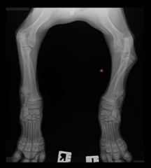
Lateral and Cr-Cd projections of both forelimbs are available for interpretation.
Describe the study
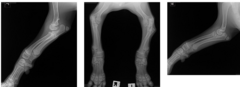
Patient positioning is adequate for interpretation.
Study technique is adequate for interpretation.
Describe patient positioning and study technique
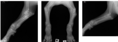
The left radial head is displaced caudo-laterally. Multiple open physes are noted, consistent with the patient's reported age. The right elbow is within normal limits. No other abnormalities are noted.
Describe findings
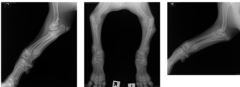
Caudo-lateral displacement of the left radial head is most consistent with Type 1 congenital elbow luxation. Traumatic luxation is less likely, as luxation of the radial head alone is rare in a traumatic situation.
Describe conclusion
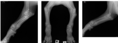
Consultation with a surgeon to discuss surgical options could be considered.
Describe next steps
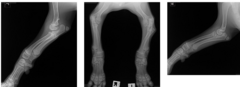
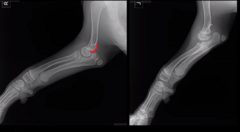
Deformation in soft tissue contour adjacent to radial head due to displacement of joint
What is the soft tissue like in the left cubital joint?
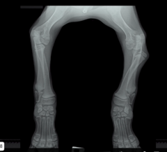
The left image which is actually the right leg
(these are both images of the cubital joint)
Which of these joints is normal?
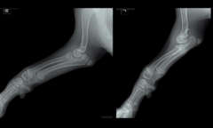
Caudal displacement of the radius from the humerus
What is the displacement like in the left leg aka the right image?
False
The best way to make conclusions about the anatomy of a joint is by only looking at the joint in question and nothing else.
TRUE OR FALSE
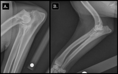
Left image = b. ununited anconeal process
Right image = a. fragmented medial coronoid process
Match the radiograph with the correct diagnosis.
a. fragmented medial coronoid process
b. ununited anconeal process
False
The best way to get good at seeing lameness in dogs is to watch as many YouTube videos on canine lameness as possible.
TRUE OR FALSE
False
CT is a very helpful modality for evaluating the elbow in dogs because it allows us to see the soft tissues of the elbow very clearly.
TRUE OR FALSE
c. Congenital Elbow Luxation
What does CEL stand for?
a. Conspicuous Elbow Lesion
b. Congenital Elbow Lesion
c. Congenital Elbow Luxation
d. None Of The Above
Medial epicondyle
What specific part of the humerus is this (outlined in red), missing on the right image?
CATEGORIES: Trauma or congenital
Congenital should be prioritized.
WHY? Trauma is unlikely because there is no soft tissue inflammation (inc. opacity like swelling/inflammation), no fractures. No pain also helps you move away from trauma.
This young puppy presents like this on radiographs. There is no pain in the gait or limping.
What are the two big disease processes you should be thinking about in an x-ray like this? What broad categories? Which one are you prioritizing and why?
xx
This is just an image for reference for a normal young dog elbow