CH5.1-5.2 Integumentary System: Epidermis
 Call Kai
 Learn
Learn Practice Test
 Spaced Repetition
 Match
Match Flashcards
 Knowt Play
1/25

Earn XP
Description and Tags
Vocabulary flashcards covering the structure, cellular components, and layers of the epidermis based on the BIOL 430 lecture notes.
Name | Mastery | Learn | Test | Matching | Spaced | Call with Kai | Chat |
|---|
No analytics yet
Send a link to your students to track their progress
26 Terms
Cutaneous Membrane
The skin, which is the largest organ of the body with a surface area of 22sq ft and weighing around 10lbs (16% of body weight)
consists of an epithelial epidermis and connective tissue dermis. Hypodermis (subcutaneous layer, superficial fascia)
Epidermis
The outermost protective layer of the skin composed of keratinized stratified squamous epithelium. Has multiple cell types and strata.

Dermis
The layer of skin deep to the epidermis composed of connective tissue, divided into a thin superficial papillary layer (areolar tissue) and a thicker deep reticular layer (dense irregular tissue).

Hypodermis
Also called the subcutaneous layer or superficial fascia, this adipose-rich tissue layer lies deep to the dermis and is technically not part of the skin.

Keratinocytes
The most abundant epidermal cells; they arise in the deepest layer (closest to CT; stratum basale), migrate superficially as they fill with keratin and glycolipids, die, and eventually slough off.
Dead keratin makes it hard and the glycolipids make it water proof.
Desmosomes hold cells together and tight junctions → it forms a protective membrane so it keeps things from getting in


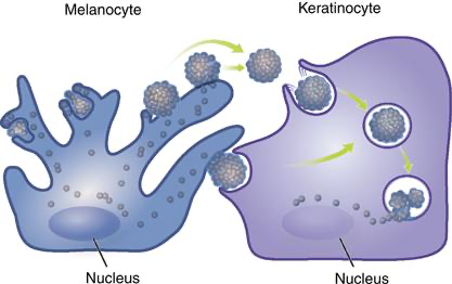
Melanocytes
Spider- or star-shaped cells in the stratum basale that produce melanin pigment and transfer melanosome vesicles to neighboring keratinocytes to protect nuclear DNA from UV radiation.
Found in stratum basale → and can be found in more superficial layers

Melanosomes
Pigment-containing vesicles synthesized by melanocytes and transferred via exocytosis/endocytosis into neighboring keratinocytes.
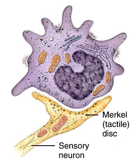
Epidermal Tactile Cells
Also known as Merkel cells, these specialized sensory cells located in the stratum basale associate with nerve endings (tactile discs) to detect light touch and fine pressure.
Dendritic Cells
Also known as Langerhans cells, these phagocytic cells in the stratum spinosum act as antigen-presenting immune cells to detect foreign pathogens and activate immune responses.


Stratum Basale
The deepest epidermal layer consisting of a single row of actively mitotic stem keratinocytes, melanocytes, and tactile cells attached to the dermis via hemidesmosomes; also called stratum germinativum.
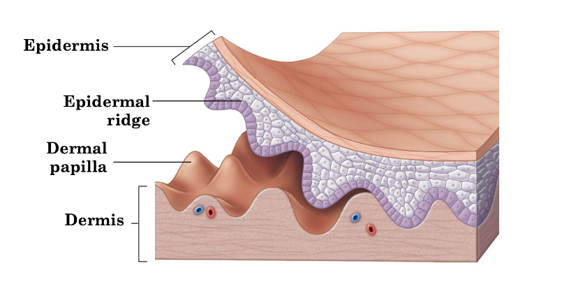
Has epidermal ridges that prevent sliding of tissue and creates fingerprints and sweat patterns


Stratum Spinosum
An epidermal layer 8–10 cell layers thick containing keratinocytes with pre-keratin intermediate filaments, melanin granules, and dendritic cells, and lamellar bodies (lipid filled).
The deeper the layer, the more mitotic activity


Stratum Granulosum
A layer 1–5 cells thick of flattened, degenerating keratinocytes containing dark-staining keratohyalin granules and lipid-filled lamellar granules.
Protein envelope inside the cell membrane


Stratum Lucidum
A thin, translucent epidermal layer of 2–3 rows of clear, flat, dead keratinocytes containing eleidin; present only in thick skin.

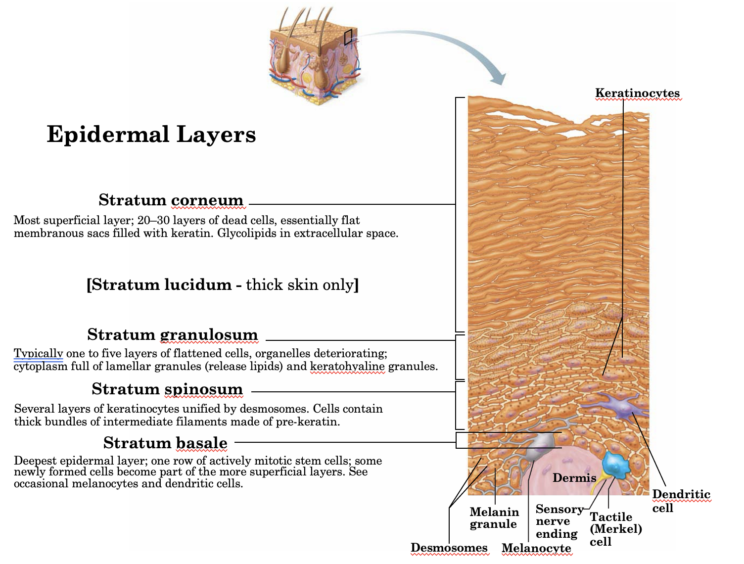
Stratum Corneum
The most superficial epidermal layer composed of 20–30 layers of dead, flat, anucleate keratinocytes packed with keratin and surrounded by extracellular glycolipids. Contains desmosomes but no nucleus.
shed and replaced every 30 days → shed about 40 pounds a lifetime
Protects against infection and dehydration
Has a thickened plasma membrane
Keratin
Lipids in between dead cells

Eleidin
A clear, translucent pre-keratin protein found within the dead cells of the stratum lucidum (thick skin).
Keratohyalin Granules
Granules formed within keratinocytes in the stratum granulosum that assist in assembling pre-keratin into mature keratin proteins.
Lamellar Granules
Lipid-filled vesicles in the stratum granulosum that release hydrophobic glycolipids into intercellular spaces to provide a water-resistant seal.
Hemidesmosomes
Specialized cellular anchoring structures that join the basal keratinocytes of the stratum basale to the underlying dermal basement membrane.
Epidermal Ridges
Downward projections of the epidermis into the dermis that interlock with dermal papillae to anchor skin layers and form fingerprints.

Dermal Papillae
Finger-like projections of the papillary dermis extending upward into the epidermis that increase surface area for adhesion and form friction ridges.
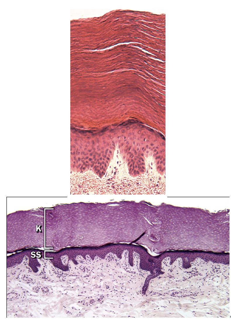
Thick Skin
Skin containing all five epidermal strata (including stratum lucidum) and a thick stratum corneum, located on palms and soles; lacks hair follicles, sebaceous glands, and apocrine sweat glands.

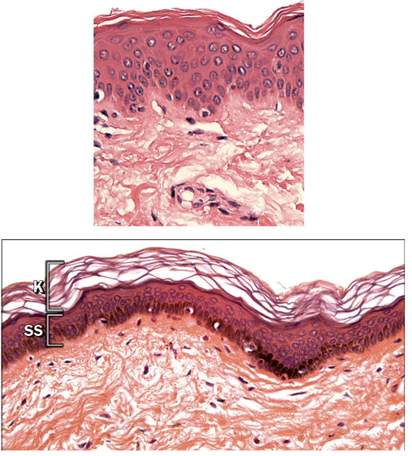
Thin Skin
Skin covering most of the body that lacks a stratum lucidum, features a thinner stratum corneum and thicker dermis, and contains hair follicles and sebaceous glands.

What is the most abundant type of cell in the epidermis?
Keratinocyte, which produces keratin and forms the bulk of the epidermal layers.
Which epidermal cell functions as part of the immune system?
Langerhans cell (or dendritic cells), which helps detect and fight off pathogens in the skin.
Identify differences between thick and thin skin.
Thick skin is located in palms and soles of feet. Has a thicker stratum corneum and thin dermis, and statum lucidium. Lacks hair follicles, no sweat/oil glands.
Thin skin covers most of the body. Has a thinner stratum corneum and thicker dermis. Includes hair follicles and sebaceous glands.
Why are there no skin cancers (abnormal cell growth) that originate from stratum corneum cells?
The stratum corneum consists of dead keratinocytes that are no longer viable, so they cannot undergo abnormal cell growth. Skin cancers typically arise from living cells in the deeper layers of the epidermis.