Neuronal Synapses & Synaptogenesis
1/20
There's no tags or description
Looks like no tags are added yet.
Name | Mastery | Learn | Test | Matching | Spaced | Call with Kai |
|---|
No analytics yet
Send a link to your students to track their progress
21 Terms
The Growth Cone
On the tips of axons to guide neurons (flattened fan shape)
Express specific receptors to sense environmental cues and transform them into steering decisions
They require
Solid substrates
Extracellular cues (attractive or repulsive) act together to ensure accurate guidance
They use intermediate targets provide guidance information and prepare for the next stage of their journey.
Neurons can extinguish their response to to cues and acquire responsiveness to others by changing their surface receptors after reaching these points.
Mechanisms Guiding Growth Cones
Long Range | Short Range |
|
|
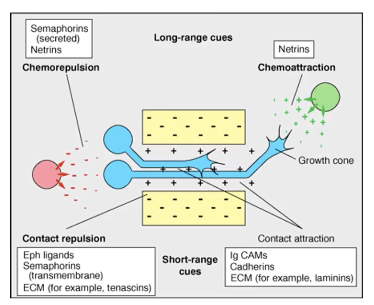
4 Major Classes of Guidance Cues
Semaphorins
Netrins
Slits
Ephrins
Morphogens
Cell adhesion molecules
Semaphorins
Can be secreted and transmembrane guidance cues for long and short range communication
20 different semaphorins contain a sema domain
Domain allows them to bind directly to plexins or the co-receptors Neurophilin-1/2.
Neurophilins will interact with a plexin receptor to form a different receptor complex to allow them to attract and repel.
Sema3A is a repellant in tissues surrounding peripheral nerves acting as a repellant so they grow around the tissue.
Netrins
Secreted and tethered to the cell surface through an G protein coupled anchor that they cleaved for short range communication.
For long range communicate the can diffuse from their source.
Attractive through DCC and repel using UNC5
Slits
Large secreted proteins
Repulsive actions that are mediated by the robo receptor family which controls axon guidance across the midline of the corpus callosum to prevent re crossing of axons.
Slit 2 can be processed and cleaved by protease to generate 2 active fragments with different receptors
N terminal fragment: Binds to robo receptors
C terminal fragment: Binds to plexin A1 (same as semaphorin)
Netrins can act as attractant or repellant depending on the netrin gradient and the receptors expressed by the neuron
Ephrins
Cell surface signaling proteins embedded in the plasma membrane that can bind to F receptors
Important role in axonal guidance and axonal branching
Two types
Class A which are tethered to the cell surface via GPI anchors
Class B are transmembrane proteins
Not active if they are released from the surface so they only do short range communication
Morphogens
Wnt - attractant & repellant
Sonic hedgehog- attractant & repellant
Transforming growth factor beta - Attractant?
Bone morphogenic protein
Cell Adhesion Molecules
Interact with guidance cues
DsCAM
attractant with DCC
repellant with Unc5 co-receptor for netrin-1
Binding of these molecles
Ligand binds to receptor
Downstream signalling causes
Regulation of cytoskeleton causing
Growth cone retraction/extension, neuronal branching or pruning, altered morphology, substrate adhesion or attachment.
Steer neuron to reach target
Laminar, Cellular, Subcellular & Synapse-type Specificity
Laminar Specificity: Axons are guided to the appropriate target area
Cellular Specificity: Within the area axons choose a particular cell as a partner
Subcellular Specificity: The portion f the target cells surface it forms a synapse with such as the dendrites, somata or axonal segments.
Synapse-type specificity: Differentiation and maturation into a specific type of synapse
Mechanisms of cellular specificity
Target recognition (lock-and-key).
Axon guidance (guidance cues).
Interactions among axons.
Inhibition of inappropriate synapses (repel).
Matchmaking by intermediate target cells to form transient contact (priming (changing receptors to ensure it responds to the correct signals)).
Elimination of inappropriate synapses (pruning only).
Death of cells (excess synapses).
Conversion of partner after forming random synapses (slow muscle fibres to fast muscle fibres when need).
Guidance by dendrites.
Delivery at stereotyped times and places
Synaptic Specificity
Target recognition is mediated by signaling molecules that help with target recognition and contact stabilization
Two main classed implicated are
Cadherins
Immunoglobin
Act as attractants and repellants
Inner plexiform layer of the retina
Axons must find their target in a very crowded space
Neurites are stratified within in the plexiform and neurons will form connections within distinct sublayers
Each neuron expresses a different immunoglobin adherent molecule
Pre and post synaptic cells express the proteins for a match up in the same sublamina
Suggests homophilic recognition tested on chicken eggs
overexpression of descam only form in layer 5 on retina and synapse on layer 2
Subcellular Specificity
Axons selective innervate certain domains on the partener
Extrinsic cues guide them to particular potentials of cells
Repellent/attracting molecules
Timing - excludes from synapsing on synapses/layers that haven’t formed yet
Interactions with partners can cause remodelling of the target cell (spines in excitatory)
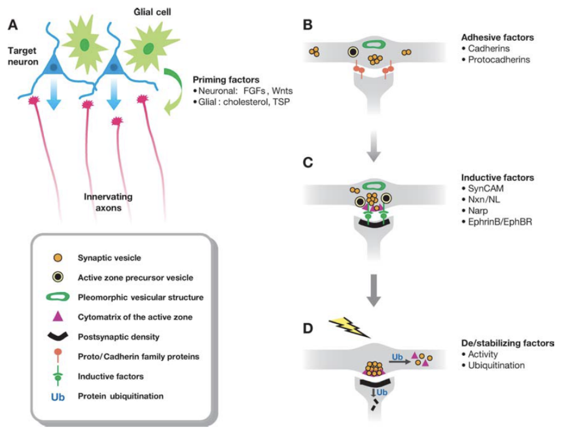
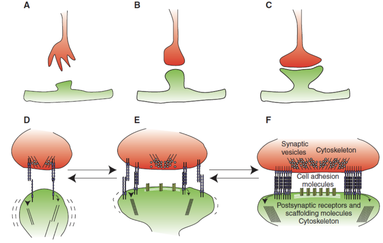
What’s involved in synaptogenesis?
Priming factors from surrounding cells that help initiate synapse formation
Diffusible molecules
From surrounding cells or glial
Induce accumulation of vesicles, promote axonal and dendritic maturation and facilitate the ability of the processes to initiate synapse formation
Adhesive factors help neurons survive and help stabilize them
Inducing factors can directly induce synapse formation
Clustering of post-synaptic proteins
Triggering formation of pre-synaptic buttons
Recruitment of receptors and vesicles
NARP and FMB1 increase synaptic clustering of AMPA or NMDA receptors and initiate presynaptic specialisation
Stabilising and destabilising factors that regulate synaptic strength and stability that help regulate neuronal activity in synaptogenesis
Neuronal activity can trigger neuronal clustering
?Can can protein degradation to promote synapse elimination
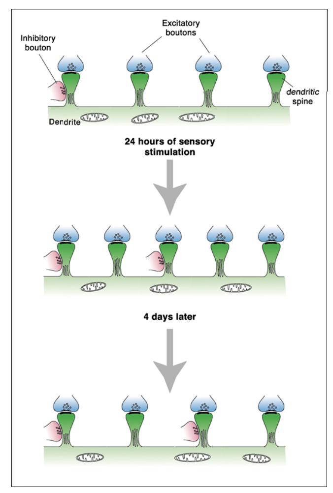
Whisker to Barrel Pathway in the Mouse
Stimulated whiskers of mouse for 24h 36% increase in synapses both excitatory and inhibitory
4 days later it returned to normal
Synaptic maturation
Recruitment of essential proteins and components to the synapse (both pre- and postsynapse).
Pre-synapse:
Vesicles deliver critical proteins
Establishment of docking and fusion sites.
Post-synapse:
Gradual accumulation of essential proteins e.g. PSD95, NMDA and AMPA receptors, scaffolding.
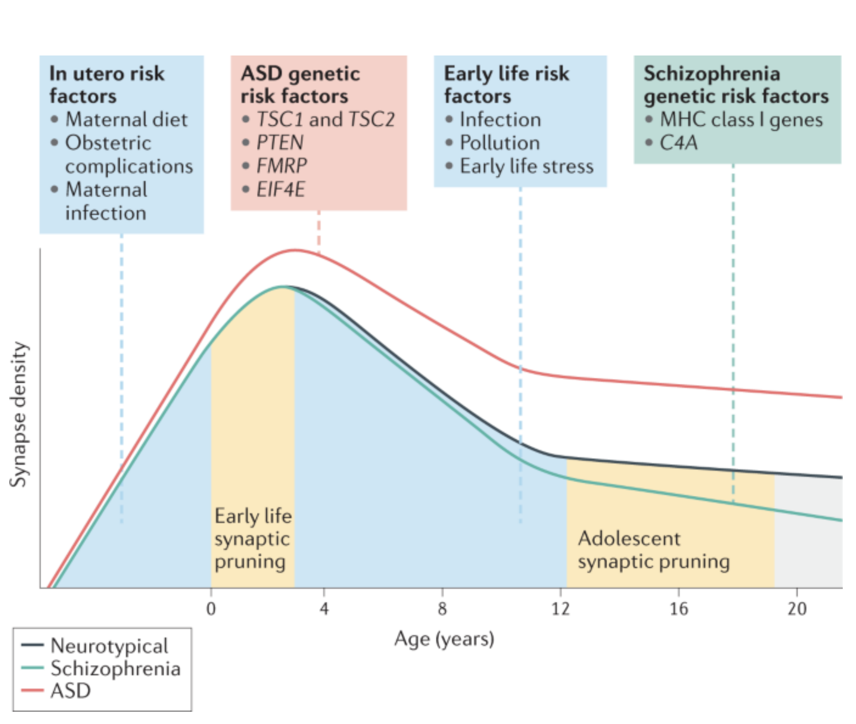
Synaptic Pruning
Starts at 2 and continues until 10 years old (around 50% removed)
Not enough pruning in autism and too much in schizophrenia
Support from astrocytes is needed to strength remaining synapses and prevent other fibres connecting
Regulation of Synaptic Pruning
Microglial cells engulf synapses in response to fractalkine secreted from the synapse
Signaling from the complement cascade can also cause pruning from micro glial
Astrocytes prune synapses in response to phagocytic receptors Megf10 and MERTK
Astrocytes regulate microglial elimination by secreting transforming growth factor beta or interleukin 33 causing complementary signaling on microglial resulting in pruning of the synapse.
Sequential and Concurrent Formation-Elimination Models
Sequential Formation-Elimination Models | Concurrent Formation-Elimination Models |
|
|
Example
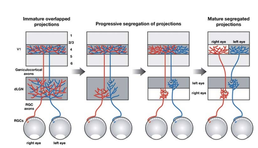
Inputs from the two eyes projecting to the same target are segregated into eye-specific adomains in the LGN.
In each projection system, the two monocular inputs are initially overlapped and gradually segregate from one another by the selective local pruning of overlapping parts of axonal arbors to form the adult pattern of connections through a competitive process driven by neural activity.