Canvas Cardiac Anatomy
1/28
There's no tags or description
Looks like no tags are added yet.
Name | Mastery | Learn | Test | Matching | Spaced |
|---|
No study sessions yet.
29 Terms
List the different borders of the heart?
Superior: RA & LA
Inferior (Diaphragm): Mostly RV & little LV
Right: RA
Left: LV & little LA
Anterior: Mostly RV & little LA & LV
Posterior: LA & LV
What is the pericardium and its layers?
Sac that houses heart and roots
Parietal and Visceral
How much serous fluid is usually in pericardium?
10-20 mL
Cardiac Skeleton (the 4 different sulcus)
IV sulcus - separates RV from LV
AV sulcus - separates LA from LV
Coronary sulcus - separates RA from RV
Sulcus terminalis - separates anterior and posterior RA
Crux - posterior heart where 4 chambers come together
What is the epicardium?
Outermost (visceral) layer of pericardium
Composed of epithelial cells
What is the myocardium?
Middle layer; involuntary muscle fibers
What is the endocardium?
Inner later; covers valves and tendons; simple squamous epithelial cells
What are the two portion of the right atrium?
Posterior smooth and Trabeculated anterior
What separates the right atrium portions internally and externally?
Internally: Crista terminalis
Externally: Sulcus terminalis
What is a Eustachian Valve?
Functional valve in fetus, in right atrium near IVC; best visualized in Right ventricular inflow view
What is a Chiari network?
Fine mobile fibers within right atrium that originates near entrance of IVC
What is the right ventricular inflow tract?
TV and apparatus
What is the RVOT and it’s alternate name?
Infundibulum
Below pulmonic valve and is smooth walled
What is the Moderator Band?
Tissue from anterior free wall of right ventricle to IVS
Quick path for conduction system to reach ventricular wall
Left Atrium two portions?
Anterior - thicker; atrial appendage; store blood
Posterior - smooth; where pulmonary veins enter
Left Ventricle?
75% of cardiac mass (main pumping chamber)
What is the Left ventricular inflow tract?
Mitral valve and apparatus
What is Left ventricular outflow tract?
Just below aortic valve
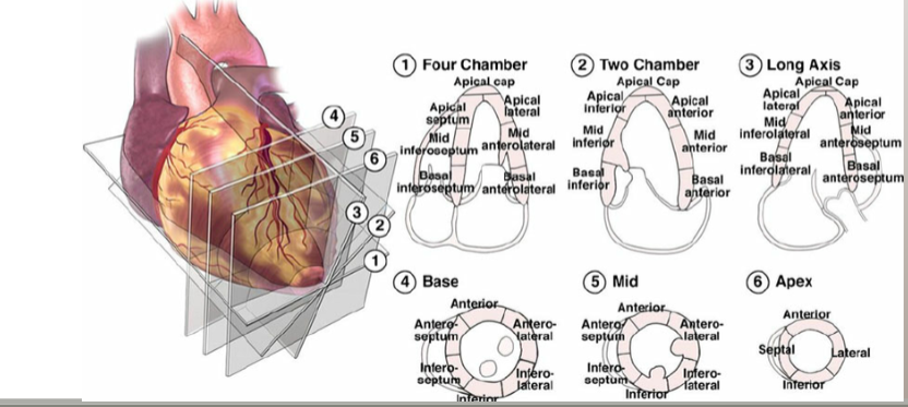
What are the LV wall segments
What are the three regions where defects occurs in the interatrial septum?
Sinus Venosus - junction of VCs with atrium
Septum secundum - mid portion
Septum Primum - at endocardial cushion level (AV valves)
What is an interatrial septal aneurysm?
bulge in septum that moves to and fro with respiration
interventricular septum two regions?
Membranous portion - btw outflow tracts; THIN
Muscular portion - membranous portion to apex
What are the two AV valves?
Mitral and Tricuspid
What do the AV valves do during dia
What is the Mitral Valve?
Between LA and LV
Bicuspid (anterior and posterior leaflet)
What is the Tricuspid valve?
Between RA and RV
Three leaflets (anterior, posterior, septal (medial)
What are papillary muscles?
Muscular columns attached to inside of ventricular walls
How many papillary muscles are attached to the valves?
Mitral: 2
Tricuspid: 3
What are Chordae Tendineae?
Fibrous strands connect papillary muscles to each leaflet