Topic 1.2 Ultrastructure of cells
0.0(0)
Studied by 3 peopleCard Sorting
1/55
Earn XP
Description and Tags
Last updated 10:49 PM on 7/7/23
Name | Mastery | Learn | Test | Matching | Spaced | Call with Kai |
|---|
No analytics yet
Send a link to your students to track their progress
56 Terms
1
New cards
What is resolution?
Making the separate parts of an object distinguishable by eye
2
New cards
Which are the two types of organisms according to their cell structure?
Eukaryotes and Prokaryotes
3
New cards
What is the main difference between eukaryotes and prokaryotes?
Eukaryotes have a compartment within the cell that contains the chromosomes called the nucleus bounded by a nuclear envelope consisting of a double layer of membrane; prokaryotes don’t have a nucleus
4
New cards
What is the cell wall?
An additional barrier on prokaryote cells outside of the cell membrane; it’s thicker and stronger than the membrane, it protects the cell by maintaining its shape and preventing it from bursting. It’s also considered to as being extracellular
5
New cards
What is the capsule?
An additional layer of a type of polysaccharide outside the cell wall which makes it possible for some bacteria to adhere to structures like teeth, skin and food
6
New cards
What are pili?
Hair-like growths on the outside of the cell wall, they can be used for attachment but their main function is to join bacterial cells in preparation for DNA transfer (sexual reproduction)
7
New cards
What are flagella?
They are longer pili with allow a cell to move
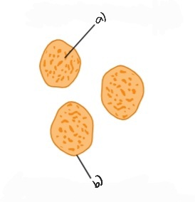
8
New cards
What is the cytoplasm?
It’s a region inside the plasma membrane where organelles are found
9
New cards
What is the cytosol?
The fluid portion of the cytoplasm around the organelles
10
New cards
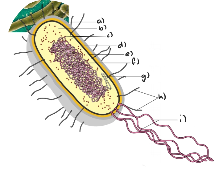
Label the following diagram of a prokaryotic cell
a) capsule
b) cytoplasm
c) ribosomes
d) cell wall
e) plasma membrane
f) nucleoid of DNA
g) plasmid
h) pili
i) flagella
b) cytoplasm
c) ribosomes
d) cell wall
e) plasma membrane
f) nucleoid of DNA
g) plasmid
h) pili
i) flagella
11
New cards
Which chemical is the cell wall made up of in prokaryotic cells?
It’s made up of peptidoglycan
12
New cards
What does prokaryotic cells not having a nucleus imply of the interior of the cell?
It’s entirely filled with cytoplasm, which is not divided into compartments by membranes
13
New cards
_____ are present in the cytoplasm of eukaryotic cells
Organelles
14
New cards
What are organelles?
Distinct structures with specialized functions inside eukaryotic cells
15
New cards
Which are the only organelles that can be found in prokaryotic cells?
Ribosomes, although they are smaller (70S) than those found in eukaryotes (80S)
16
New cards
What is the nucleotide?
The lighter section of the cytoplasm in a prokaryotic cell where the DNA is contained in the form of one circular DNA molecule
17
New cards
How do prokaryote cells divide?
By binary fission, which is used for asexual reproduction
18
New cards
Briefly, how does binary fission work?
The single circular chromosome is replicated and the two copies of the chromosome move to opposite ends of the cell; division of the cytoplasm follows and each daughter cell contains one copy of the chromosome so they are genetically identical
19
New cards
Outline all of the differences between prokaryotic cells and eukaryotic cells
* Prokaryotes have DNA in a ring form without protein; eukaryotes have DNA with proteins as chromosomes/chromatin
* In prokaryotes, the DNA is free in the cytoplasm (nucleoid region); in eukaryotes, the DNA is enclosed within a nuclear envelope (nucleus)
* There are no mitochondria in prokaryotic cells; in eukaryotic cells, mitochondria is present
* Ribosomes in prokaryotic cells are 70S; in eukaryotes, they’re 80S
* There is no internal compartmentalization to form organelles in prokaryotes; eukaryotes have internal compartmentalization present to form many types of organelles
* Prokaryotic cells measure less than 10μm; eukaryotic cells measure more than μm
20
New cards
Which are the similarities between both prokaryotic and eukaryotic cells?
* Both cells have some sort of outside boundary, which always involves a plasma membrane
* Both types of cell carry out all the functions of life
* DNA is present in both cell types
* Both types of cell carry out all the functions of life
* DNA is present in both cell types
21
New cards
How is the cytoplasm of eukaryotic cells?
It’s compartmentalized, meaning that they are divided up by partitions into compartments
22
New cards
How are the compartments in the cytoplasm known?
They’re known as organelles
23
New cards
Which is the most important compartment in eukaryotic cell’s cytoplasm?
The nucleus
24
New cards
Which are the advantages of the cytoplasm of eukaryote cells being compartmentalized?
* Enzymes and substrates can be much more concentrated
* Harmful substances can be kept inside the membrane of an organelle
* Conditions (pH, temperature, etc.) can be maintained at an ideal level for particular processes
* Organelles can be moved around the cell
* Harmful substances can be kept inside the membrane of an organelle
* Conditions (pH, temperature, etc.) can be maintained at an ideal level for particular processes
* Organelles can be moved around the cell
25
New cards
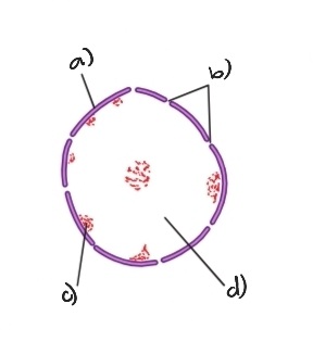
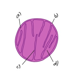
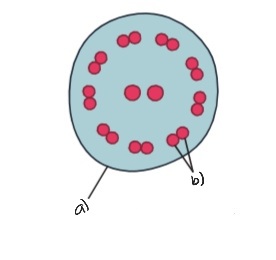
Label the following diagram of the nucleus
a) double nuclear membrane
b) nuclear pores
c) dense chromatin
d) chromatin
b) nuclear pores
c) dense chromatin
d) chromatin
26
New cards
Describe briefly the structure and function of the nucleus
It has a double membrane with pores through it called the nuclear envelope; it contains the chromosomes, which consist of DNA associated with histone proteins.
Uncoiled chromosomes are spread through the nucleus and are called chromatin, which often densely stain the edges of the nucleus.
The nucleus is where DNA is replicated and transcribed to form mRNA, which is exported via the nuclear pores to the cytoplasm
Uncoiled chromosomes are spread through the nucleus and are called chromatin, which often densely stain the edges of the nucleus.
The nucleus is where DNA is replicated and transcribed to form mRNA, which is exported via the nuclear pores to the cytoplasm
27
New cards
What are histones?
Strands of DNA and proteins that form chromatin
28
New cards
What is a nucleosome?
Eight spherical histones with a strand of DNA wrapped around them and secured with a ninth histone, producing a structure that resembles a string of beads
29
New cards
What is a chromosome?
A highly coiled structure of many nucleosomes
30
New cards
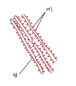
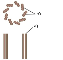
Label the following diagram of the rough endoplasmic reticulum
a) ribosomes
b) cisterna
b) cisterna
31
New cards
Briefly describe the structure and function of the rough endoplasmic reticulum
Consists of flattened membrane sacs called cisternae; outside of these, ribosomes are attached.
They synthesize protein for secretion from the cell, which passes into the cisternae and is then carried by vesicles, which bud off and move on to the Golgi Apparatus
They synthesize protein for secretion from the cell, which passes into the cisternae and is then carried by vesicles, which bud off and move on to the Golgi Apparatus
32
New cards
Briefly describe the structure and function of the soft endoplasmic reticulum
It doesn’t have any ribosomes on its exterior surface but has unique enzymes embedded into its surface.
It produces membrane phospholipids and cellular lipids, sex hormones, detoxifies drugs from the liver, stores calcium ions in muscle cells, transports lipid based compounds, and helps the liver release glucose into the bloodstream
It produces membrane phospholipids and cellular lipids, sex hormones, detoxifies drugs from the liver, stores calcium ions in muscle cells, transports lipid based compounds, and helps the liver release glucose into the bloodstream
33
New cards
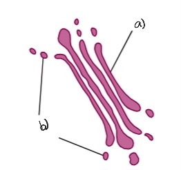
Label the following diagram of the Golgi apparatus
a) cisternae
b) vesicles
b) vesicles
34
New cards
Briefly describe the structure and function of the Golgi apparatus
Consists of flattened membrane sacs called cisternae, however they are curved, don’t have ribosomes attached and have many vesicles nearby.
It processes proteins brought in vesicles from the rER, which are then carried in vesicles to the plasma membrane for secretion
It processes proteins brought in vesicles from the rER, which are then carried in vesicles to the plasma membrane for secretion
35
New cards
How does the movement of vesicles across the Golgi apparatus occur?
Through the *cis* side, the Golgi apparatus receives products from the ER which move into the cisternae of the Golgi apparatus which are discharged at the opposite side, the *trans* side
36
New cards
Label the following diagram of lysosomes
a) digestive enzymes
b) lysosome membrane
b) lysosome membrane
37
New cards
Briefly describe the structure and function of lysosomes
Approximately spherical with a single membrane, and are formed from Golgi vesicles; the interior environment of a functioning lysosome is acidic.
They contain high concentrations of proteins, and also contain hydrolytic enzymes which can be used to break down ingested food in vesicles or organelles in the cell
They contain high concentrations of proteins, and also contain hydrolytic enzymes which can be used to break down ingested food in vesicles or organelles in the cell
38
New cards
Label the following diagram of the mitochondrion
a) inner membrane
b) outer membrane
c) crista
d) matrix
b) outer membrane
c) crista
d) matrix
39
New cards
Briefly describe the structure and function of the mitochondrion
It’s surrounded by a double membrane, the inner of these being invaginated forming crista; the shape is usually spherical or ovoid. They also have their own DNA
They produce ATP for the cell by aerobic cell respiration; here fat is digested if its used as an energy source in the cell
They produce ATP for the cell by aerobic cell respiration; here fat is digested if its used as an energy source in the cell
40
New cards
Briefly describe the structure and function of free ribosomes in the cytoplasm
They appear as dark granules and aren’t surrounded by a membrane, they have a 80S size and are constructed in a region of the nucleus called the nucleolus. They’re always composed of a type of RNA and protein and are always composed of two subunits
These ribosomes synthesize protein, releasing it to work in the cytoplasm as enzymes or in other ways
These ribosomes synthesize protein, releasing it to work in the cytoplasm as enzymes or in other ways
41
New cards
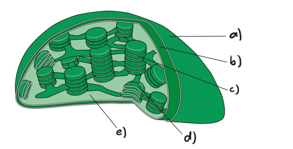
Label the following diagram of a chloroplast
a) outer membrane
b) inner membrane
c) granum (pile of thylakoids)
d) one thylakoid
e) stroma
b) inner membrane
c) granum (pile of thylakoids)
d) one thylakoid
e) stroma
42
New cards
Briefly describe the structure and function of the chloroplast
A double membrane surrounds the chloroplast and inside are stacks of thylakoids (flattened sacs of membrane); they usually have a spherical or ovoid shape and starch grains may be present inside of them.
They produce glucose and a wide variety of other organic compounds by photosynthesis. They’re capable of reproducing independently of a cell
They produce glucose and a wide variety of other organic compounds by photosynthesis. They’re capable of reproducing independently of a cell
43
New cards
What is a granum?
It’s made up of numerous thylakoids stacked like a pile of coins; the thylakoids are flattened membrane sacs with components necessary for the absorption of light
44
New cards
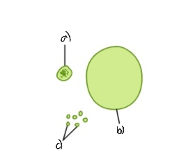
Label the following diagram of the vacuoles and vesicles
a) vacuole containing food
b) large vacuole
c) vesicles
b) large vacuole
c) vesicles
45
New cards
Briefly describe the structure and function of vacuoles and vesicles
Consist of a single membrane with fluid inside.
Some animals absorb foods from outside and digest them inside vacuoles; some unicellular organisms use them to expel excess water. They also allow cells to have higher surface area to volume ratios
Vesicles are very small vacuoles used to transport materials inside the cell
Some animals absorb foods from outside and digest them inside vacuoles; some unicellular organisms use them to expel excess water. They also allow cells to have higher surface area to volume ratios
Vesicles are very small vacuoles used to transport materials inside the cell
46
New cards
Label the following diagram of microtubules and centrioles
a) triple
b) microtubules
b) microtubules
47
New cards
Briefly describe the structure and function of microtubules and centrioles
Microtubules are small cylindrical fibres that can move chromosomes during cell division.
Centrioles are two groups of nine triple microtubules; they form an anchor point for microtubules during cell division and inside cilia and flagella. It’s usually located at one end of the cell close to the nucleus
Centrioles are two groups of nine triple microtubules; they form an anchor point for microtubules during cell division and inside cilia and flagella. It’s usually located at one end of the cell close to the nucleus
48
New cards
Label the following diagram of cilia and flagella
a) plasma membrane
b) double microtubule
b) double microtubule
49
New cards
Briefly describe the structure and function of cilia and flagella
Whip-like structures projecting from the cell surface, containing a ring of nine double microtubules plus two central ones, usually a large flagella is present as in sperm; cilia are smaller and many are present.
Cilia and flagella can be used for locomotion; cilia can also be used to create a current in the fluid next to the cell
Cilia and flagella can be used for locomotion; cilia can also be used to create a current in the fluid next to the cell
50
New cards
Outline all of the differences between plant and animal cells
* In plant cells, the exterior of the cell includes an outer cell wall with a plasma membrane just inside; in animal cells, the exterior of the cell only includes a plasma membrane without a cell wall
* In plant cells, chloroplasts are present in the cytoplasm are; in animal cells, there aren’t chloroplasts
* In plant cells, large centrally located vacuoles are present; in animal cells, vacuoles aren’t usually present or are small
* In plant cells, carbohydrates are stored as starch; in animal cells, carbohydrates are stored as glycogen
* Plant cells don’t contain centrioles within a centrosome area; animal cells contain centrioles within a centrosome area
* Because a rigid cell wall is present, plant cells have a fixed, often angular, shape; without a cell wall, animal cells are flexible and more likely to be a rounded shape
51
New cards
What is the outermost part of bacteria made up of?
Peptidoglycan
52
New cards
What is the outermost part of fungi made up of?
Chitin
53
New cards
What is the outermost part of yeasts made up of?
Glucan and mannan
54
New cards
What is the outermost part of algae and plants?
Cellulose
55
New cards
What is the extracellular matrix?
In many animal cells, it’s composed of collagen fibres plus a combination of sugars and proteins called glycoproteins, which form fibre-like structures that anchor the matrix to the plasma membrane, strengthening it.
It also allows cell-to-cell interactions and directs stem cells to differentiate
It also allows cell-to-cell interactions and directs stem cells to differentiate
56
New cards
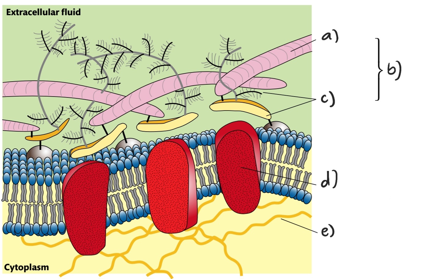
Label the following diagram
a) collagen fibre
b) extracellular matrix
c) glycoprotein
d) integral protein in plasma membrane
e) microfilaments
b) extracellular matrix
c) glycoprotein
d) integral protein in plasma membrane
e) microfilaments