Gram Negative Anaerobes
 Call Kai
 Learn
Learn Practice Test
 Spaced Repetition
 Match
Match Flashcards
 Knowt Play
1/60

There's no tags or description
Looks like no tags are added yet.
Name | Mastery | Learn | Test | Matching | Spaced | Call with Kai | Chat |
|---|
No analytics yet
Send a link to your students to track their progress
61 Terms
What are the general characteristics of Gram-negative anaerobes?
Gram-negative obligate anaerobes; most are elongated rods (some spherical); grow only in low-oxygen environments.
Where are Gram-negative anaerobes normally found?
Normal flora of the alimentary tract (GIT) and mucous membranes.
What type of infections do Gram-negative anaerobes usually cause?
Endogenous opportunistic infections following tissue injury or reduced oxygen supply.
What body sites favor anaerobic infections?
Body sites with low oxygen tension or devitalized tissue.
Are anaerobic infections usually monomicrobial or polymicrobial?
Usually polymicrobial with synergistic interactions between facultative and obligate anaerobes.
Why do anaerobic infections often have a foul odor?
Production of volatile fatty acids.
What are the key characteristics of Fusobacterium necrophorum?
Gram-negative obligate anaerobic long filamentous rod with variable hemolysis.
Where is Fusobacterium necrophorum normally found?
Alimentary tract (especially the rumen) and mucous membranes.
What are infections caused by Fusobacterium necrophorum collectively called?
Necrobacillosis.
What virulence factors does Fusobacterium necrophorum possess?
Endotoxin, leukotoxin, and a polysaccharide capsule.
Which virulence factor of Fusobacterium necrophorum kills phagocytes?
Leukotoxin.
Is Fusobacterium necrophorum a primary or secondary pathogen?
It can act as both a primary and secondary pathogen.
What lesions are characteristic of Fusobacterium necrophorum infection? (3)
Necrosis, abscess formation, and foul-smelling lesions due to volatile fatty acid production.
Can Fusobacterium necrophorum become systemic?
Yes; localized infections may become septicemic and spread to multiple organs.
What major diseases are caused by Fusobacterium necrophorum? (4)
Necrotic liver abscesses, calf diphtheria (necrotic laryngitis), foot rot in cattle, bovine interdigital necrobacillosis, and equine thrush.
Which animals commonly develop rumenitis-liver abscess complex? (be specific)
Grain-fed cattle.
What predisposes cattle to rumenitis-liver abscess complex?
Ruminal acidosis (bc grain fed) causing ulceration of the rumen wall.
How does Fusobacterium necrophorum reach the liver?
Colonizes ruminal ulcers → forms ruminal abscesses → travels through the hepatic portal vein → liver abscesses.
Which bacterium works synergistically with Fusobacterium necrophorum in rumenitis-liver abscess complex?
Trueperella pyogenes.
How does Trueperella pyogenes assist Fusobacterium necrophorum?
Removes oxygen and makes iron available for anaerobic growth.
How is rumenitis-liver abscess complex commonly diagnosed?
Discovery of liver abscesses at the abattoir (slaughterhouse).
What organism causes calf diphtheria (necrotic laryngitis)?
Fusobacterium necrophorum.
Which calves are most commonly affected by calf diphtheria?
Calves younger than 3 months.
What predisposes calves to calf diphtheria?
Teeth eruption, rough feed, improper dosing gun use, or concurrent disease causing mucosal injury.
How is Fusobacterium necrophorum transmitted in calf diphtheria?
Invades injured oral or laryngeal mucosa.
What is the major lesion in calf diphtheria?
Necrosis of the oral and laryngeal mucosa.
What fatal complication may develop in calf diphtheria?
Aspiration pneumonia resulting in necrotizing pneumonia.
What are common clinical signs of the oral form of calf diphtheria? (3)
Swollen cheek, salivation, foul-smelling oral ulcers, fever, coughing, dysphagia, depression, and pneumonia.
What are common clinical signs of the laryngeal form of calf diphtheria? (3)
Painful cough, dyspnea, swollen larynx, fever, depression, airway obstruction, and rapid death.
What organism causes equine thrush?
Fusobacterium necrophorum.
What part of the hoof is affected in equine thrush?
The frog.
What predisposes horses to equine thrush?
Moist, dirty environments and poor hoof hygiene.
What lesions develop in equine thrush?
Degeneration of the frog and infection of the central and collateral sulci.
What are the clinical signs of equine thrush?
Thick foul-smelling discharge, pain during hoof cleaning, and lameness.
What are the key characteristics of Dichelobacter nodosus?
Large Gram-negative obligate anaerobic rod that is barbell- or club-shaped and possesses fimbriae.
What is the habitat of Dichelobacter nodosus?
Obligate parasite of the digital epidermis of ruminants.
What major disease is caused by Dichelobacter nodosus?
Ovine contagious foot rot.
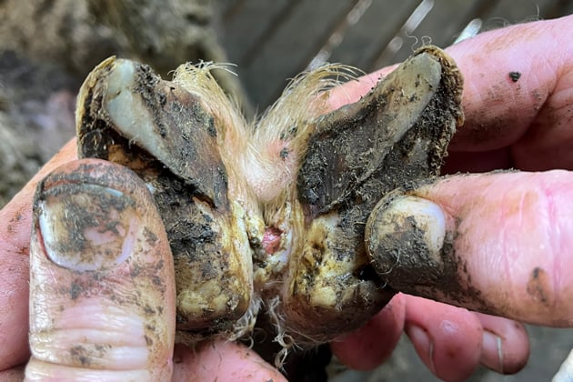
Which organism is the primary pathogen in ovine foot rot?
Dichelobacter nodosus.
Which organism commonly acts as the secondary invader in ovine foot rot?
Fusobacterium necrophorum.
What environmental condition predisposes sheep to ovine foot rot?
Moist environmental conditions.
What must occur before Dichelobacter nodosus can establish infection?
The integrity of the skin must be breached.
How does Dichelobacter nodosus cause tissue damage?
Produces proteases that destroy the epidermis.
What lesions develop during ovine foot rot?
Inflammation, necrosis, foul-smelling tissue, and severe pain.
What are the key characteristics of Prevotella melaninogenica?
Gram-negative slender rod or coccobacillus.
What pigment does Prevotella melaninogenica produce?
Hematin, producing black colonies.
What fluorescence characteristic helps identify Prevotella melaninogenica?
Brick-red fluorescence under UV light.
Which organism commonly works together with Prevotella melaninogenica?
Fusobacterium necrophorum.
What disease commonly involves Prevotella melaninogenica?
Bovine foot rot.
What predisposes cattle to bovine foot rot?
Moist environmental conditions.
How does Prevotella melaninogenica contribute to bovine foot rot?
Produces proteolytic and collagenolytic enzymes that destroy connective tissue and tendons.
How does Fusobacterium necrophorum contribute to bovine foot rot?
Produces leukotoxin leading to abscess formation.
What are the clinical signs of bovine/ovine foot rot?
Swelling of the interdigital space and coronary band, foul odor, lameness, fever, anorexia, weight loss, and decreased milk production.
How is foot rot treated?
Debridement of necrotic tissue, foot baths, and systemic antibacterial therapy.
A Gram-negative obligate anaerobic long filamentous rod causing necrotic liver abscesses and calf diphtheria.
Fusobacterium necrophorum.
A Gram-negative obligate anaerobe with leukotoxin that kills phagocytes.
Fusobacterium necrophorum.
A Gram-negative obligate anaerobe associated with grain-fed cattle and liver abscesses.
Fusobacterium necrophorum.
A Gram-negative obligate anaerobe causing equine thrush.
Fusobacterium necrophorum.
A Gram-negative obligate anaerobe that is barbell- or club-shaped with fimbriae and causes ovine foot rot.
Dichelobacter nodosus.
A Gram-negative anaerobe producing black colonies and brick-red fluorescence under UV light.
Prevotella melaninogenica.
Which Gram-negative anaerobe produces proteolytic and collagenolytic enzymes in bovine foot rot?
Prevotella melaninogenica.
Which Gram-negative anaerobe is usually the secondary invader in ovine foot rot?
Fusobacterium necrophorum.