Embryology - Part 1 (The Retina)
1/77
There's no tags or description
Looks like no tags are added yet.
Name | Mastery | Learn | Test | Matching | Spaced |
|---|
No study sessions yet.
78 Terms
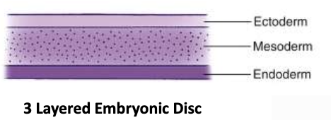
When do the 3 primary germ layers form during embryonic development?
3rd week
→ ectoderm, mesoderm & endoderm form the embryonic plate
Which germ layers contribute to developing ocular structures?
only the ectoderm and mesoderm

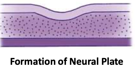
How is the Neural plate formed?
Ectoderm thickens to form the neural plate, which gives rise to → CNS + ocular structures
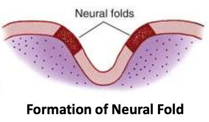
How is the Neural fold formed?
optic groove forms down the middle of the neural plate
ridges on each side become neural folds
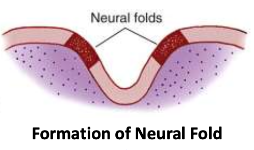
When are the Neural groove + Neural folds formed by?
Day 18
When does the first indication of eye development appear?
Day 22
How is the Neural tube formed?
Groove expands, causing these folds to grow towards each other
Neural crest cells (cells on the crest of each neural fold) separate from the ectoderm
Folds fuse to form the neural tube
Forebrain forms as part of the anterior neural tube
Neural crest cells migrate & form discrete “islands” of cells in the mesoderm
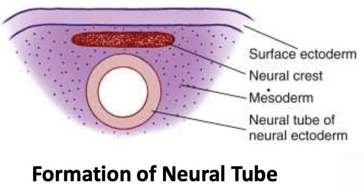
What are the tissues called after the Neural tube forms?
Neural tube tissue = Neural ectoderm
Surface layer above neural tube = Surface ectoderm
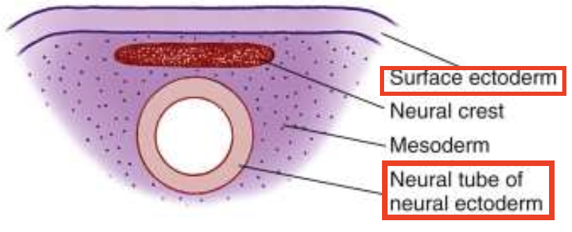
When is the neural tube formed by?
Day 22
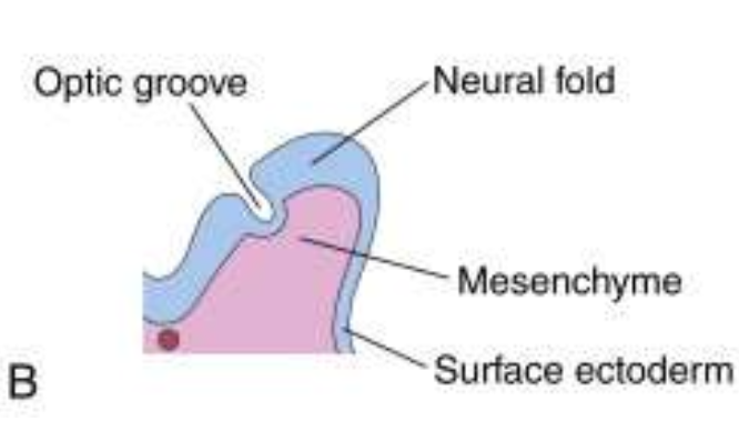
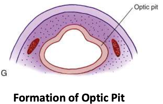
How is the Optic Pit formed?
Forebrain evaginates → optic pit
What ocular structures does the Surface Ectoderm give rise to?
Lens
Epithelium of eyelids, cilia, meibomian, Zeis & Moll
Epithelium lining nasolacrimal system
Corneal epithelium
Conjunctival epithelium
What ocular structures does the Neural Ectoderm give rise to?
Iris sphincter & dilator muscles
RPE
Optic nerve fibers
Neural retina
Neuroglia
Epithelium of ciliary body
Epithelium of Iris
What ocular structures does the Neural Crest give rise to?
Corneal stroma (which gives rise to Bowman’s layer)
Uveal pigment cells
Sclera (most)
Trabecular structures
Uveal CT
Meninges of optic nerve
Corneal endothelium (which gives rise to Descemet’s membrane)
Vascular pericytes
Ciliary muscle
By day 28, what’s most visible?
Forebrain + its covering layers (mesenchyme and surface ectoderm)
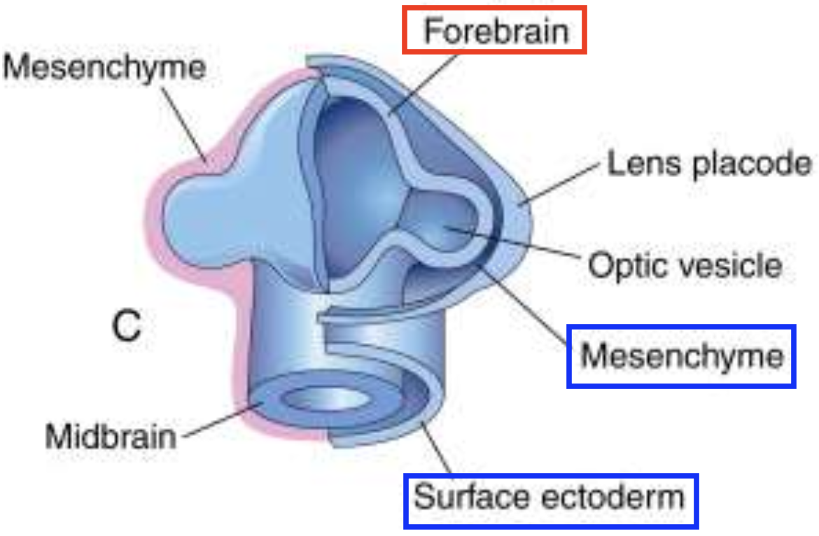
What 2 structures begin forming after the forebrain coverings develop? When does this happen?
Day 28-32
Optic cup
Lens vesicle
Lens
What is the sequence of lens formation?
Note: This occurs after the forebrain coverings.
Lens placode → Lens pit → Lens vesicle (which closes to form the lens)

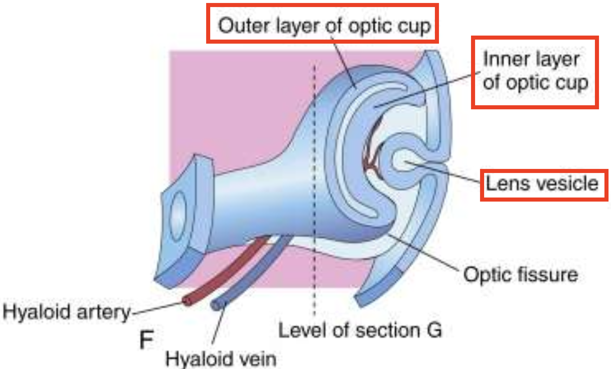
Optic Cup
What are the 2 layers of the optic cup, and what do they form?
Note: This occurs after the forebrain coverings.
Outer layer → future RPE
Inner layer → future neural retina

When does the optic cup first appear externally?
32 days
Optic Cup
What is the optic (choroidal) fissure? What vessels enter through the optic fissure?
→ groove along the inferior surface of the optic cup & optic stalk that allows blood vessels (hyaloid artery & vein) to enter and supply the newly formed lens
Note: Optic fissure must close
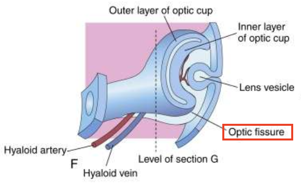
By when does the Optic fissure normally close?
6th week
We know that the Optic fissure must close. What happens if it fails to close?
Iris coloboma develops
Optic Cup
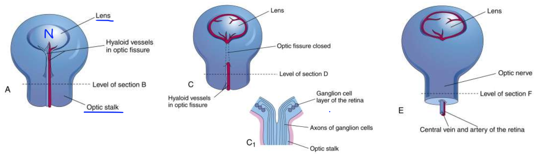
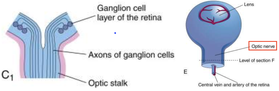
How is the Optic nerve formed?
1) Neural retina starts forming from the inner layer of the optic cup, where RGCs develop
2) RGCs send out axons towards the brain
3) To reach the brain, they must pass the optic stalk
4) As more axons grow through the optic stalk, the lumen of the stalk disappears (involution)
5) Bundled RGC axons → optic nerve fibers
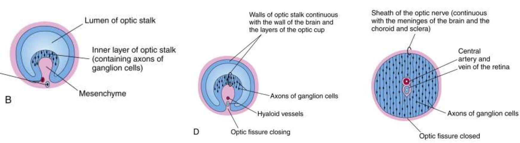
What do the hyaloid artery & vein develop into? What do these new vessels supply and drain?
CRA & CRV → supply & drain the inner retina
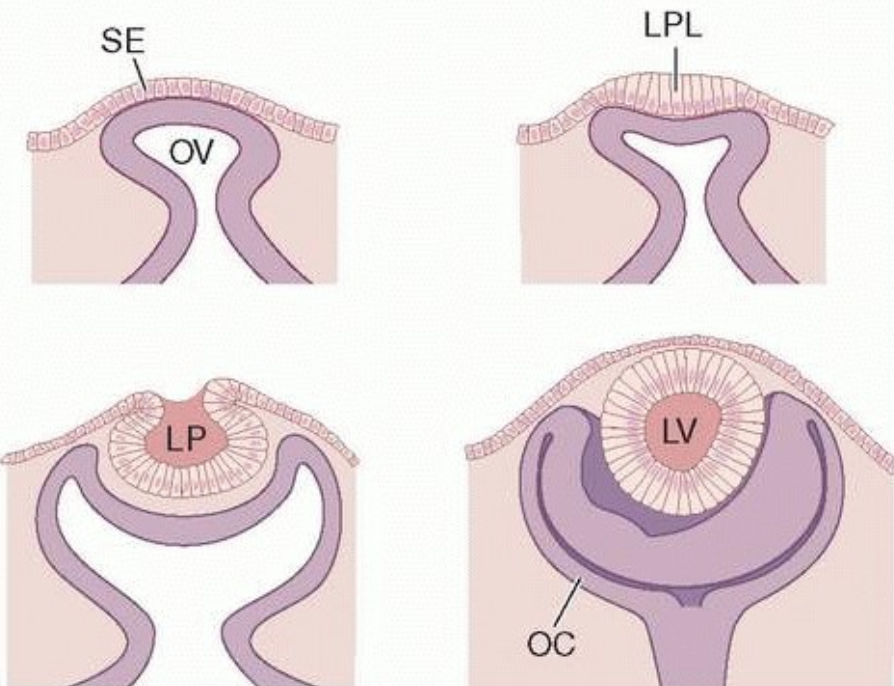
Lens
Recall: Lens placode → Lens pit → Lens vesicle (which closes to form the lens)
In detail, how is the Lens vesicle formed?
1) Surface ectoderm (SE) adjacent to the optic vesicle thickens → lens placode (LPL) forms
Thickening occurs due to:
ectodermal cells elongate
↑ regional cell div
2) SE progressively invaginates, forming → Lens pit (LP) & Lens vesicle (LV)
Lens
What happens to the posterior cells of the Lens vesicle (LV) during early lens development?
Elongate → 1er lens fibers
fibres fill up the lens vesicle’s lumen → embryonic nucleus formed (small dots)

Lens
What is the lens bow? What’s the typical bow angle?
→ curved line formed by the cell nuclei of elongating 1er fibers
120–130°
Lens
What happens to the Anterior cells of the Lens vesicle (LV) during early lens development?
1) Anterior epithelium stays in place, while posterior epithelium elongates
2) Undergo mitosis near the equator to form “new epithelial cells”
Lens
How is the 2er lens fiber formed?
1) New cells at the equator (formed anterior epithelium mitosis) elongate anteriorly and posteriorly
2) Elongated cells → 2er lens fibers
3) 2er lens wrap around the embryonic nucleus in concentric layers → growing lens mass
4) Lens are formed where the fiber ends meet.
When are the secondary lens completed?
Week 7
Zones of Development of Lens Epithelium
Describe the Central zone in the lens epithelium.
→ occupies 80% of the front surface of the lens, sitting right under the lens capsule (not part of the lens fibers yet)
Cells are arrested (non-dividing)
Exposed to incoming light via the pupillary aperture
only zone not protected by the iris from DNA-damaging UV/visible light
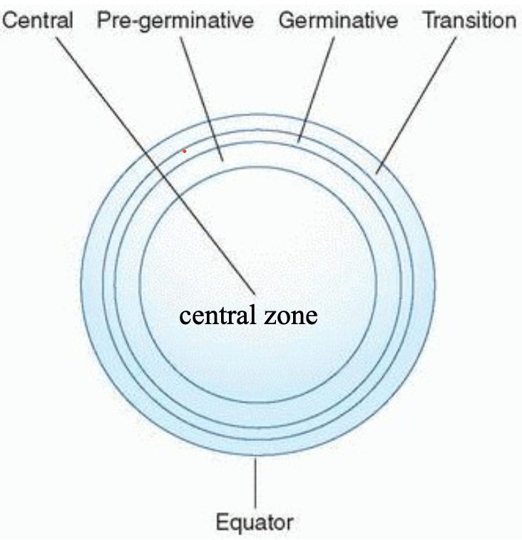
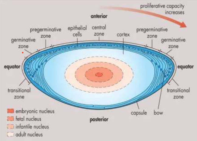
Zones of Development of Lens Epithelium
Describe the Pre-Germinative Zone in the lens epithelium.
→ Lies concentric to the central zone
Cells divide to produce new anterior epithelial cells as the lens surface increases in size with age
these are a source of new anterior epithelial cells for the Central zone
Zones of Development of Lens Epithelium
Describe the Germinative Zone in the lens epithelium.
→ Zone of active mitosis
cells divide and migrate toward the lens equator
protected from DNA-damaging light since they’re behind the iris
Zones of Development of Lens Epithelium
Describe the Transition Zone in the lens epithelium.
→ divided anterior epithelial cells → 2er lens fibers
2er lens fiber are non-mitotic + have no nuclei
What do the ends of the lens fiber meet in? Where are they found?
Upright Y-suture (posterior to the anterior epithelium)
Inverted Y-suture (anterior to the posterior capsule)
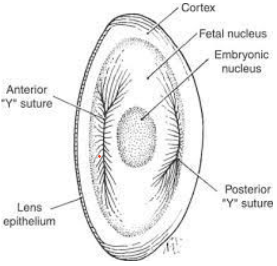
When are Y-sutures visible?
3rd month
How does the lens shape change as fibers are added? How does this affect vision?
Spherical → ellipsoidal as new fibers are added
Spherical lens = better for near vision
Elliptical lens = better for distance vision
How is the Lens capsule formed?
from secretions of the lens epithelium
When is the lens capsule evident by?
5 weeks
Compare the following nuclei:
Embryonic
Fetal
Infantile
Adult
Embryonic: formed from the original cavity of the lens vesicle; filled by 1er lens fibers
Fetal: has Y-sutures + all fibers formed before birth
Infantile: has fibers formed from birth to puberty
Adult: has fibers formed from post-puberty to adulthood
Describe the development of Zonule Fibres.
Origin: near the ora serrata
→ Extend from valleys b/w ciliary processes to attach to the lens capsule (near the lens equator)
form a 90o with the Marginal bundle of Drualt
aka: 3er vitreous fibers (b/c arise within the vitreous)
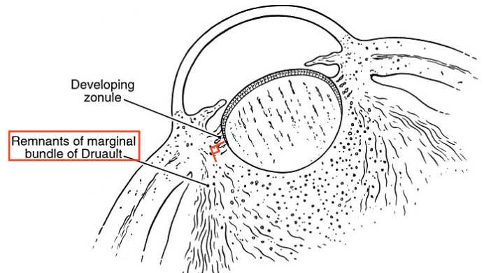
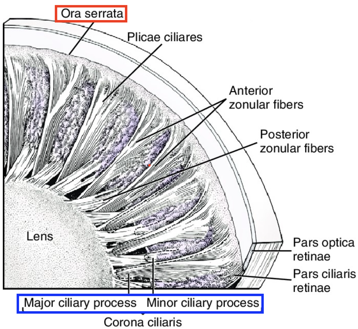
When are Zonules formed by?
7th month
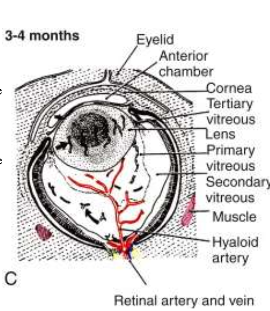
How does the Hyaloid artery develop?
Branch of the internal carotid artery enters the optic cup via the fetal fissure → forms hyaloid artery
How is the Posterior tunic (“blood supply to back of lens”) formed?
Hyaloid forms a highly branching vascular network that fills the vitreous cavity (behind the lens) → Posterior vascular tunic
vessels + branches make up most of the space b/w the lens & neural ectoderm (developing retina)
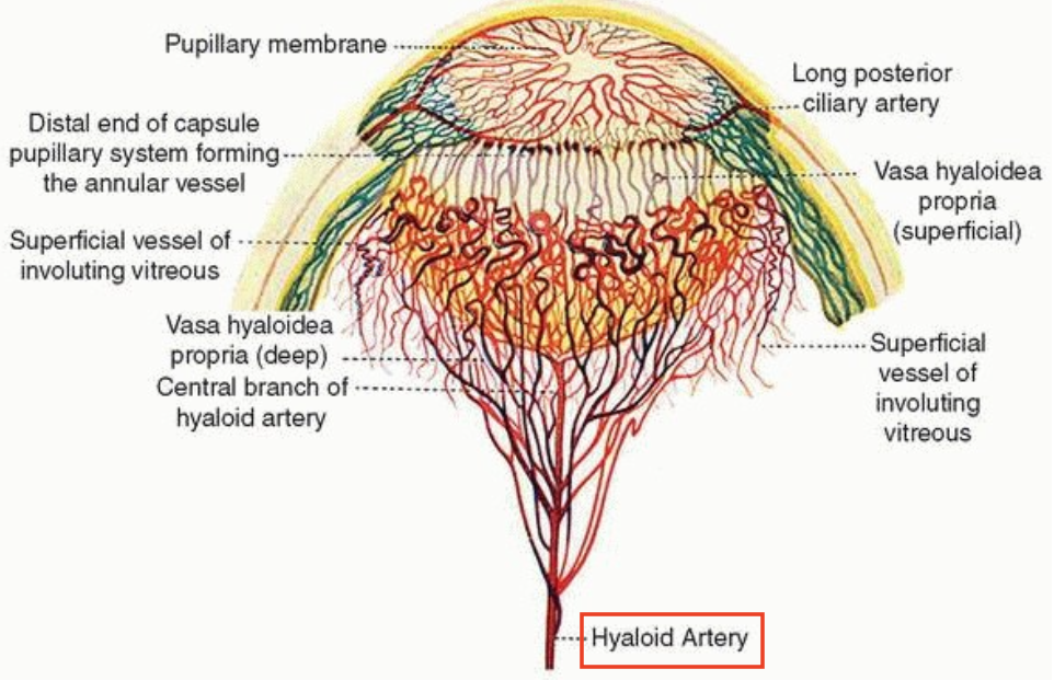
How is the Anterior vascular tunic?
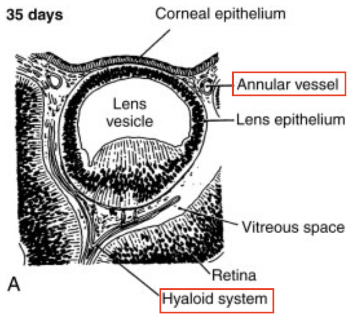
1) Branches of hyaloid artery near the lens equator anastomose with the annular vessel at the margin of the optic cup
2) Annular vessel sends loops forward onto the anterior surface of the lens → forms the anterior vascular tunic
3) These vascular networks carry nutrients to the developing lens until the aq humor begins to form
4) Hyaloid & annular vessels regress as the eye matures
Where do the Anterior tunic vessels (hyaloid + annular vessel) drain, and what structures are found there?
drain into a network found in the region that will become the ciliary body
When is the Anterior Vascular tunic formed?
7th week
When is the hyaloid vasculature fully formed?
End of 2nd month
→ Entire hyaloid system = posterior & anterior vascular tunic fully formed by then
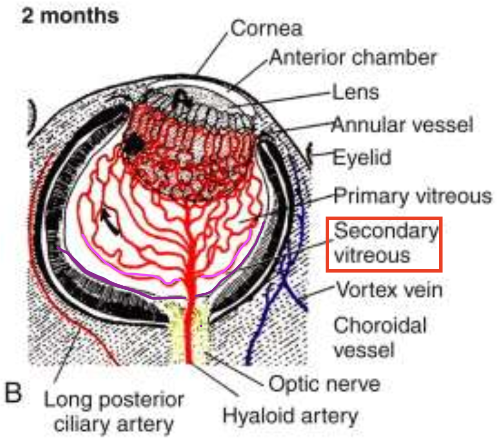
When does the vascular primary vitreous reach its greatest extent, and what happens in the vitreous at this stage?
By 2nd month, 1er vitreous becomes fully vascularized by the hyaloid system
Avascular 2er vitreous begins to form
has a thinner/finer fibrillar composition
creates a narrow zone b/w the peripheral hyaloid vessels & retina
What key changes occur in the vitreous during the 4th month of eye development?
Hyaloid vessels regress
Zonular fibers (3er vitreous) start to stretch from the growing ciliary region toward the lens capsule
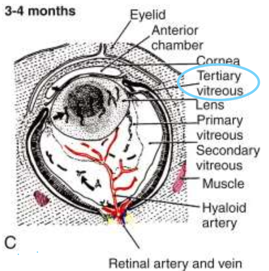
Compare the 1er, 2er, 3er vitreous.
1er vitreous → vascular (contains hyaloid system)
2er vitreous → avascular, more fibrillar
3er vitreous → includes zonules (zonular fibers)
What vascular changes occur as the hyaloid system regresses?
1) Hyaloid vessels enter through the optic nerve + connect with 1er vitreous, forming small loops into the developing retina
2) As the hyaloid system regresses:
loops shrink and disappear
CRA & CRV enter
What happens to the hyaloid system by the 7th month of development?
No blood flow remains in the hyaloid vasculature
Hyaloid system is fully reabsorbed
CRA & CRV take over as permanent retinal vessels
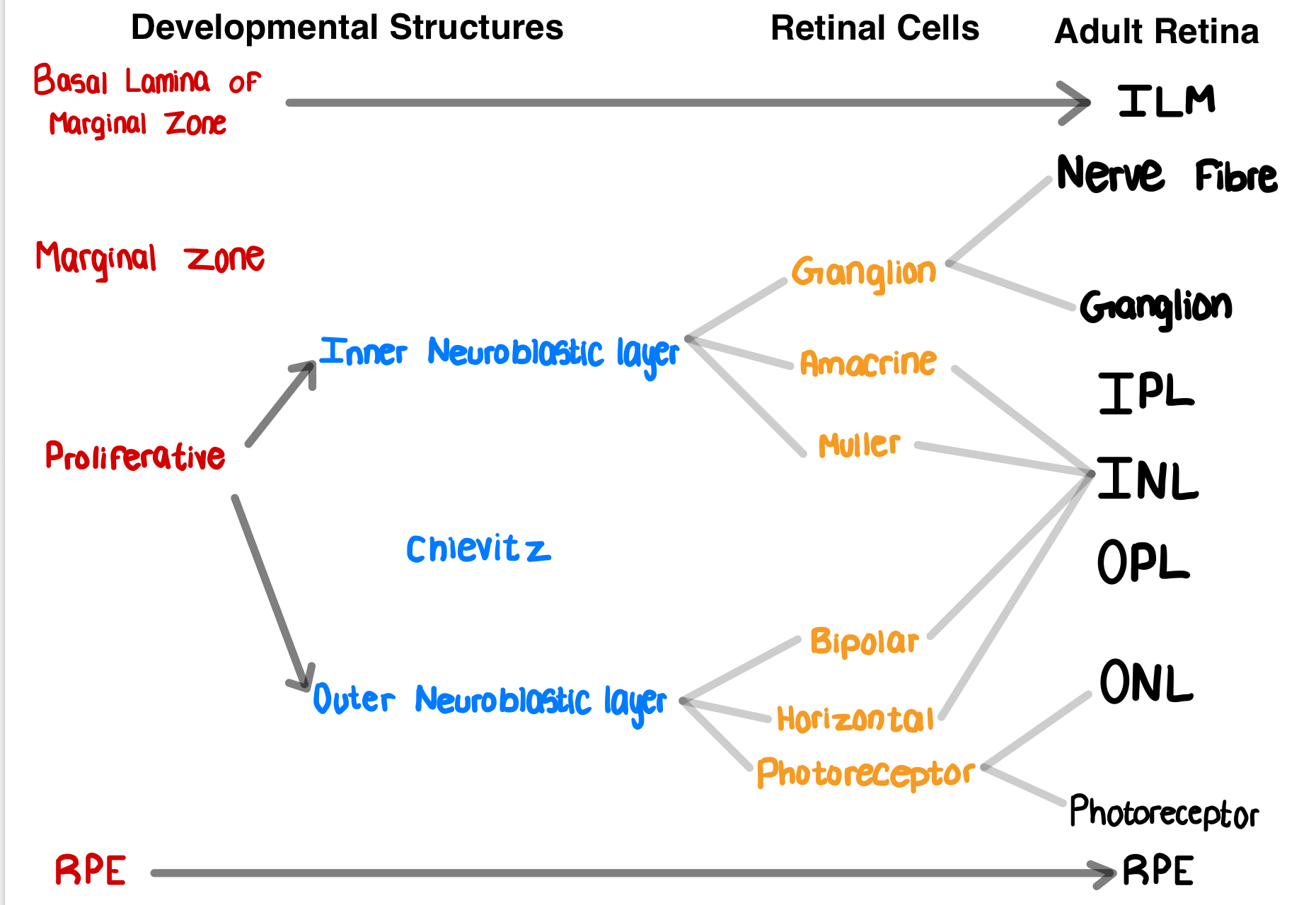
What is the first retinal layer to differentiate?
RPE
Development of Neural Retina
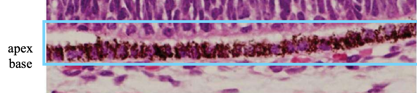
Describe the development of the RPE.
1) Cellular structures and melanosomes begin to appear in the outer layer of the optic cup
Melanosomes: melanin-producing cells inside RPE
2) After week 6, the RPE is 1 cell thick, where they’re cuboidal to columnar
Base of each RPE cell = external toward the developing choroid
Apex of each RPE cell = internal toward the inner layer of the optic cup
Development of Neural Retina
When does the pigmentation of the RPE occur? What is this known as?
Weeks 3 - 4
“Earliest pigmentation evident in the embryo”
Development of Neural Retina
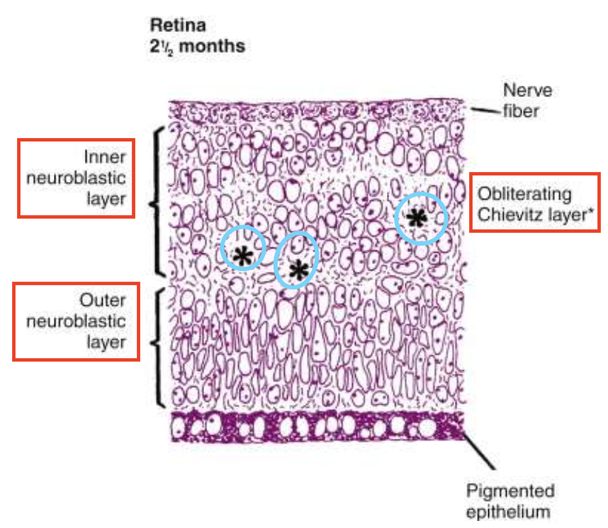
When do the inner and outer neuroblastic layers form in eye development?
Week 7: Cells from the neural ectoderm migrate to form the Inner & Outer neuroblastic layer
By month 3: Formation of neuroblastic layers (inner and outer) is complete
neural retinal cells will begin to differentiate in the central retina and proceed to the periphery

Development of Neural Retina
What is the fiber layer of Chievitz?
Transient, uneven, nucleus-free area that lies b/w inner & outer neuroblastic layers
Development of Neural Retina
At 2.5 months, what do the following cells differentiate with?
Uppermost cells (above inner neuroblastic layer)
Below Chievitz layer
Outer neuroblastic layer

Uppermost cells → RGCs (form the optic nerve)
Below Chievitz layer → Amacrine & Müller cell
b/w shifted nuclei of amacrine & Müller cell → Inner plexiform
Outer neuroblastic layer → photoreceptor, bipolar & horizontal cells
Development of Neural Retina
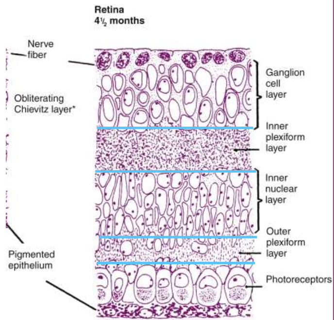
What happens to retinal layers around midterm (4.5 months)?
Retinal lamination is complete
Development of Neural Retina
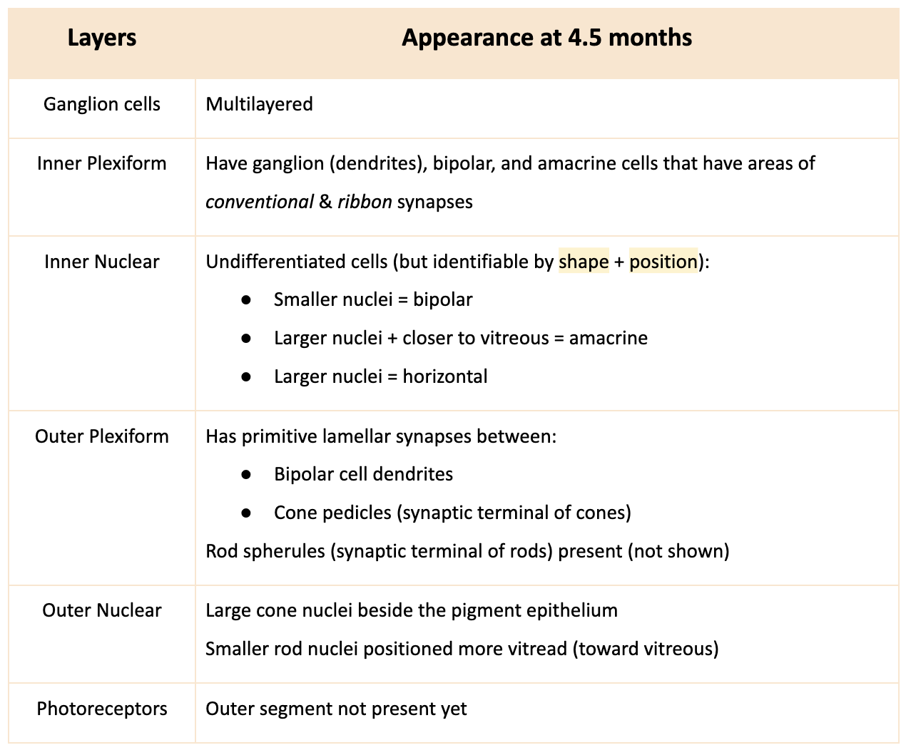
Describe the changes in the retinal layers at 4.5 months.
RGC layer
Inner Plexiform
Inner Nuclear
Outer Plexiform
Outer Nuclear
Photoreceptors
Development of Neural Retina
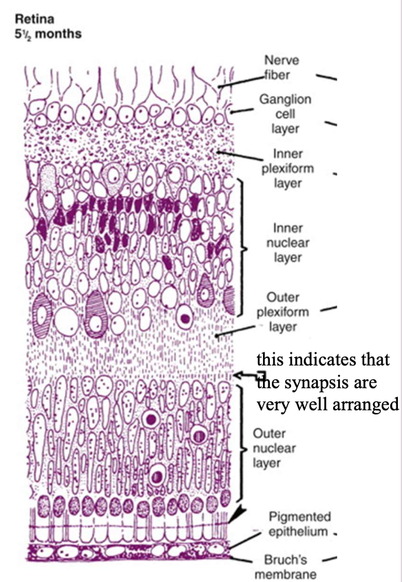
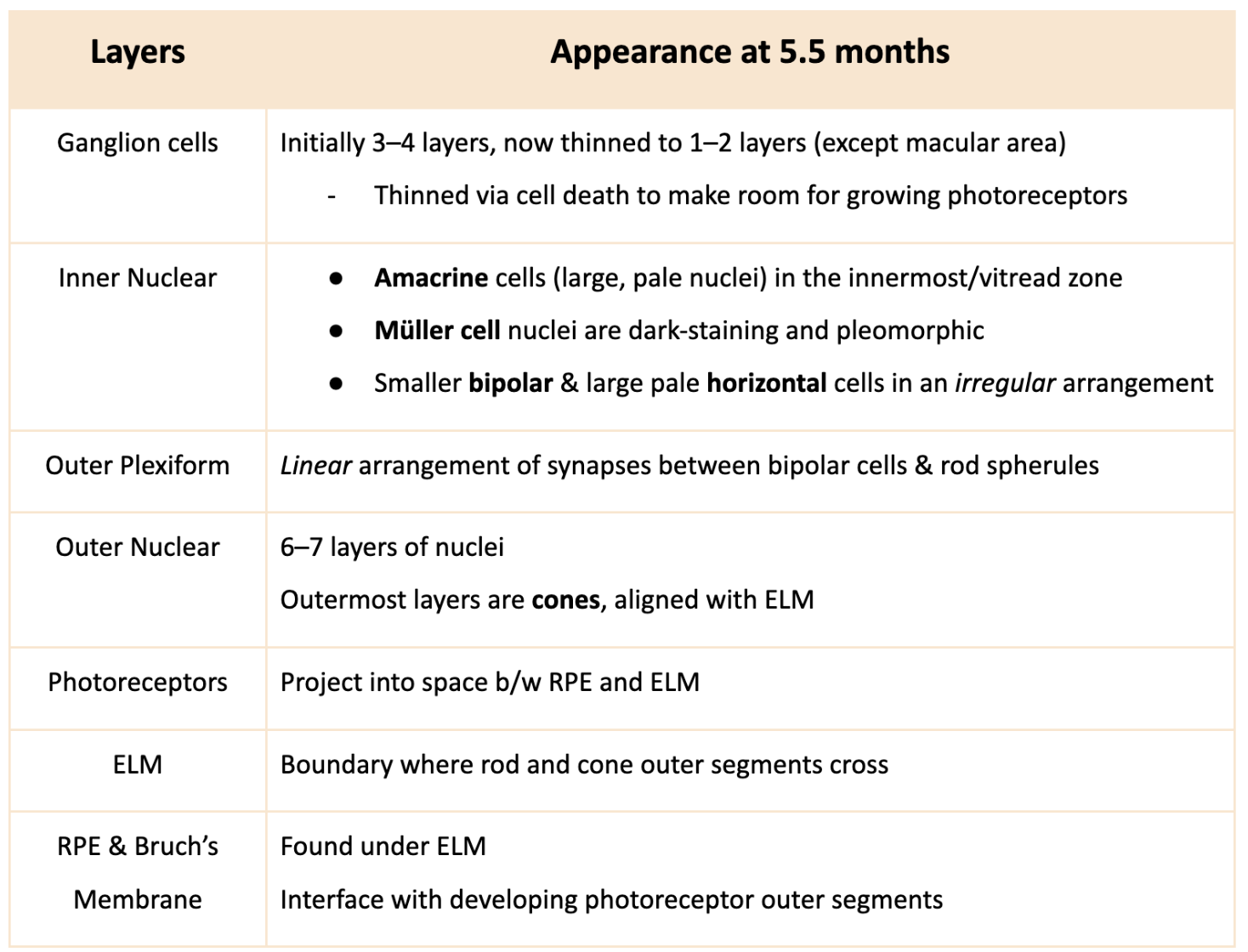
Describe the changes in the retinal layers at 5.5 months.
Ganglion cells
Inner Nuclear
Outer Plexiform
Outer Nuclear
Photoreceptors
Development of Neural Retina
Describe the Newborn retina.
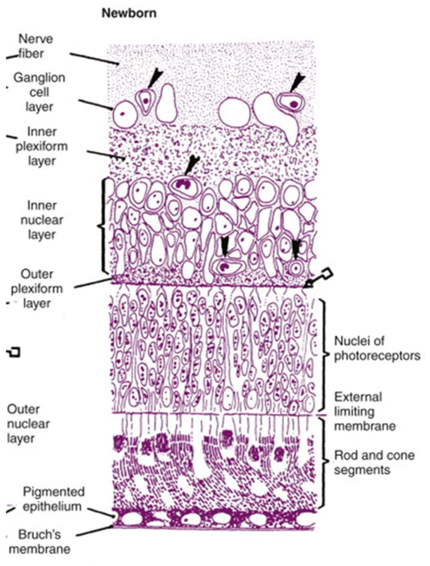
→ has adult configuration + fully layered
Nerve fiber layer: forms from RGC axons
Vascularization extends to outer limits of the INL (arrowheads)
OPL
thinner than adult retina → will thicken by ~4 years
well-established synapses
Development of Neural Retina
Describe the photoreceptor outer and inner segments in the Newborn retina.
fully developed
its tips contact RPE → outer segment renewal
Old outer segment discs are phagocytosed by RPE
New discs form at the base and migrate outward
Draw out the Retinal Development Flow chart. Mention the developing structures, retinal cells and the layers they form in the adult retina.
List the 3 Foveal Development Stages.
1) Displacement of inner retinal components → forms foveal depression
2) Migration of photoreceptors toward the center → ↑ cone packing
3) Maturation of photoreceptors
Foveal development
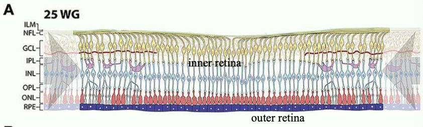
Describe Foveal development at 25 Weeks Gestation.
CRA nourishes inner retina
Choriocapillaries nourish the outer retina
No foveal pit formed yet
Foveal development
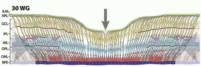
Describe Foveal development at 30 Weeks/ 7th month Gestation.
Depression forms at the center of the retina → foveal pit
occurs due to displacement of inner retinal neurons (ganglion + bipolar cells)
Ganglion cell layer and INL move towards macular periphery
Cone packing increases at the center → maximum cone density
Foveal development
Describe Foveal development at 40 Weeks WG to 6 months PG.
Foveal depression present BUT not fully deep at birth (deepens more at 6 mons)
1 interrupted row of ganglion cells remains across foveal depression
1–2 layers of bipolar cells still extend across the fovea
OPL (Henle’s fiber layer) is wide and well-developed
Elongated cone inner and outer segments
Parafoveal area has large accumulation of ganglion and inner nuclear cells → indicates mature macula

What is the Foveola?
→ central part of fovea (site of sharpest visual acuity)
last retinal area to mature
Describe the Foveola before birth.
Large rod-free area
As cone density increases as they migrate towards the centre → rod-free zone narrows
Describe the Foveola after birth.
1 week old: cone population is ~18 cones (diameter of rod-free zone ≈ 100 μm)
4–5 years: cone population increases to ~42 cones (adult level) (diameter shrinks)
Perifovea and parafovea begin to show rod presence, but the foveola stays rod-free

Retinal Vessels
What is the pre-cursor of the CRV?
branch of the primitive maxillary vein within the optic stalk
Retinal Vessels
When do retinal vessels (CRV & CRA) begin developing?
Early 4th month
Retinal Vessels
How do retinal vessels (CRV & CRA) begin developing?
Primitive retinal vessels emerge from the hyaloid artery near the optic disc
These vessels enter the developing nerve fiber layer
Retinal vessels form arterioles, venules, and capillary beds
Note: Nasal retinal vessels develops before the temporal retinal (nasal matures first)
Retinal Vessels
When is the full vascular structures of the retina arterioles, venules & capillary beds developed by?
~3 months after birth
What signals and direct the retinal vessels? What do they signal and direct?
Guidance molecules signal and direct:
growth and path of neurons
growth and branching of retinal vessels
What is induction?
→ influence of one developing structure on another via signaling molecules
occurs via a series of biochemical steps, not a single event
Describe the role of Induction in Embryological Development.
→ Development depends on differentiation and interaction b/w tissues
Some tissues develop only when near another developing structure at the right time
Ex: Lens release signaling molecules that induce corneal development
Contact or proximity b/w tissues allows biochemical signaling