Higher order visual processing
1/21
There's no tags or description
Looks like no tags are added yet.
Name | Mastery | Learn | Test | Matching | Spaced | Call with Kai |
|---|
No analytics yet
Send a link to your students to track their progress
22 Terms
What is the ventral pathways responsible for
Form and colour processing - identifying ‘what’ the object is
Where does the ventral pathway project to
From V1 to The inferior temporal lobe (IFC)
What is the dorsal pathways responsible for?
Motion, spatial location and perception in low light - identifying ‘where’ the object is
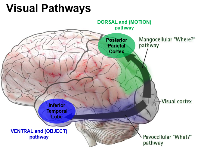
Where does the dorsal pathway project to?
The posterior parietal cortex
What types of V1 pathways project to V2 stripes
V2 has thick, thin and interstrip regions
blobs in V1 → thin stripes
interblobs in V1 → thick stripes
segregation of pathways is maintained in V2
What do the parvo, magno and koniceullar channels form in V2
Magnocellular (4ca) → motion/where pathway
Parvocellular (4Cb) and Koniocellular (blobs) → colour'/what pathway
What is V2 cortex known as
Pre-striate cortex/ Brodmann area 18
How is visual space represented in V2
as a complete topographic map, preserving spatial relations from V1
What are V2 cells sensitive to
Orientation, spatial frequency (motion) and colour
How many times are V2 receptive fields larger than V1
they are 2/3 times larger
What is V3A primarily involved in
Coherent motion and depth - part of the dorsal stream
thick stripes in V2 project to dorsal of V3
What is V3 more focused on
shape and form - part of the ventral stream (blobs in v1 project to interstripes in V2 then project toV3)
what does the MT/V5 do?
processes motion direction - requires moving stimuli for activation
what can damage to MT/V5 cause
Akinetopsia (motion blindness)
What does MST (medial superior temporal) process?
Linear motion, radial motion and circular motion → supports navigation and motion perception
more complex as not just direction but complex understanding as where object is moving
What is the role of the area v4 (parvocellular) in the visual system
Ventral stream: receives input from blobs and regions of the striate cortex
colour processing, orientation and object sensitivity, summation of edges and corners (summate several V2 and V1 neurons)
What can lesions in the V4 lead to
Achromatopsia (partial or full colour blindness)
How does V4 differ from V1
V4 has larger receptive fields and is sensitive to geometric features
What is the key functional difference between V4 and V5
V4 is slower and processes colour whereas V5 processes motion quicker
Why does visual processing increase in complexity as you go from V1 to V5/V4
Due to summation and integration of receptive fields - a hierarchal structure
What would happen if you had lesions in V1
complete visual loss as not further projections to V2