Microbiology Exam 1
 Call Kai
 Learn
Learn Practice Test
 Spaced Repetition
 Match
Match Flashcards
 Knowt Play
1/253

There's no tags or description
Looks like no tags are added yet.
Name | Mastery | Learn | Test | Matching | Spaced | Call with Kai | Chat |
|---|
No analytics yet
Send a link to your students to track their progress
254 Terms
What is microbiology?
The study of microorganisms. Cellular and acellular, too small to be seen with the naked eye
What is Bacillus? What about the Genus? What kind of microbe?
Bacillus stands for a shape of bacteria (rod). the genus Bacillus is a bacteria that is rod shaped, gram positive, and capable of forming endospores (tough structures that help bacteria in harsh conditions). Cellular.
What is Anabaena? What kind of microbe?
A type of cyanobacterium (blue-green bacterium) that can perform photosynthesis. It is a filamentous cyanobacterium, meaning its cells form long chains/filaments. They have heterocysts. Cellular.
What is a heterocyst? What kind of microbes are they specific to?
Heterocysts are nitrogen fixing cells. They are exclusive to certain types of filamentous cyanobacteria
What is Bacteriophage T4? What kind of microbe?
A virus that infects bacteria, especially E. coli. It performs the lytic cycle, infecting the bacteria and making more until it eventually lyses (bursts) the bacterial cell. Acellular.

What is this?
This is a virus. Pox virus, causes skin lesions, blisters, or bumps.

What is this
Green algae cells. They can preform photosynthesis and play a big role in the aquatic environment.
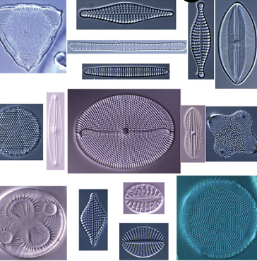
What is this
Diatom shells. They look translucent because they are dead. Diatoms are responsible for roughly 20% of global carbon fixation through photosynthesis.

What is this
Trypanosoma. A genus of parasitic protozoa. They are typically found in blood tissues of their hosts snd cause things like African sleeping sickness and chugs disease.
What is Gram Positive Bacteria
They have thick layer of peptidoglycan making up their outer shell. they stain purple or blue because the dye Geta trapped under thick peptiglycan.
What is Gram Negative Bacteria
Have a thin layer of peptidoglycan however they have an outer membrane. They stain pink or red bc dye does not get trapped underneath. They are more resistant to antibiotics. Their outer membrane acts as a protective shield, so its hard for immune cells and antibodies to get in
What type of microscope was the Human Mouth taken with? How do you know?
It was taken with an SEM microscope. You can tell because you can see very precise details of the surface (texture and everything) but not in the cell like TEM.
What kind of microscope was the Lake taken with? How do you know?
Phase contrast light microscope. You can tell because of the different light intensity and contrast on it
What kind of microscope is the sewage sludge taken with? How do you know?
Fluorescent light microscopy. You can tell because of the dark background and glowing cells. Glow sticks
What are the types of light microscopes that we learned about?
Compound, Fluorescence
What are the types of electron microscopes we learned about?
Transmission (TEM), Scanning (SEM)
What does a compound microscope picture look like?
Compound looks like normal cells under a microscope and are normally stained and simple looking.
What does a fluorescent microscope picture look like?
Fluorescence glows and the cells are neon against a dark background
What does a SEM microscope picture look like?
SEM is detailed outer surface, 3-D looking and you see the surface.
What does a TEM microscope picture look like?
A TEM is detailed and very flat looking and you can see the inside of cell.
Give examples of microbe extremes
Antartica (cold) and the desert (hot and lack of water). Less microbes live in extremes. Their membranes have to adapt.
Know about the relationship between gamma rays and microbes.
- Gamma rays are a type of high energy radiation that can damage microorganisms by damaging their DNA and other cellular components. Gamma rays are used for sterilization too and other things. Some microbes are resistant to radiation.
What is Deinococcus Radiodurans?
- not harmful to animals, it is known for breaking down organic compounds in environments for its energy and lives in places like soil, dust, water, and other nutrient poor environments. It survives extreme radiation because it can repair damages DNA and can survive gamma rays that would kill YOU.
When did the earth arrive?
4.6 bya
When did bacteria and arches arrive
3.8 bya
When did phototrophic bacteria arrive
3.4 bya
When did the atmosphere transition to oxygenated
2.4 bya
When did cyanobacteria come about
2.4 bya
When did oxygen become present
2 bya
When did eukarya come about
2 bya, derived from archea
When did animals come around
500 mya
When did vascular plants come around
420-430 mya
When did mammals come around
225 mya
When did humans come around
300,000 years ago
Know LUCA and the deriving from Luca.
· LUCA around 4.2 bya, bacteria and archaea diverged from it 3.8 bya, and eukarya derived from archaea 2 bya
What are the 3 domains of cellular organisms?
- Bacteria, Archaea, Eukarya
What products are made during fermentation?
Ethanol, lactic acid, proponic acid, and acetic acid
What is fermentation?
What is used when there is not oxygen available. It is a process where cells break down sugar to make energy without using oxygen.
What 2 ways can glucose ferment?
- Become lactic acid
becoming ethanol and carbon dioxide
What is pickling?
A process of preserving food by putting it in an acidic solution, usually vinegar or saltwater (vinegar is popular)

What can you conclude from this?
· 1900s infectious diseases were leading cause of death, now nonmicrobial diseases.

What can you conclude from this?
We know more about bacteria then anything, then fungi, then protozoa, then filterable viruses
Louis Pasteur
Developed the germ theory of disease and pasteurization, and helped create vaccines.
Robert Koch
Proved that specific microbes cause specific diseases and developed Koch’s postulates.
Julius Petri
nvented the Petri dish, used to grow microorganisms.
Paul Enrlich
Developed the idea of selective drugs that target harmful microbes and helped develop treatments for infections.
Alexander Fleming
Discovered penicillin, the first widely used antibiotic.
Robert Hooke (1665)
First described and named “cells” after observing cork under a microscope.
Van Leeuwenhoek
First person to observe and describe bacteria and other microscopic organisms.
- Cells are super tiny. How can you see them?
With a microscope

Type of microscope each was taken on?
Middle left electron microscope. Others light microscopy
Micrscopy allows structures to be seen, and that’s how you can even tell cells apart. What is the pink and green?
Pink- Cell
Green - endospores

- What kind of stain is this? What is this?
Mycobacterium tuberculosis. Acid fast stain
What is the purpose of an acid fast stain?
To identify bacteria that have a waxy coating around their cell wall
What is the purpose of a gram stain?
To identify and classify bacteria based on the structure of their cell walls
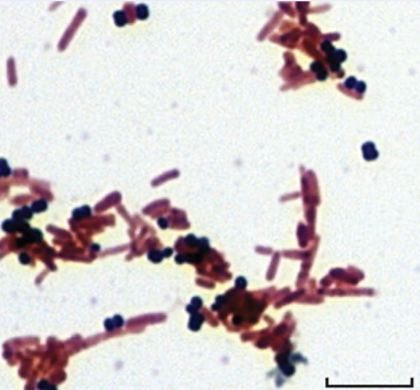
What kind of stain is this?
A gram stain
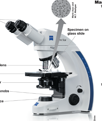
Label from top to bottom, left to right. Kind of microscope?
Compound light microscope. Ocular lens, objective lens, stage, condenser, focusing knobs, light source, specimen on glass slide
How do you figure out the magnification (total magnification)?
Depending on the objective lens you use, you multiply that by the magnification of the ocular lens (which is always 10x).
How do you use the compound light microscope?
- Focus the ocular lens at 10x. Then turn the objectives to 100x. Dab oil to be the gap between the stage and objective.
What is the maximum magnitication a compound light microscope can use?
- 1000x
What is a compound light microscope used for?
- Light microscopy
What is the compound light microscopes limit of resolution?
0.2 um
Put the compound light microscope into perspective.
Imagine a millimeter. You can see that. Take that millimeter, divide into 1,000 pieces. One of those pieces is a micron. E. coli is 1 micron! But you can see E. coli under the microscope. if you magnify it 1000x, it will look like a millimeter under the microscope.
1 micron magnified 1000x = 1 millimeter
- What is brightfield microscopy?
What we use in lab. Simplest microscopy. normally to see brightfeild you have to stain with specimen. When you stain cells it kills them, whether that be heat or the stain.

Label top to bottom
Visualized image, eye, ocular lens, intermediate image, objective lens, specimen, condenser lens, light source
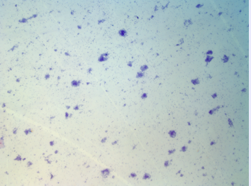
What were these cells stained with? magnification? type of microscopy?
Crystal violet. 10x objective = 100x total magnification (10 ocular)
brightfeild light microscopy

What were these cells stained with? magnification? type of microscopy?
Crystal violet. 100x objective = 1000x total magnification (10 ocular)
brightfeild light microscopy
What is phase contrast?
Type of light microscopy. It is used for microbes like biofilms. A shadow is cast and its a little glowy or glowy looking on okay background
What is darkfeild?
Glowy on a dark background. Black
Is staining needed for phase contrast and darkfeild?
No

Are the samples alive or dead?
Alive

Label these.
Brightfeild, phase contrast, darkfeild

What is this
Darkfeild

What is this and how do you know
- Phase contrast looks almost a little 3 dimensional, like they are coming out of the screen. It is used for microbes like biofilms.
What is fluorescent microscopy?
It requires a UV light source to be attached to the scope.
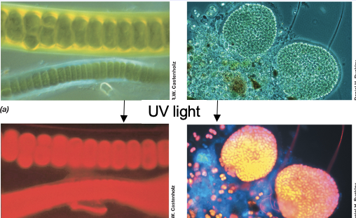
- With fluorescent microscopy, what happens when you view a cell that has chlorophyll?
Anything with chlorophyll with fluorescent microscopy will look red when hit with UV light. Sometimes you have to clear samples with chlorophyll to be able to see

What is DAPI?
A blue dye we use for fluorescent microscopy.

What is Viability Staining?
The way you can tell living cells from dead cells. The green are living and the red are dead.
How it works: Syto 9 dye stains all cells green. And then the propidium iodide stains the dead cells red. It masks the Syto 9 dye. These are dyes in fluorescent microscopy. And this staining.
What is the maximum magnification electron microscopes can use?
200,000x
What is the limit of resolution electron microscopes can use?
- 0.2-0.4 mm

- What kind of microscope is this?
SEM

What is going on?
Anytime you see dividing or cross sections in a cell, you can see the cytoplasmic membrane

What kind of microscope is this?
SEM

What kind of microscope is this
TEM
- There is no guaranteed way of cutting a cell and seeing your sample imbedded in the resin perfectly with perfect view. Why is that?
You have no control when you imbed your sample with resin how the specimen is going to lie. So you just end up cutting it and seeing whatever is there

This is Mycobacterium Leprae under a SEM microscope and a TEM microscope. Talk about it
This bacteria causes Leprosy, which is now called Hansens disease. Mycobacterium Leprae’s host is armadillo
Most bacteria and archaea are how big in diameter?
0.2 um - 700 um
When do you for sure know you’ll need an electron microscope
when you hit the nm range (like viruses)
What range is visible to you
Down to 1 mm (1,000 um)
To what range can you use a light microscope
Down to 0.2 um.
To what range can you use an electron microscope
See until 0.1-1 nm
Explain relationship in measurements.
1 mm → 1,000 um → 1,000,000 nm
mm is a unit. a thousand of um makes a mm. a million of nm makes 1 mm.

What size is this? Label it top to bottom, left to right.
100 nm. Circovirus, Tobacco mosaic virus, Flagells, Cell envelope, Cytoplasm, proteins, ribosome, nucleoid

What size is this? Label it top to bottom, left to right.
1 um. Pandoravirus, mycoplasma, Influenza A virus, T4 bcteriophage, escherichia coli, Pelagibacter

What size is this? Label it top to bottom, left to right.
10 um. Nucleus, yeast, nano flagellates, Ignicoccus, Nanoarchaeum, Haloquadratum, Mathanosarcina, spirochaetes, flexibacter

What size is this? Label it top to bottom, left to right.
100 um. Yeast, diatoms, giganthauma, Magnetoglobus, Paramecium, nucleus, epulopiscium, cyanobacteria
What is big about the relation between high surface area-to-volume ratio in cells?
- It makes it very efficient for cells to exchange waste and nutrients w/ the environment. Smaller cells also grow faster and replicate faster.
Talk about ultramicrobacteria
- Is less then 0.1 um. There are some cells we can’t see but we know its there by its DNA sequence.
Are viruses alive?
- No, they depend on life but do not have life.
What are the basic components of all microbial cells?
- DNA, cytoplasm, ribosomes, cytoplasmic membrane