Structure and function of the eye and retina - need to watch ninja nerd
1/87
There's no tags or description
Looks like no tags are added yet.
Name | Mastery | Learn | Test | Matching | Spaced |
|---|
No study sessions yet.
88 Terms
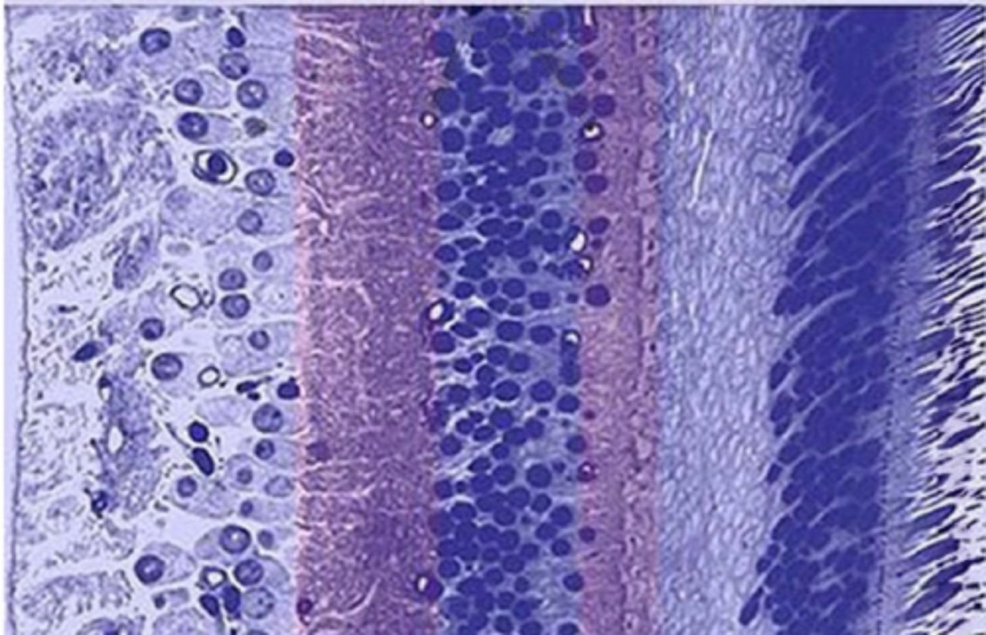
Describe the layers of the retina
1. ganglion cell layer
2. inner plexiform layer
3. inner nuclear layer
4. outer plexiform layer
5. photoreceptor layer
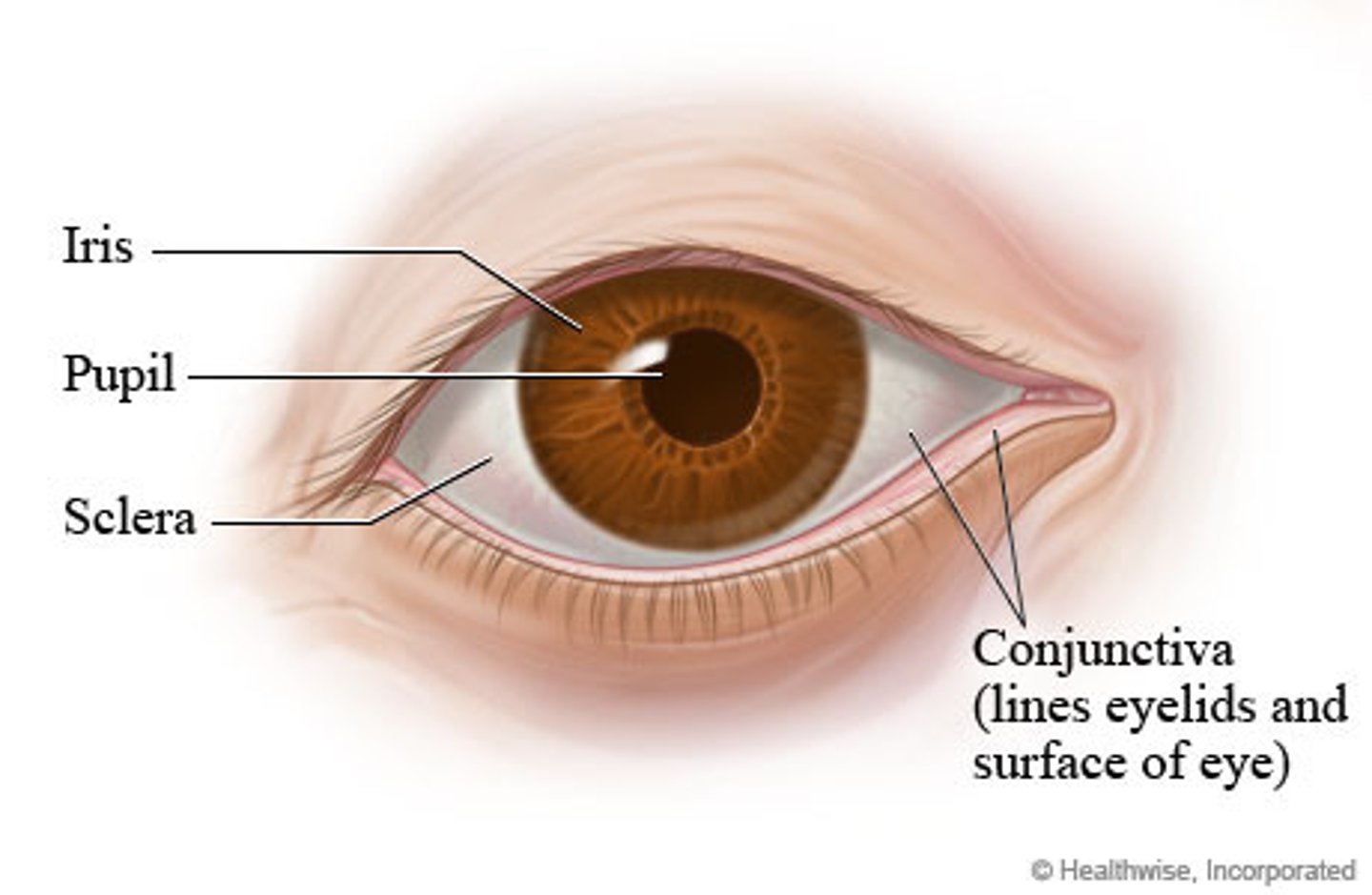
Describe the pupil
hole in the center of the iris
- allows light to enter the eye and reach the retina
- dark due to light-absorbing pigments in the retina
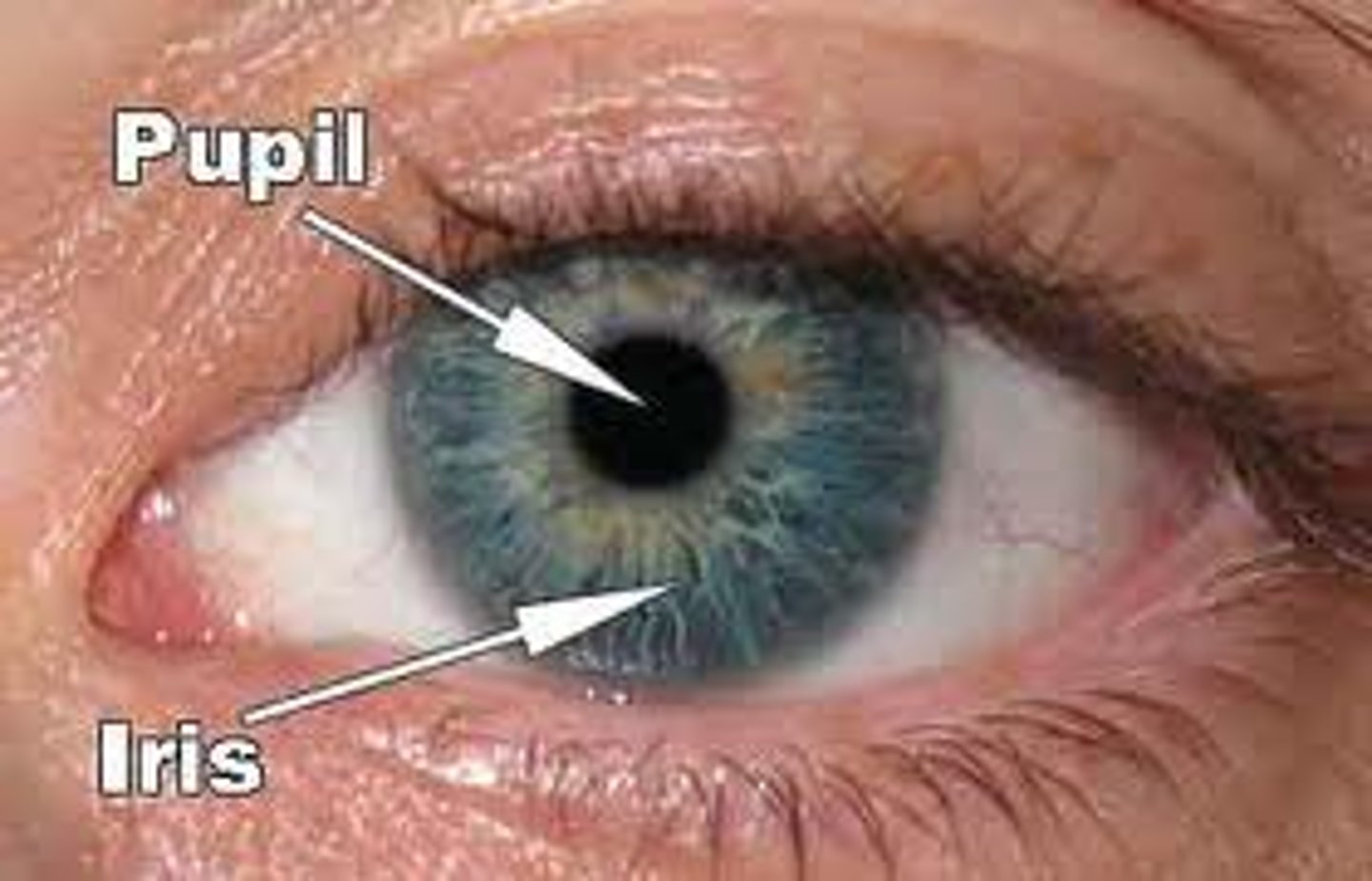
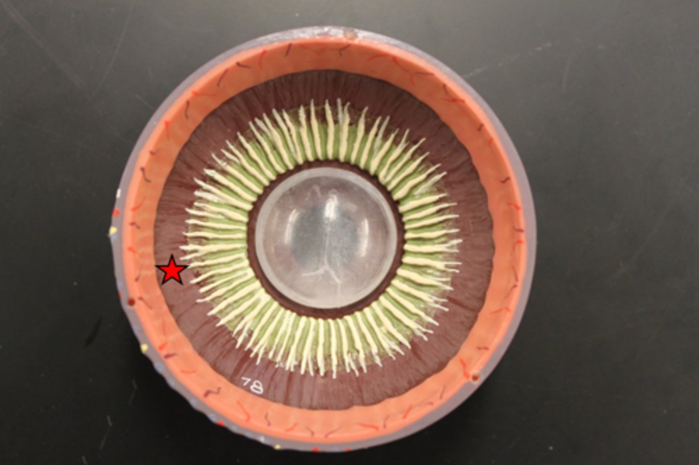
Describe the iris
- surrounds the pupil and is pigmented part of the eye
- sphincter muscles cause constriction and dilation
- controls pupillary light reflex
- post to cornea and anterior to lens
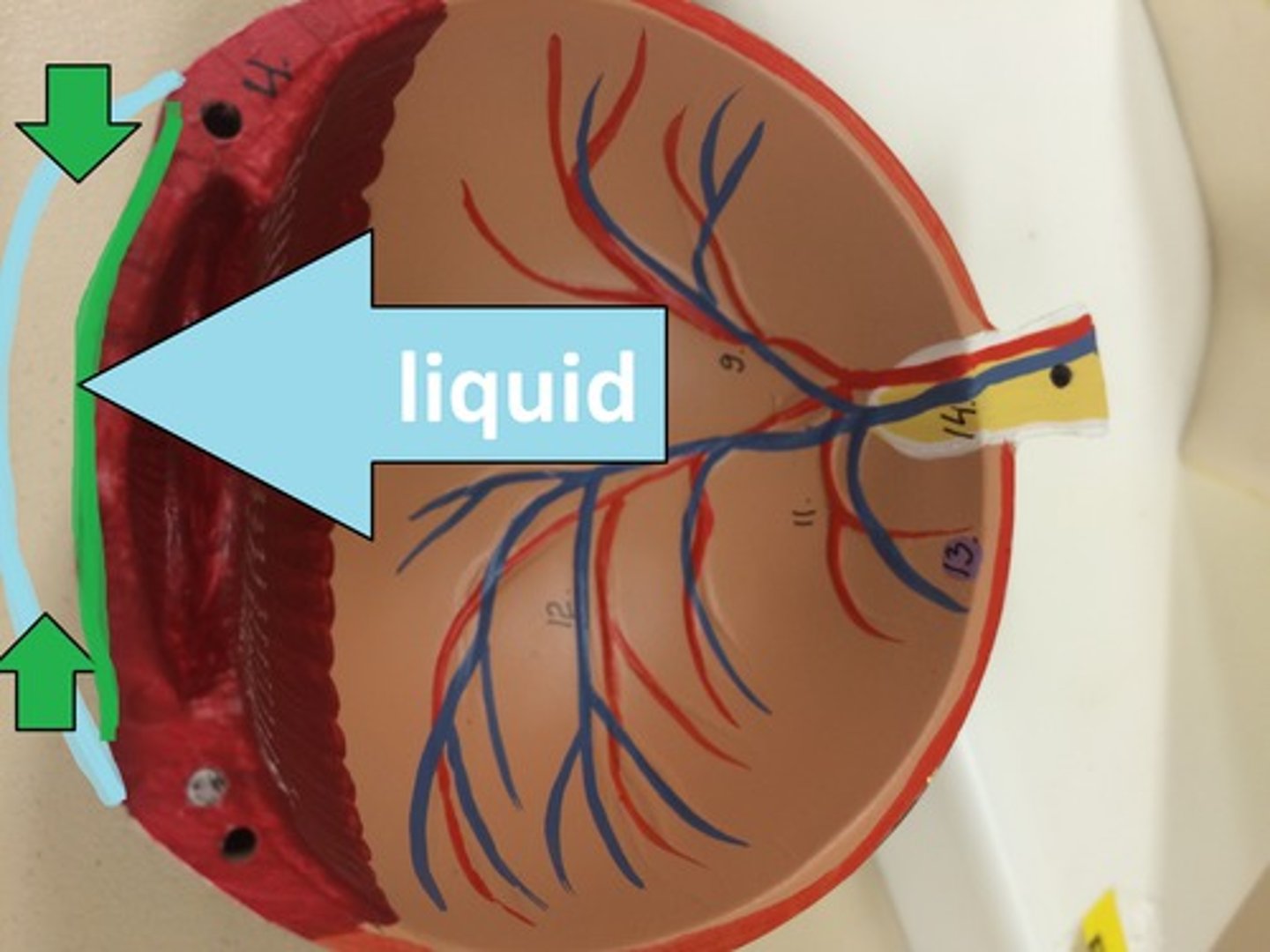
Which liquid surrounds iris
aqueous humour
When does sphincter muscle contract
In bright light
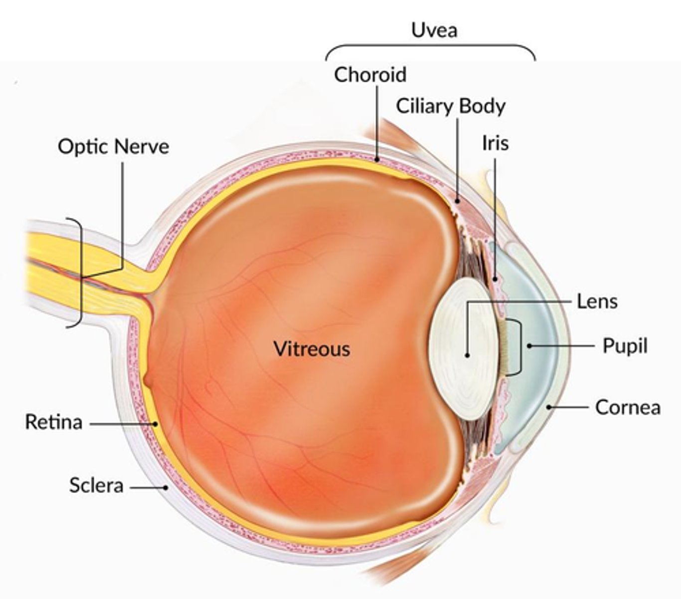
What is the cornea?
the transparent layer forming the front of the eye
- covers pupil and iris
- no blood vessels
- very sensitive
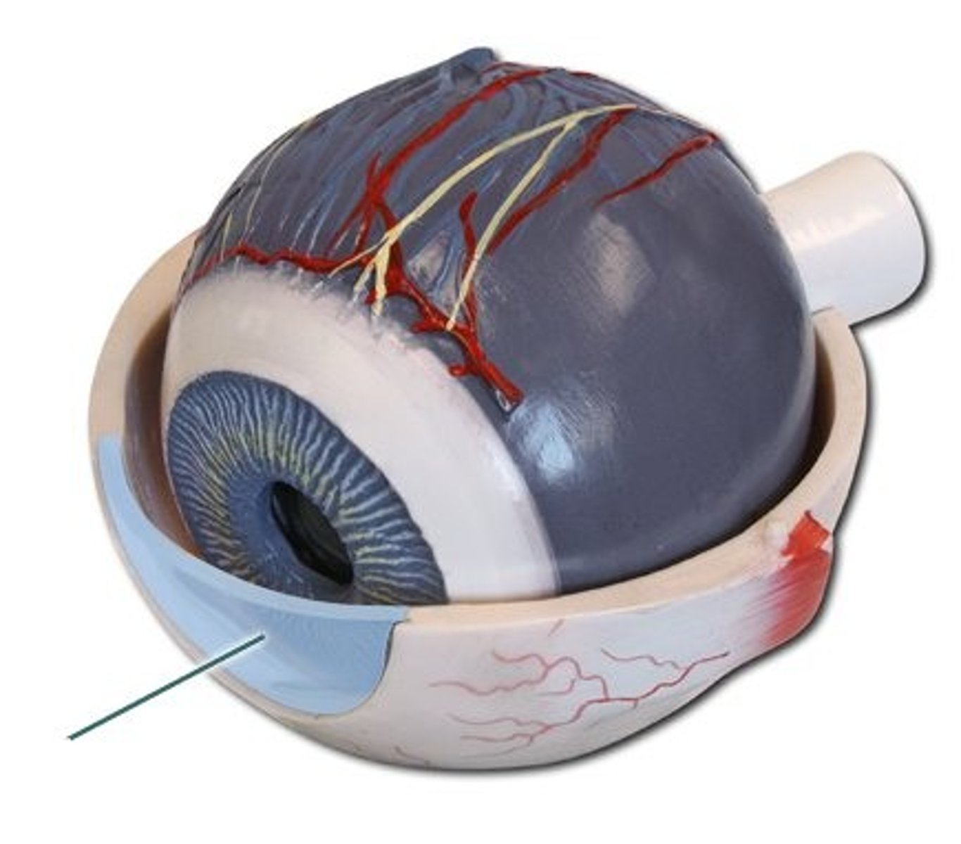
What is the sclera of the eye?
white of the eye
continuous with the cornea
(outermost fibrous layer)
What is the conjunctiva?
a thin, clear membrane that protects your eye. It covers the inside of your eyelid and the white of your eye (the sclera). The conjunctiva creates the mucus layer that forms part of your tears.
What is the optic nerve?
Carries axons from the retina, exits back of eye, passes through optic formamen and reaches base of brain near the pituitary gland where is dessicates at the optic chiasma
- ends at primary visual cortex in the occipital lobe
How does the retina work
- photoreceptors turn light into electrical signals
- These electrical signals travel from the retina through the optic nerve to the brain. Then the brain turns the signals into the images you see.
What is the macula
area of retina with high cone density
- yellow spot on retina
- surrounds the fovea
What is the fovea
the fovea is the region of the retina with highest density of cones
- small indentation at the centre of the macula
What are rod cells
Cells in the retina that are sensitive to low light intensity (dim light)
What are cone cells
Cells found in the retina that are sensitive to high light intensity (bright light) and can detect different colours.
What is the lens
the transparent structure behind the pupil that changes shape to help focus images on the retina
- thin transparent capsule
- refracts incoming light
- cataract is when lens becomes cloudy
What is the choroid
Middle layer of eye
- between retina and sclera
- contains a pigment that absorbs excess light
- prevents blurring of vision
What is the ciliary body
the part of the eye that connects the choroid to the iris.
What is in the ganglion cell layer
cell bodies of ganglion cells
- the ganglion bundle together to form the optic nerve
- where output neurons of the retina are found
What is in the photoreceptor layer
rod and cones
input layer to the retina
makes connections with neurons in the inner nuclear layer
Describe how information is transmitted to the brain
- photoreceptors translate light into a biological signal
- inner nuclear layer extracts visual information
- retinal ganglion cells form the optic nerve and transmit that signal to the brain
Describe retinal vasculature
supplies inner retina
can be disrupted in glaucoma
describe choroidal vasculature
supplies photoreceptors
disrupted by retinal detachment
What is photopigment found in rods
rhodopsin
found in rods which absorbs light and forms a biological signal.
- retinaldehyde absorbs light - from vitamin A
- exists in different isoforms
- all trans and 11cis retinal are important
Describe the different isoforms of retinal
- vitamin A is all trans retinol
- in the dark it is 11-cis retinal
- upon light exposure it becomes all-trans retinal
What are plexiform layers
Layers between cells
Describe photoreceptors
detect light
- At the top of the photoreceptor, there are many discs and invaginations which increase the SA and amount of plasma membrane for that cell - more rhodopsin can be stored and more light can be absorbed.
What is a chromophore?
pigment which asorbs light
Describe opsin protein
GPCR
7 transmembrane
- amplifies isomerisation of retinal into a signal
- determines the wavelengths that retinal absorbs
How is 11cis retinal an inverse agonist
in the dark is keeps opsin in an inactive state and stops signalling
Describe the phototransduction cascade
1. photon absorption
2. Gprotein (transducin) dissociation
3. Conversion of cGMP --> GMP
4. Closes cGMP gated NA+ channels
5. Less Na+ influx into cell
6. Cell is hyperpolarised and turns off
7. Bipolar cells turn on
- each individual step can be amplified
-The transduction process is the same in rods and cones
- Photoreceptors DO NOT generate actional potentials, they generate receptor potentials instead.
When are photoreceptors depolarised and hyperpolarised
They are depolarised in the dark and hyperpolarised in the light
- more neurotransmitter is released in the dark
- Photoreceptors respond to light exposure with graded hyperpolarisation.
- This results in a reduction in glutamate release at their synaptic terminals.
What is the neurotransmitter of photoreceptors
glutamate
- released in the dark
differences in rods and cones
- rods are more sensitive than cones and have larger signal amplification
- rods are bigger
- cones adjust sensitivity (used during daytime)
- fovea is all cones
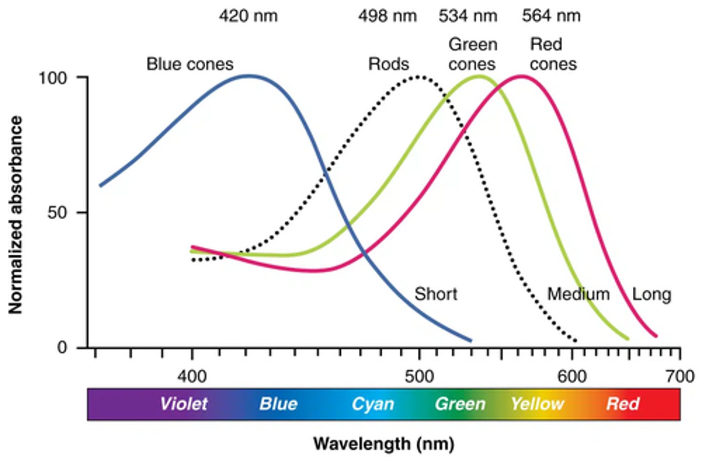
How many opsin cone genes do we have
3 cone genes
- red = 564 nm
- green = 533 nm
- blue = 433 nm
Summarise photoreceptors
Absorb light using 11-cis retinaldehyde (retinal) bound to opsin protein
Photoisomerisation to all-trans retinaldehyde induces structural change in opsin
Activates G-protein signaling cascade
Hyperpolarise and reduce release of glutamate
Rods and cones differ in distribution across retina, opsins expressed, morphology and sensitivity to light
How is a signal conveyed from cones to retinal ganglion cells
Via bipolar cells
What are the 2 types of retinal ganglion cells
on cells are excited by light and fire APs
off cells are inhibited by light and excited by darkness
Describe on and off bipolar cells
On - depolarised by flash
Off - hyperpolarised by flash
this is due to synapse between bipolar cells and RGCs
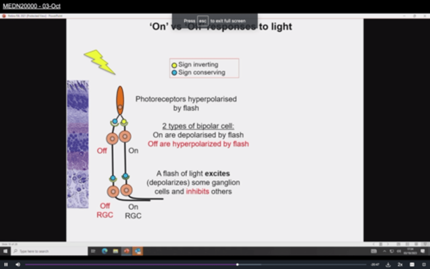
How does an on bipolar cell transmit signals
The on cell has a sign inverting synapse
- hyperpolarisation of the photoreceptors depolarises an on bipolar cell
- this causes depolarisation of the ganglion cell
- this causes firing of action potentials
Which receptors are in sign conserving synapses (off cells)
Ionotropic glu receptors
- cation channels opened by glu
Which receptors are in sign inverting synapses (on cells)
Metabotropic glu receptors
- glu activates signalling cascade closing ion channels
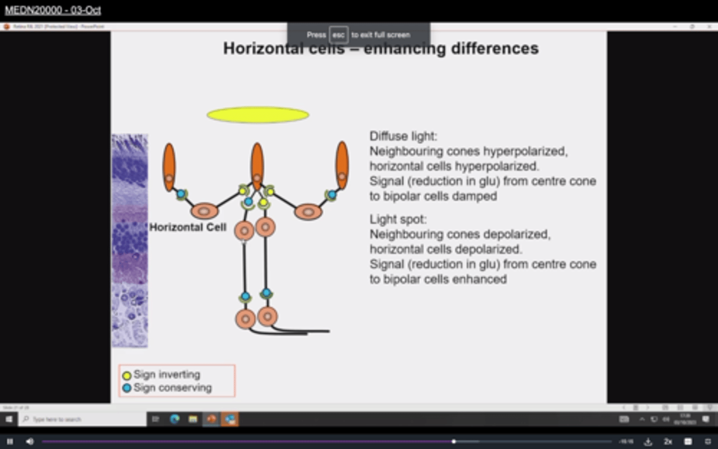
What are horizontal cells
- link cones horizontally in a region of the retina
- inputs from local cones are sign conserving and hyperpolarised by light
- outputs which are sign inverting antagonise the light response
What are horizontal cells?
-cells that extend across the outer portion of the retina at the level of the synapses between photoreceptors and bipolar cells
-can facilitate or inhibit communication between photoreceptors and ganglion cells, thereby altering the sensitivity of the retina
- horizontal cells are much more responsive when there is light in the middle
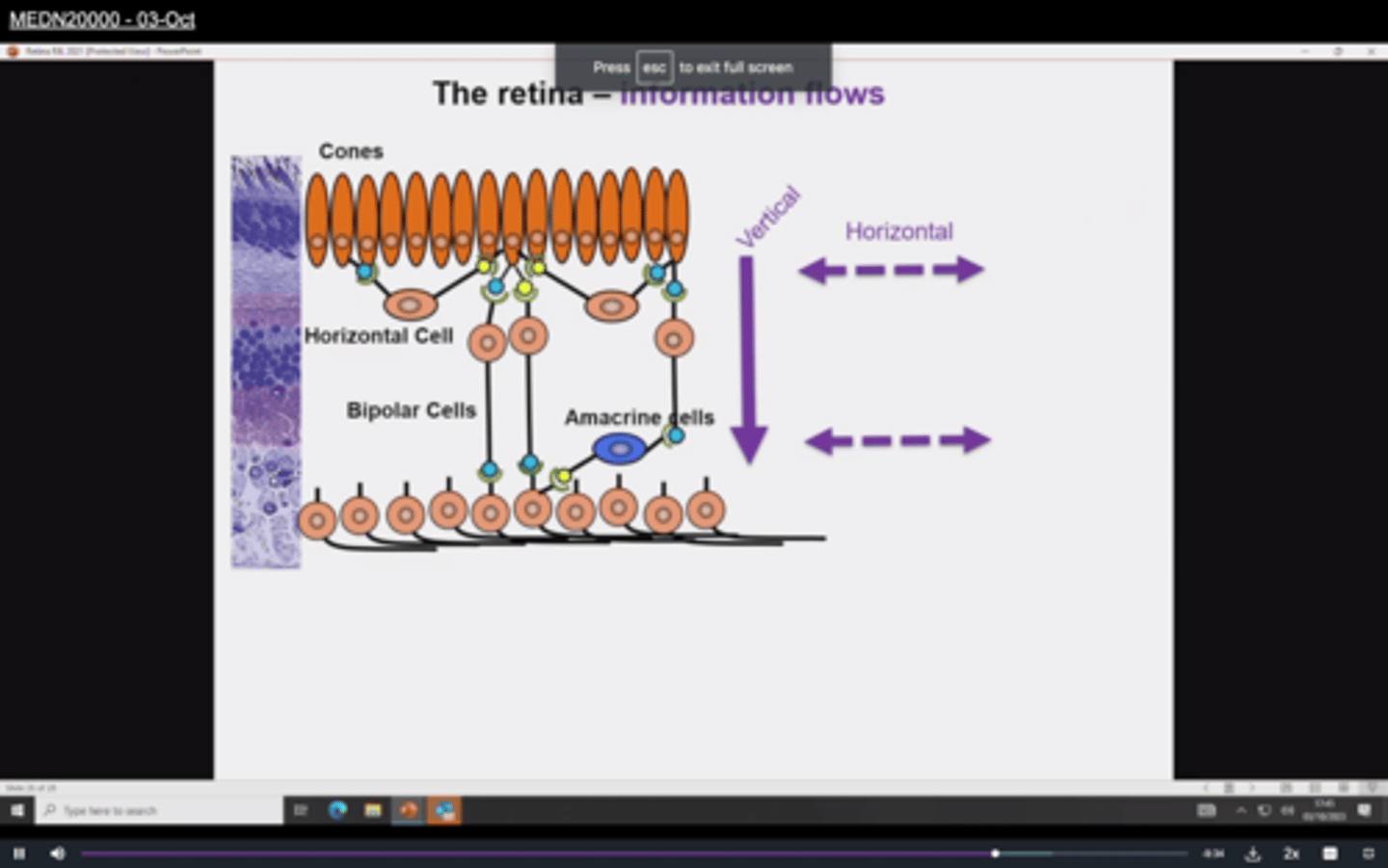
What are amacrine cells
- Where bipolar cells synapse with ganglion cells
- inhibitory link causes response modulation at the inner plexiform layer (bipolar to ganglion cell synapse)
What causes modulation at outer plexiform layer
Horizontal cells
modulates info from photoreceptor to bipolar cell synapse
Overview of flow of information in the retina
How does the optic nerve send signals to the brain
sends multiple simplified versions of the same scene
Where are photoreceptors located
neural layer of the retina (at the back)
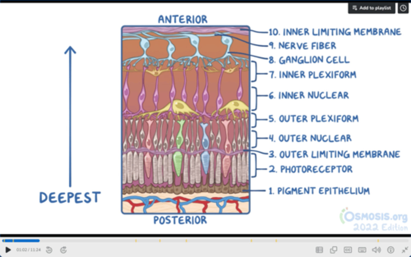
name the layers of the retina from posterior to anterior
1. Pigment epithelium
2. Photoreceptors (rods and cones)
3. Outer limiting membrane
4. Outer nuclear layer
5. Outer plexiform layer
6. Inner nuclear layer
7. Inner plexiform layer
8. Ganglion cell layer
9. Nerve fibre layer
10. Inner limiting membrane
Which layer of the retina absorbs the light
Pigmented layer
Function of outer limiting membrane
Provides mechanical support to retina - helps to maintain structure
What does outer nuclear layer consist of
cell bodies and nuclei of photoreceptor cells
What does outer plexiform layer consist of
Axons of photoreceptors synapse with bipolar cells
What is the inner nuclear layer composed of
Cell bodies of inter neurons which connect outer and inner plexiform layer
(bipolar cells)
What happens in inner plexiform layer
Axons of interneurons synapse with dendrites of ganglion cells
What happens in ganglion cell layer
Ganglion cell bodies reside
What is in the nerve fibre layer
Axons of ganglion cells are located
- axons travel to posterior centre of retina
- exit eye through optic disc
- form optic nerve (CN II)
- carries visual info to the brain
What is the purpose of internal limiting membrane
Separates retina from vitreous humour of eyeball
What are rods and cones
photoreceptors
- modified neurons
- hyperpolarise upon light stimulus to deliver impulse to brain
Structure of photoreceptors
3 segments
- Synaptic terminal: connects to interneurons such as bipolar cells
- Inner segment: cell body
- Outer segment: Detects light
Which vision does rhodopsin allow
black and light
What is the area of the eye with highest visual acuity
fovea
- high density of cones
Which vision does photopsin allow
- colour
- bright light vision
- photopsin is very sensitive to light
What are the different types of cones
S cones - blue
M cones - green
L cones - red
Why are rods considered low acuity
Multiple rods converge onto one ganglion cell
- makes the image fuzzy and low res
Why are cones high acuity
each cone connects to its own ganglion cell
- high acuity
Which cells have rhodopsin and which have photopsin
Rhodopsin = rods
Photopsin = cones
Both proteins are
- G-protein coupled receptors
Which layer of retina contains rod cell bodies
Outer nuclear layer
Which layer of retina contains bipolar cells
inner nuclear layer
Red (L) cones are sensitive to which wavelengths
Sensitive to long-wave light
Green (M) cones are sensitive to which wavelengths
medium-wave light
Blue (S) cones are sensitive to which wavelengths
Short wave light
Function of pigment epithelium
adjacent to the choroid, absorbs light to reduce back reflection of light onto the retina
What is in the outer plexiform layer
synapses between axons of photoreceptors and dendrites of intermediate neurons
What is in inner nuclear layer
Cell bodies of bipolar, horizontal, and amacrine cells and muller cells
What are muller cells
Müller cells
- glial cell in retina
- homeostatic and metabolic support of retinal neurons
What is in ganglion cell layer
cell bodies of ganglions
What is in optic nerve fibre layer
axons of ganglion cells
Describe fibrous layer of eye
cornea and sclera
avascular
Describe uvea/middle/vascular layer
iris, ciliary body, choroid
- drained by sup/inf ophthalmic veins into cav sinus
2 layers of retina
- pigmented layer (outer)
- neural layer
Visual pathway steps
SOOLOP
Second cranial nerve
Optic chiasm
Optic tract
LGN (thalamus)
Optic radiation
Primary visual cortex
Which nuclei controls eye reflexes
superior colliculi in brainstem
Is contraction sympathetic or parasymoathetic
- Contraction upon exposure to light - parasympathetic
- Dilation in dark - sympathetic
Describe pupillary light reflex
- fibres synapse in superior colliculi of brainstem
- fibres travel from nasal and temporal sides
- reach pretectal nucelus of superior colliculus and decussate
- travel to EDW nucleus
- PNS fibres arise from EDW nucleus to ciliary ganglion
- short ciliary nerve arises and causes constriction via CN3
What is functional model of disability
caused by deficits and limits ability to do functional activity
Social model
disability is caused by barriers of social environment
Medical model disability
Disability caused by health conditon