Digestive Microanatomy, Model, & Experiment
1/18
There's no tags or description
Looks like no tags are added yet.
Name | Mastery | Learn | Test | Matching | Spaced |
|---|
No study sessions yet.
19 Terms
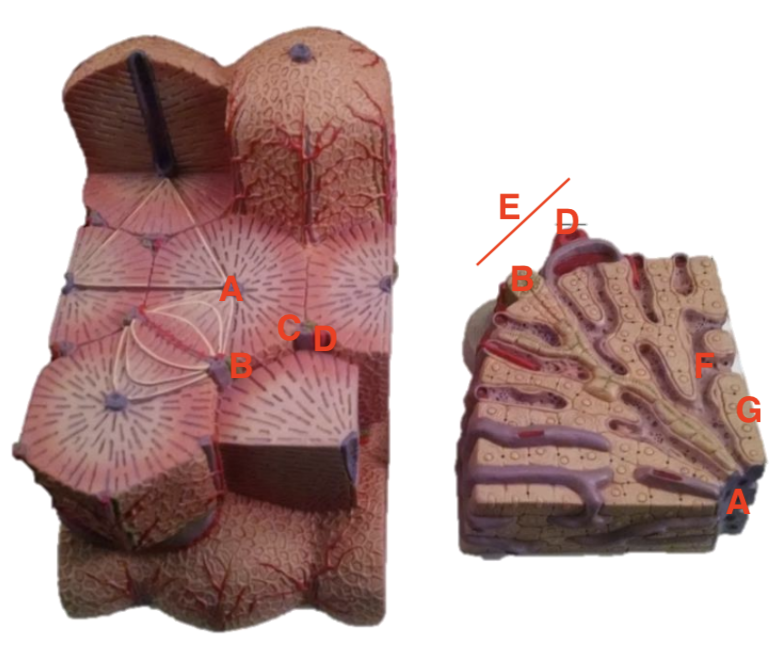
Label parts of a hollow organ. [mucosa, submucosa, muscularis externa (circular & longitudinal layers), serosa, muscularis mucosa, peyer’s patch]
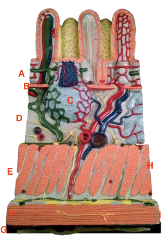
a = mucosa
b = muscularis mucosa
c = peyer’s patch
d = submucosa
e = muscularis externa
f = X
g = serosa
h = circular layer
i = longitudinal layer
Label parts of a hollow organ. [villus, lacteal, goblet cell, intestinal crypt, intestinal gland, enteric neuron]
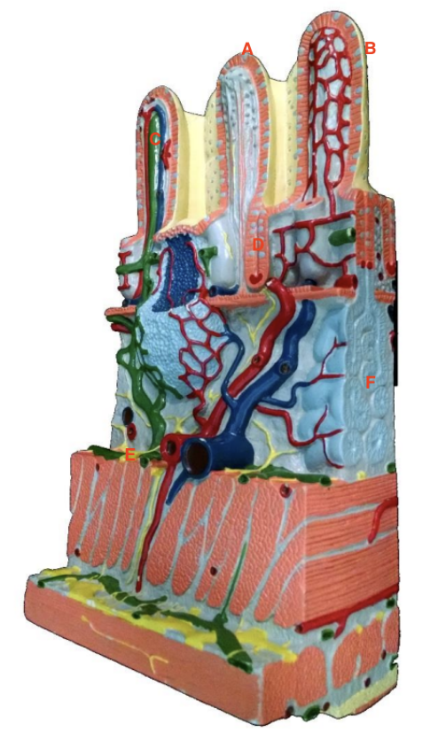
a = goblet cell
b = villus
c = lacteal
d = intestinal gland
e = enteric neuron
f = intestinal crypt
Label components of the oral cavity. [uvula, gingiva, oral vestibule, hard palate, soft palate]
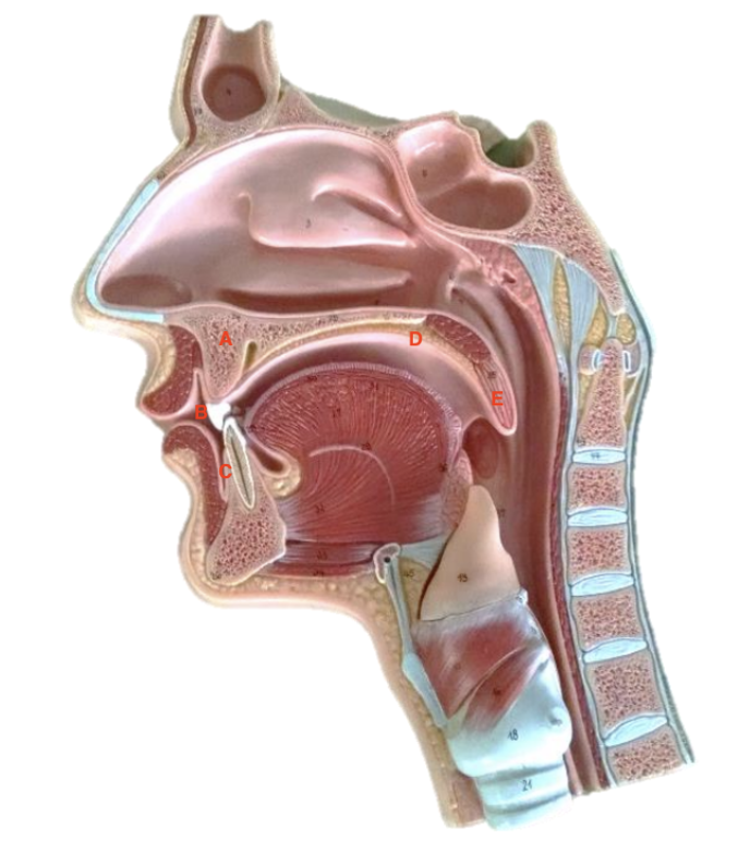
a = hard palate
b = oral vestibule
c = gingiva
d = soft palate
e = uvula
Label parts of a tooth. [enamel, dentin, cementum, pulp cavity, apical foramen, root canal, crown, root]
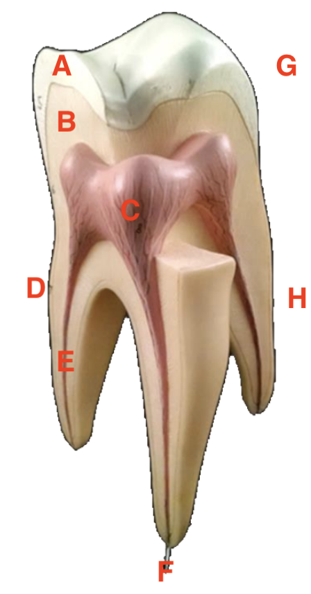
a = enamel
b = dentin
c = pulp cavity
d = cementum
e = root canal
f = apical foramen
g = crown
h = root
Label the salivary glands.
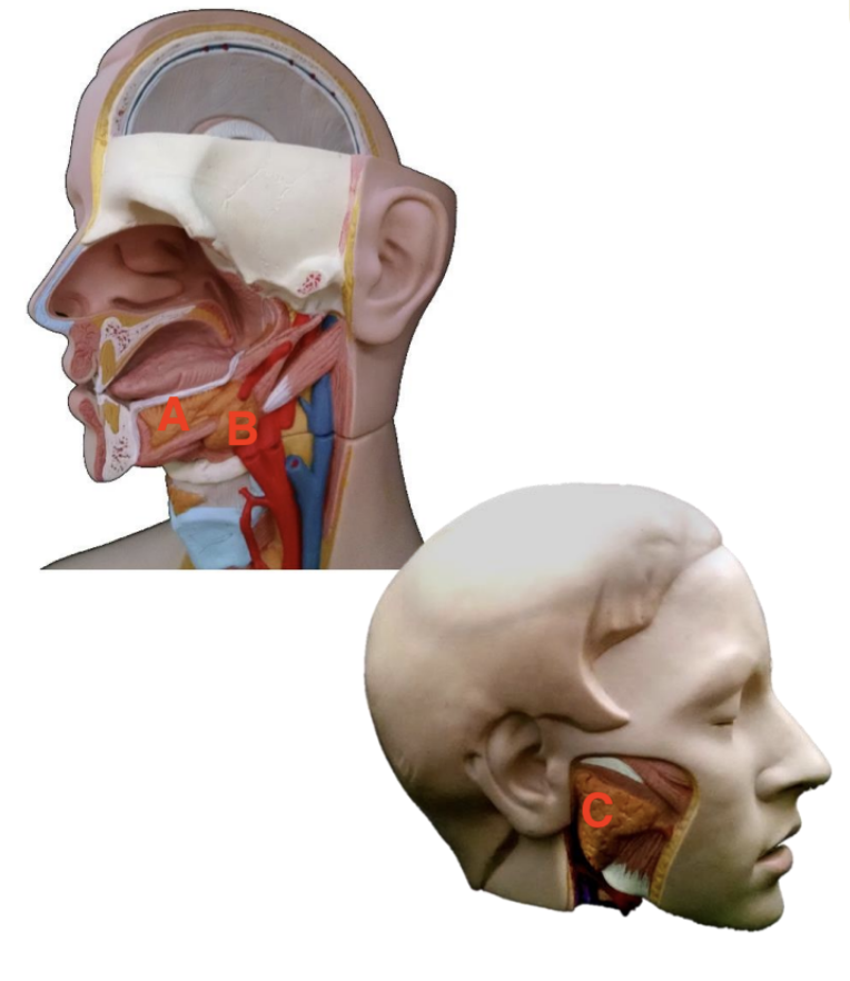
a = sublingual
b = submandibal
c = parotid
Label parts of the stomach. [fundus, body, cardiac region, pyloric canal, pyloric antrum, pyloric sphincter, cardiac sphnicter, lesser curvature, greater curvature]
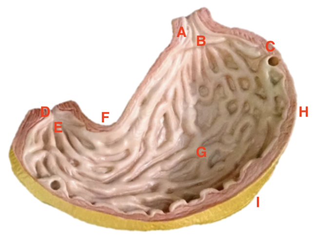
a = cardiac sphincter
b = cardiac region
c = fundus
d = pyloric sphincter
e = pyloric canal
f = lesser curvature
g = pyloric antrum
h = body
i = greater curvature
Label the 4 types of teeth.
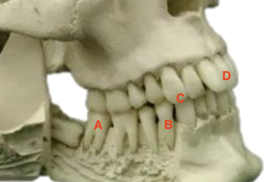
a = molar
b = premolar
c = canine
d = incisors
Label parts of the liver. [right lobe, left lobe, gall bladde, hepatic portal vein, hepatic artery, inferior vena cava, falciform ligament]
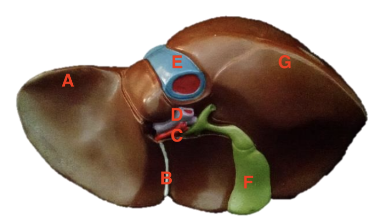
a = right lobe
b = falciform ligament
c = hepatic artery
d = hepatic portal vein
e = inferior vena cava
f = gall bladder
g = left lobe
Label path of bile and pancreatic secretions. [right hepatic duct, left hepatic duct, cystin duct, common bile duct, common hepatic duct, pancreatic duct, accessory pancreatic duct, hepatopancreatic ampulla, pancreas, duodenum, gall bladder]
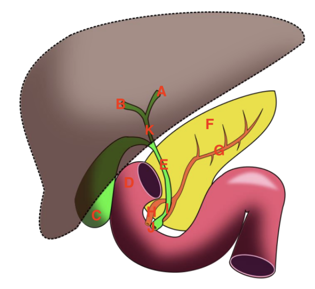
a = left hepatic duct
b = right hepatic duct
c = gall bladder
d = duodenum
e = cystic duct
f = pancreas
g = pancreatic duct
h = accessory pancreatic duct
i = common bile duct
j = hepatopancreatic ampulla
k = common hepatic duct
Label the lower abdomen. [stomach, duodenum, jejunum, ileum, colon]
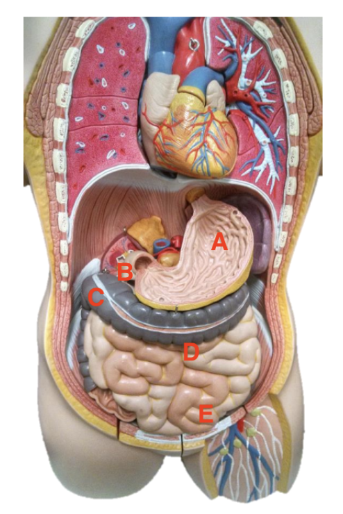
a = stomach
b = duodenum
c = colon
d = jejunum
e = ileum
Label parts of the colon. [cecum, ascending colon, transverse colon, descending colon, sigmoid colon, appendix, mesocolon, hepatic flexure, splenic flexure, haustra, teniae coli]
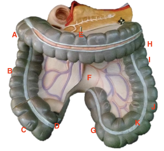
a = hepatic flexure
b = ascending colon
c = cecum
d = appendix
e = transverse colon
f = mesocolon
g = sigmoid colon
h = splenic flexure
i = descending colon
j = haustra
k = teniae coli
Label parts of the rectum. [rectum, anus, rectal valve, external anal sphincter, anal columns]
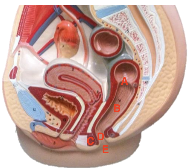
a = rectal valve
b = rectum
c = external anal sphincter
d = anal columns
e = anus
Label histology of the liver. [hepatic triad, central vein, hepatic artery branch, hepatic portal vein branch, bile ductile, hepatocyte, sinusoid]
a = central vein
b = bile ductile
c = hepatic portal vein
d = hepatic artery branch
e = hepatic triad
f = sinusoid
g = hepatocyte
Label histology of the liver. [hepatic triad, central vein, hepatocyte, sinusoid]
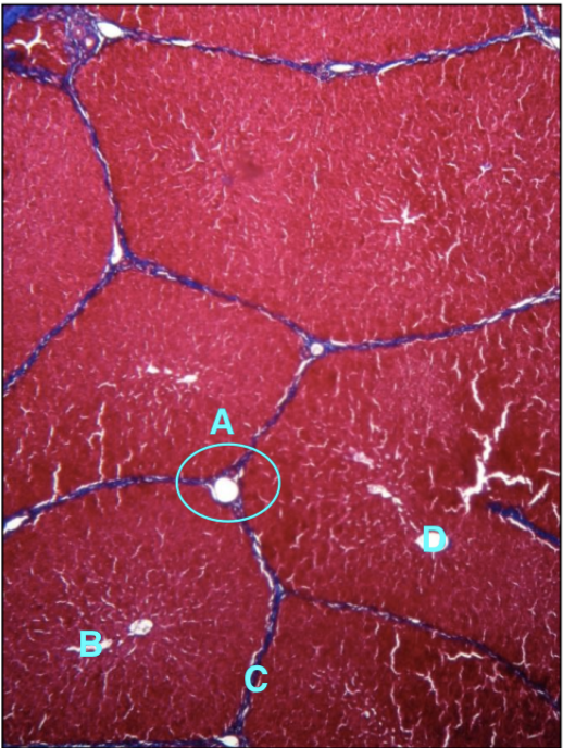
a = hepatic triad
b = sinusoid
c = hepatocytes
d = central vein
Identify and label the tissue.
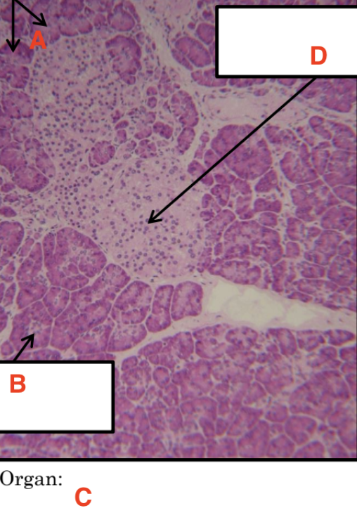
a = lobules (glandular epithelial cells)
b = duct
c = pancreas
d = islets of langerhans
Identify and label this gland.
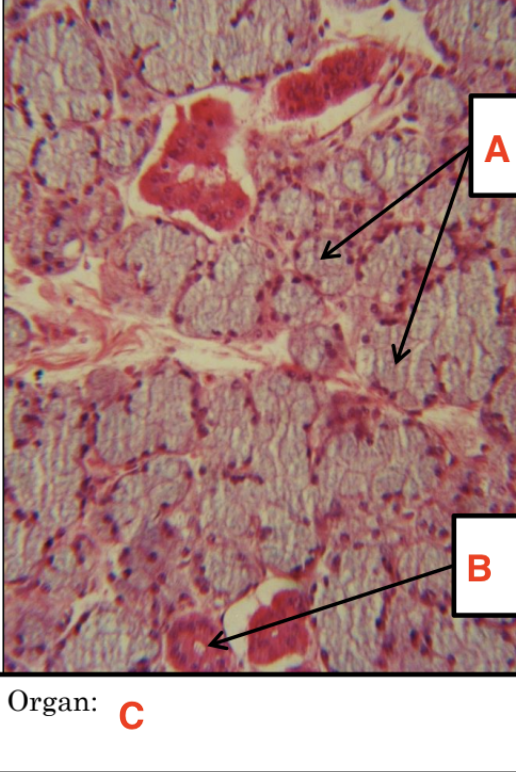
a = mucous acini
b = striated duct
c = submandibular gland - salivary gland
Environment of digestive enzymes
temp. 37 C
< 60 C = denature, no longer functioning properly
> 0 C = decrease to no activity
pH sensitivity
indicates where within pathway of digestion enzyme primarily functions
Environment of digestive enzymes
temp. 37 C
< 60 C = denature, no longer functioning properly
> 0 C = decrease to no activity
pH sensitivity
indicates where within pathway of digestion enzyme primarily functions
Carbohydrate digestion
Polysaccharide
starch
enzyme
amylase
Disaccharide
maltose/dextrose
Monosaccharide
glucose
Carbohydrate digestion
Polysaccharide
starch
enzyme
amylase
Disaccharide
maltose/dextrose
Monosaccharide
glucose
Digestion results
IKI color test = blue/black
starch present = didn’t breakdown!
Benedict’s solution = orange/yellow/green
reduced sugar = breakdown occurred!
Digestion results
IKI color test = blue/black
starch present = didn’t breakdown!
Benedict’s solution = orange/yellow/green
reduced sugar = breakdown occurred!