Higher Cortical functions
 Call Kai
 Learn
Learn Practice Test
 Spaced Repetition
 Match
Match Flashcards
 Knowt Play
1/33

There's no tags or description
Looks like no tags are added yet.
Name | Mastery | Learn | Test | Matching | Spaced | Call with Kai | Chat |
|---|
No analytics yet
Send a link to your students to track their progress
34 Terms
Define:
Aphasia:
Paraphasia:
Neologism
Prosody
Anomia
Definitions:
Aphasia:
acquired disturbance of language function resulting from damage to CNS
Paraphasia:
production of incorrect word
via substitution of phonemes or words w/ correct word
Neologism
paraphasic error where a novel word is produced
Prosody
melodic contour of speech
Anomia
difficulty in accessing names of objects
Describe the relationship between Language Dominance vs Hand dominance:
Anatomical Proof?
Laboratory Proof?
Language Dominance vs Hand dominance:
Right handed: 99% Left Hemi Dominance
Left Handed: 70% Left Hemi Dominance
Anatomical Proof:
Seen in size differences the two Planum temporale
Laboratory Proof:
Wada Procedure:
Sodium amytal injection into 1 (R or L) carotid artery -> put hemisphere to sleep
Test Pts. Language capabilities
Which ever hemisphere provoke language difficulties = language dominance
Also, Which ever hemisphere provokes Contralateral paralysis/sensory loss
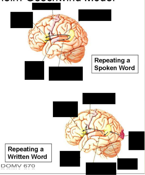
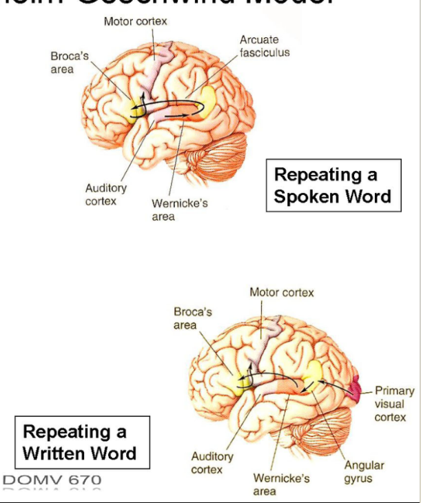
List the Neurological Locations of Language and Function
Neurological Locations of Language:
Perisylvian structures
Broca's area
motor speech
Wernicke's area
language comprehension
arcuate fasciculus
Links the two
Angular Gyrus:
Involve in comprehension of Written speech
Info transferred from visual cortex
Draw out the Wernicke-Lichtheim Schema

List the Exact Locations of language implementation
Broca’s
Wernicke's
Other
List the Neural Substrates for conceptual knowledge
What is Conceptual Knowledge:
Locations that mediate language implementation/conceptual systems:
Exact Locations of language implementation :
Broca’s:
Inferior frontal operculum
BA 45, 44
Wernicke's
left posterior superior temporal gyrus
BA 22 (posterior)
Other regions in Left Perisylvian
Inferior parietal lobule
insula
subadjacent white matter
arcuate fasciculus
Neural Substrates for conceptual knowledge
What is Conceptual Knowledge:
Association of word forms w/ meaning
Locations that mediate language implementation/conceptual systems:
Posterior sensory association cortices,
left prefrontal cortex,
inferotemporal region
temporal pole
What are the 3 factors for determining aphasias:
3 factors for determining aphasias:
Fluency
Comprehension
Verbatim repetition
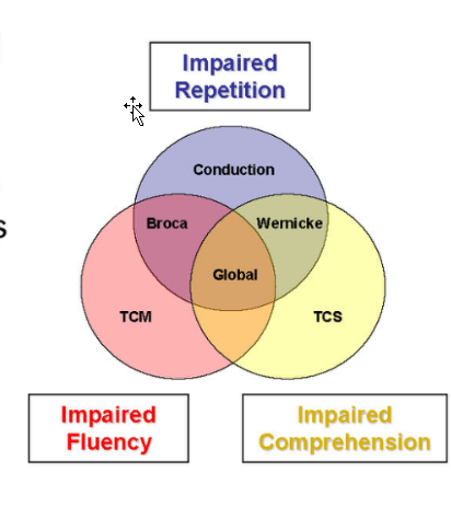
Describe Expressive Aphasia (Broca's aphasia)
Symptoms
Areas Invovled
Expressive Aphasia (Broca's aphasia)
Symptoms:
Impaired speech production (fluency) + repetition
Decreased and labored output
Intact auditory comprehension
Pts aware of disability + frustrated -> use inappropriate or vulgar language
Agraphia
Understanding complex instructions
Hemiparesis + upper motor neuron signs (facial weakness)
Areas involved:
Broca's area (BA 44/45)
left frontal operculum,
underlying white matter
anterior insula.
Describe Receptive Aphasia (Wernicke's aphasia)
Symptoms
Locations
Receptive Aphasia (Wernicke's aphasia)
Symptoms:
impaired auditory comprehension, naming, and repetition
“Everyone seems to mumble”
Fluent speech w/ paraphasia + neologisms
Alexia
Agraphia
Less aware of deficit -> not as frustrated
Contralateral homonymous hemianopia (or quadrantanopia)
Via lesions of Meyer’s loop + Optic Radiation
Locations:
Wernicke's area
nearby regions in temporal and lower parietal cortices
Describe Conduction aphasia
Symptoms
Locations
Conduction aphasia
Symptoms:
Impaired naming and repetition w/ intact comprehension
Fluent paraphasic speech
Locations:
Arcuate Fasciculus
Damage to either supramarginal gyrus (BA 40) in inferior parietal lobe or to primary auditory cortices with extension into insular cortex and subadjacent white matter
Describe Global aphasia
Symptoms
Locations
Describe Transcortical aphasia:
What causes/spares impaired repetition?
Describe Transcortical Motor/Sensory Aphasia
Global aphasia
Symptoms:
all major functions of language are impaired
Poor recovery
Location:
Extensive destruction of left perisylvian region
Transcortical aphasia:
What causes Impaired repetition:
Impaired repetition: damage to perisylvian region
Repetition spared: Damage anterior to Broca's area or posterior to Wernicke's area
Transcortical Motor Aphasia
Resembles Broca's aphasia but repeat verbatim ability is preserved
Transcortical Sensory Aphasia
Resembles Wernicke's aphasia but verbatim repetition is spared
Damage to temporal-occiptal-parietal junction — angular gyrus
Describe Anomic Aphasia
What is it?
Location
List the Verb/Noun retrieval Locations:
List the specific noun locations:
Proper
Common
Animal?
Tools?
Anomic Aphasia
What is it?
impairment of confrontation naming (anomia)
Location:
Left inferotemporal or anterior temporal regions
Noun/Verb retrieval Locations:
Verb:
left premotor/prefrontal region
Noun:
left inferior and anterolateral temporal regions
Specific Noun Locations:
Proper nouns (names)
left temporal polar region (BA 38)
Common nouns
inferotemporal region (BA 20/21; anterior 37)
Animal Names: Ant. Left
Tools: Post. Left
Describe Aprosadia
What is it?
Symptoms?
Location?
Aprosadia
What is it?
Prosody w/o aphasia
Motor or Sensory
Symptoms:
Monotone voice,
inability to express emotion in speech (anger, distress, etc)
Inability to understand prosody of speech, e.g., "You're going home?"
Location:
right hemisphere in areas corresponding to language areas of the dominant hemisphere

Broca’s
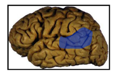
Wernicke’s

Conduction Aphasia

Global Aphasia
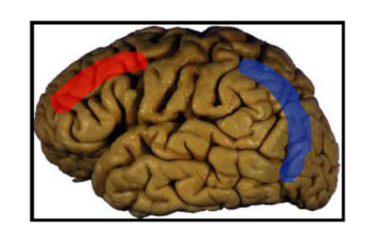
Red = transcortical motor aphasia
blue = transcortical sensory aphasia

Blue = verb retrieval
red = proper nouns
yellow = common nouns
Describe Alexia + Agraphia:
Location
Describe Alexia w/o agraphia
What is it?
Location?
Alexia + Agraphia:
Location:
Dominant Angular Gyrus
Alexia w/o agraphia
What is it?
Pure word blindness
Writing = normal, but pt cannot read it.
disconnection between bilateral visual system and unilateral language system (angular gyrus)
Right hemianopia
color agnosia
Location:
Left visual cortex and splenium of corpus callosum

Alexia + agraphia
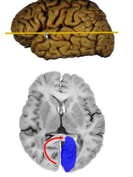
Alexia w/o agraphia
Describe the Magnocellular Stream
Pathway
Function?
Lesions Symptoms
Blindsight?
Magnocellular Stream
Pathway:
Involves lateral geniculate + pulvinar.
Info from RGCs (mostly rods) w/ large receptive fields
Function:
"where" objects are
position,
Motion (objects moving + perception of body moving)
Moving edges detection
Appreciation of motion
depth, and other spatial information.
Higher level processing:
Navigation
Directing eye movement
Motion perception
Suppression of vision during saccades
Lesions Symptoms:
Cannot appreciate object motion
“not being able to cross the street because cars seemed at first far away and then suddenly very near.
People in a room would also suddenly appear in different places.
Blindsight:
In pts w/ qeniculostriate pathways lesions
Lose all conscious vision but can demonstrate perception of movements or changes in light
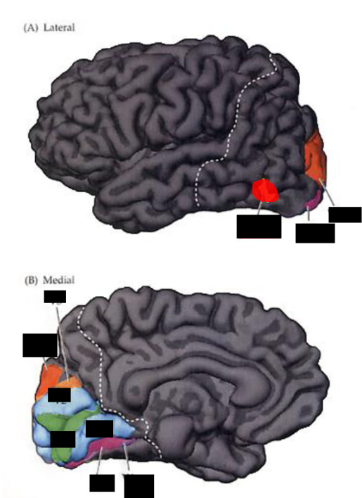


Describe the P stream
Pathway
Function
Lesions/Locations:
Prosopagnosia
cerebral achromatopsia
The P stream (parvocellular, X, blob zone)
Pathway:
Parvocellular (Cone) area of LGN -> V2 -> V4 (A19) -> inferior temporal region -> A 20, 21, 37 of temporal cortex
Function:
“What”
color
Shape
(high) resolution aspects of form
@ higher levels:
Face Recognition
(inferior temporal lobe)
Lesions/Locations:
Bilat lesions of Inferior temporal + fusiform:
Prosopagnosia
Inability of facial recognition
May be associated w/ autism
V4 lesions + adj. Area (occipitotemporal gyrus; fusiform gyrus)
cerebral achromatopsia
World = shades of gray
May affect quadrants (upper), Hemi field, or complete Field
Associated w/ visual field defects or prosopagnosia

Prosopagnosia

cerebral achromatopsia
Describe Balint syndrome
Cause
Symptoms
Balint syndrome
Cause:
Bilat lesions involving parieto-occipital junction
Symptom: triad:
optic apraxia
poor control of eye movements
optic ataxia
poor visually guided movement
ability to point to or reach for an object
Simultapnosia
inability to perceive visual field as a whole

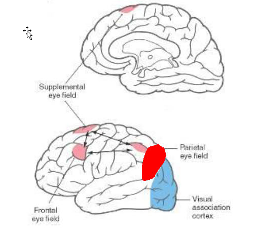
Describe Non dominant Parietal Lesions
Function/ Lesion -
Symptoms
Non dominant Parietal Lesions
Function:
Dominant in directing attentive processing
Lesion = disruption of spatial relationships and selective attention
Symptoms:
contralateral neglect (left spatial/visual field) svndrome
unaware of objects/events in left half of his surrounding space, including own body (asomatoapnosia)
dressing apraxia
“staff get the ‘other person" out of the bed
Constructional apraxia
Inability to successfully manipulate objects in space or to duplicate a simple block construction
Note:
Dom. parietal = language
Describe Prefrontal Cortex lesions
Function
dx difficulty
Old ways to treat?
Specific locations/functions:
Dorsolateral prefrontal region
Ventral medial Region (includes? )
Superior mesial region (includes? )
Frontal Lobe and emotion:
Left vs right lobes
Clinical Associations:
Prefrontal Cortex lesions
Function:
human intellectual traits
Judgment. Foresight, purpose, responsibility, social propriety
Diagnosis difficulty:
Subtle lesions hard to detect
Old ways to treat:
Prefrontal lobotomy
Ice pick procedure
Specific locations/functions:
Dorsolateral prefrontal region
Working memory;
intellectual capacities;
creative, flexible thinking;
fluency:
L — verbal, R - nonverbal
Ventral medial Region (includes medial orbitofrontal cortex)
Judgment,
planning,
decision making,
social conduct
Superior mesial region (includes anterior cingulate gyrus)
emotional behavior
Basic drive states
associated w/ motivation and arousal
Frontal Lobe and emotion:
Left vs right lobes
Left = positive affects;
Right = Negative affects
Clinical Associations:
Depression incidence + severity = higher in pts w/ left anterior lobe lesion
Right lesion = inappropriately cheerful
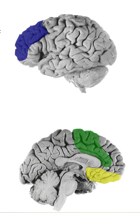
Blue: Dorsolateral prefrontal region
Yellow: Ventral medial Region (includes medial orbitofrontal cortex)
Green: Superior mesial region (includes anterior cingulate gyrus)
Describe Function in Split Brain patients
Stereognosis ability
R vs L Visual Fields
Function in Split Brain patients
Stereognosis ability
Right Hand:
Pt can name object
Left Hand:
Can indicate object’s use
Right vs Left Visual field
Right:
Read word or ID picture verbally
Left:
Show emotional response to humorous object