GI 2 - Hepatic doppler
 Call Kai
 Learn
Learn Practice Test
 Spaced Repetition
 Match
Match Flashcards
 Knowt Play
1/25

There's no tags or description
Looks like no tags are added yet.
Name | Mastery | Learn | Test | Matching | Spaced | Call with Kai | Chat |
|---|
No analytics yet
Send a link to your students to track their progress
26 Terms
Normal flow direction in portal vein and hepatic artery
Hepatopetal, toward liver
Normal flow pattern in portal vein
Just above baseline, gently undulating but overall monophasicitt undulerende, men monofasisk.
Normal flow pattern in hepatic artery
Typical arterial peaks with lowest pressure during end-diastole.
Normal RI in hepatic artery
RI 0.55-0.7
Normal velocity in portal vein
16-40 cm/s
Normal flow pattern in levervenene? Describe/draw it
When above baseline: retrograde / hepatopetal / toward usg probe.
When below baseline: anterograde / toward heart/hepatofugal / away from usg probe.
A-wave: Atrial contraction, retrograde/hepatopetal flow
S-wave: Ventricular systole, large volumes of blood return to atrium. Anterograde flow.
V-wave: Atrial overfilling, followed by tricuspid valve opening (peak V-wave) and early diastolic filling of right ventricle. Peak should be just below baseline.
D- wave: Continued fillig of right ventricle during ventricular diastole, some blood flows retrograde to right atrium.
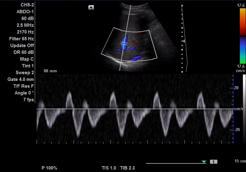
Pattern in portal vein. What do you suspect?
Pulsatile flow in portal vein is always pathologic and should rise suspicion of right-sided heart failure and/or tricuspid regurgitation. This picture is of tricuspid regurgitation.
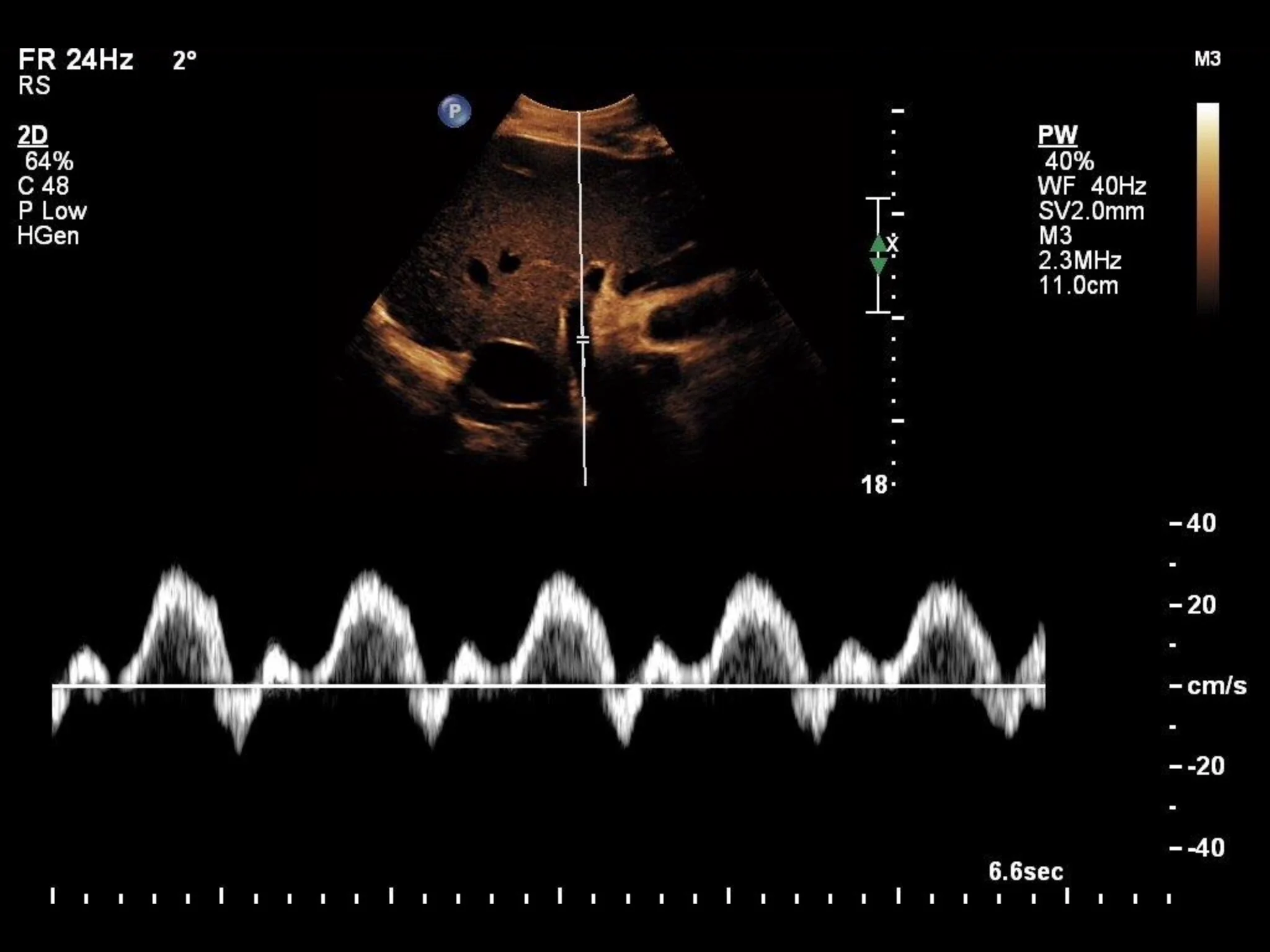
Portal vein. What do you suspect?
Prominent diastolic flow and weak systolic flow. Differential includes right sided heart failure and tricuspid regurgitation. In this case, tricuspid regurgitation.
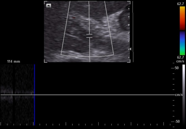
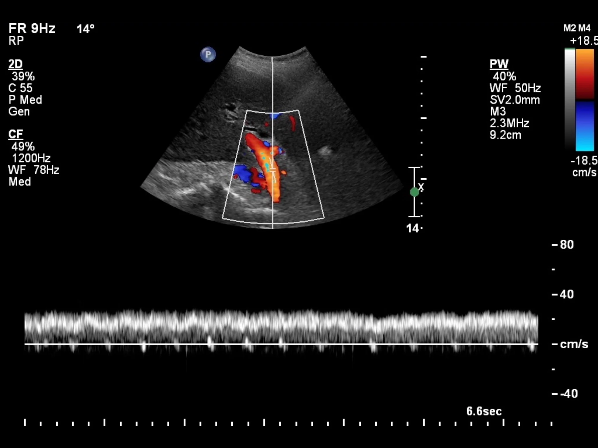
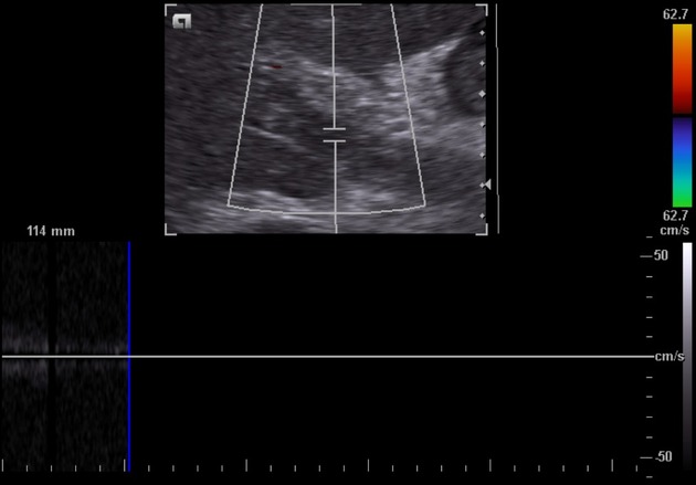
Portail vein. Describe findings and what you suspect
Absent flow. Portal vein thrombosis.
Common etiologies of portal vein thrombosis in INFANTS
Dehydration and omphalitis
Differential diagnosis in abscent/weak flow portal vein
Portal hypertension may mimic portal vein thrombosis due to absent/stagnant flow!
How to differentiate pneumobilia from portal venous gas?
Pneumobili: Tends to lay centrally
Portal venous gas: tends to involve peripheral segments. Color doppler will reveal numerous spikes.
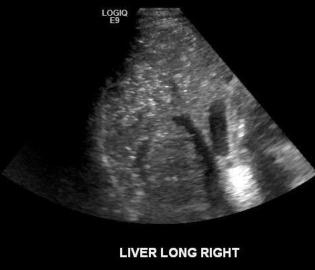
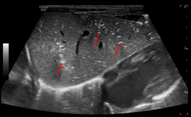
What do you suspect, and what is an important etiology to keep in mind?
Portal venous gas. OBS! Bowel ischemia/infarction.
Definition of portal pressure
Direct portal venous pressure of >5 mm Hg, although not measured directly.
Describe mechanism of portal hypertension
When portal pressure is higher than forward pressure, the portal flow may decrease below 16 cm/s or reverse (hepatofugal). This is diagnostic for portal hypertension.
How to classify portal hypertension
According to location relative to hepatic capillary bed
Pre-sinusoidal: proximal to liver parenchyma, e.g. portal vein thrombosis
Sinusoidal: Hepatic insult, e.g. cirrhosis
Post-sinusoidal: Budd-Chiari og VCI thrombosis.
Sonographic findings in portal hypertension
Portal vein flow below 16 cm/s
Hepatofugal flow in portal vein
Indirect signs of portal hypertension
Dilated portal vein > 13 mm, varices, splenomegaly, Gamma-Gandy bodies, MULTUPLE portosystemic shuntes (astroesophageal is the most common, paraumbilical is pathognomonic, splenorenal also possible, + more).
Treatment for portal hypertension
TIPS. Transjugular intrahepatic portosystemic stent, artificial shunt from branch of portal vein to branch of hepatic veins.
Normal velocity in TIPS
90-190 cm/s
Normal flow direction in TIPS?
Flow in TIPS - toward hepatic veins/VCI.
Flow in main portal vein - hepatopetal, towards TIPS
Flow in right and left portal veins - hepatofugal, towards TIPS
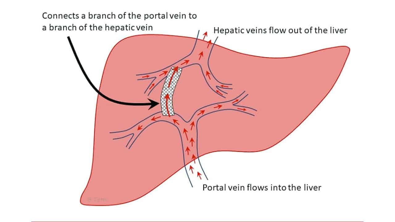
Findings suspicipus of TIPS stenosis
Velocity in portal vein <30 cm/s
Change in baseline velocity in TIPS ± >50 cm/s
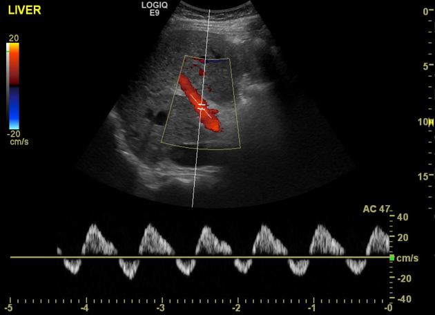
Pathology in INCREASED pulsatility hepatic veins.
Right sided heart failure and tricuspid insufficiency
Pathology in DECREASED pulsatility of hepatic veins
Stenosis, compression due to cirrhosis, Budd-Chiari, hepatic VOD
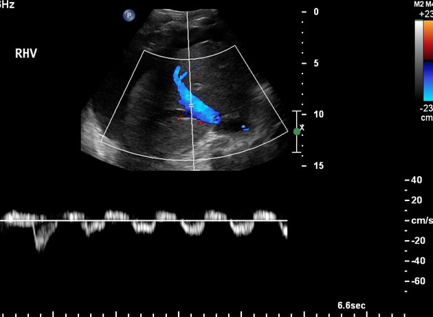
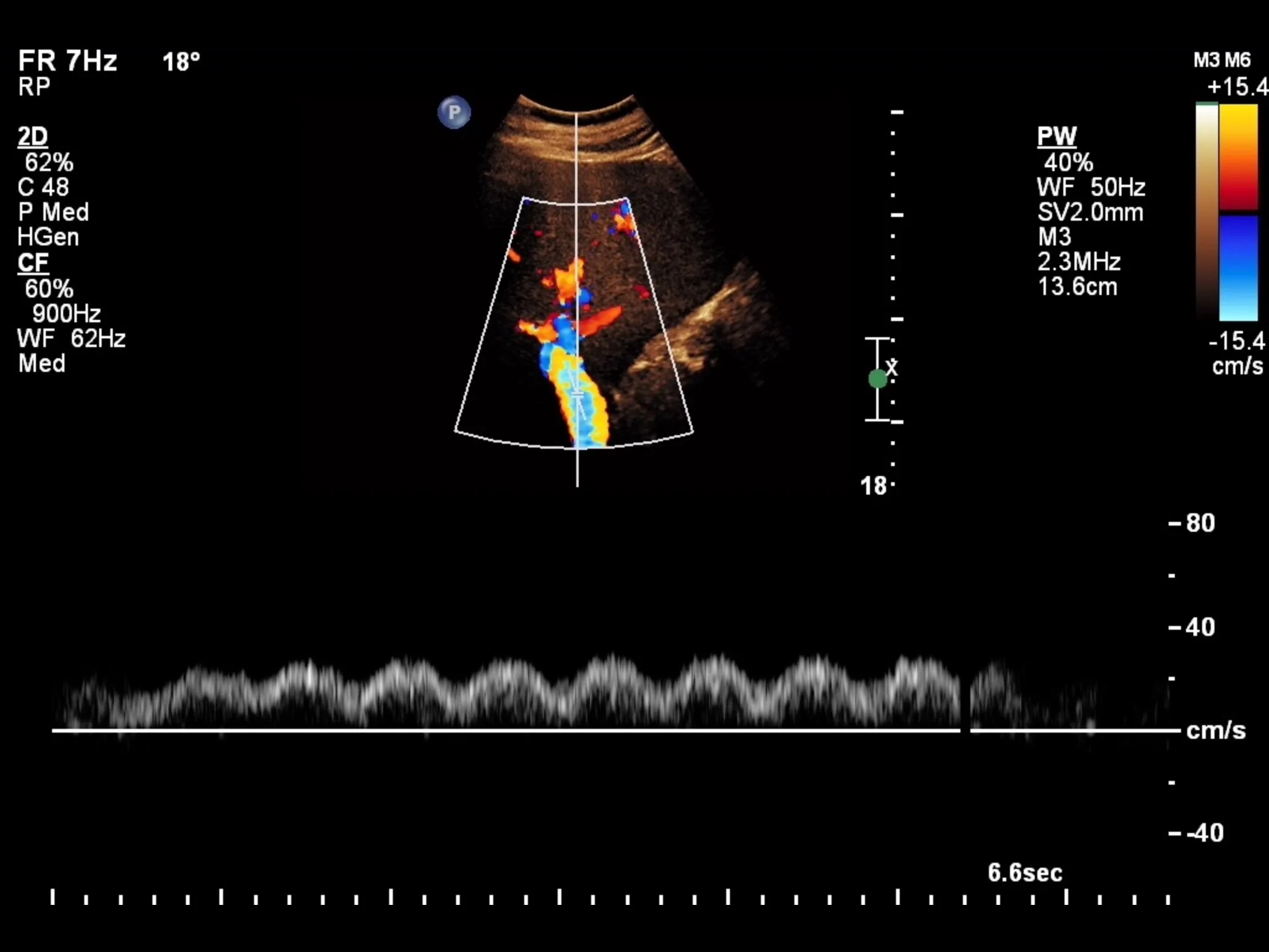
Imaging of hepatic veins. What are the findings and suspected diagnoses.
Reduced pulsatility in hepatic veins. Diff. diagnosis incl. stenosis, cirrhosis, Budd-Chiari, hepatic VOD
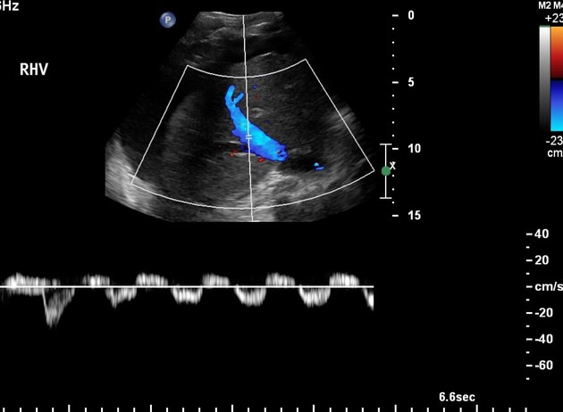
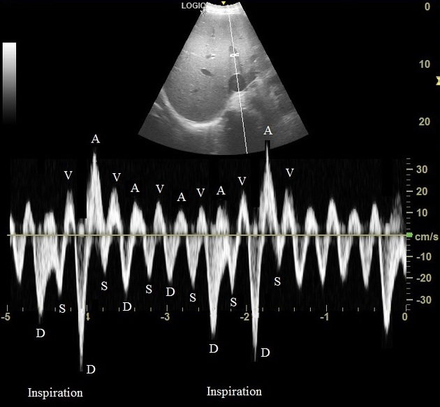
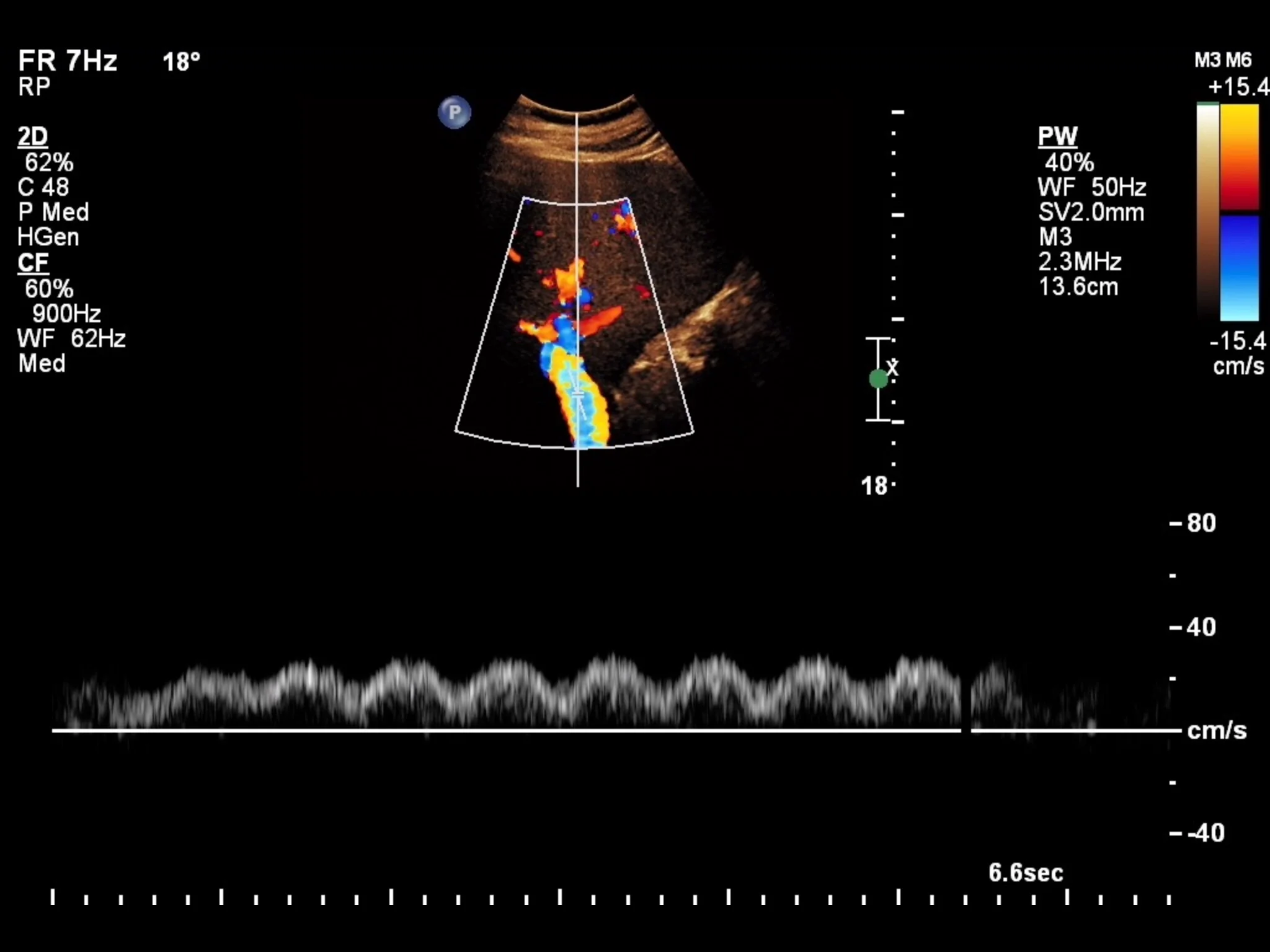
Imaging of hepatic veins. What are the findings and suspected diagnoses.
Increased pulsatility in hepatic veins. Diff. diagnosis incl. right sided heart failure and tricuspid insufficiens