MIC 102
1/71
Earn XP
Description and Tags
MD #1 Content
Name | Mastery | Learn | Test | Matching | Spaced | Call with Kai |
|---|
No analytics yet
Send a link to your students to track their progress
72 Terms
Light Microscopy
Magnifies up to 1000x ; resolve structures around 200 nm apart ; Brightfield, Phase contrast, and Fluorescence are common techniques
Brightfield
To view pigmented or stained specimens (High Contrast)
Phase Contrast
To view non-pigmented (low contrast) specimens
Fluorescence
To view cells/ structures labeled with a fluorochrome
Electron Microscopy
Magnifies up to 500,000x; can resolve structures, estimated 0.1 nm apart; including Scanning, Transmission
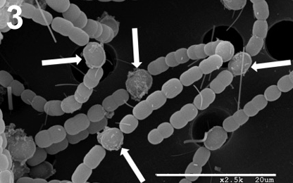
Scanning EM
Scans the exterior of a specimen to reveal the topography and fine details; requires processing of the specimen
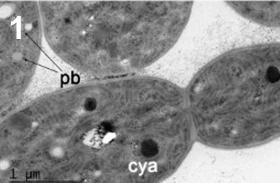
Transmission EM
Used to view the internal structures of (embedded, sliced, stained) specimens or view small specimens
How did diverse life forms arise?
Mutations, Horizontal gene transfer (between same generation), vertical gene selection(inheritance, parent, to offspring), selection
Overview of Life Forming Chart
Hadean| Archean| Proterozoic| Phanerozoic
Anaerobic bacteria and archaea ( O2 is toxic for organisms)
Cyanobacteria producing O2 → buildup of it
O2 in the atmosphere because of a buildup→ killing organisms
Aerobic bacteria→ Selection to be able to survive oxygen
Unicellular eukaryotes: significantly diverse and able to survive/ use oxygen
Multicellular eukaryotes- Plants and animals, hominidsand
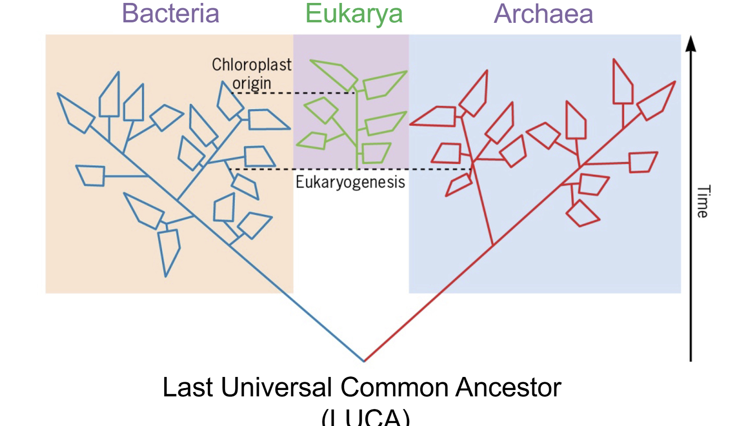
Phylogenetic “ Tree of life” with 3 domains
Bacteria , Archaea, and Eukarya which diverted from archaea with the engulfment of bacteria
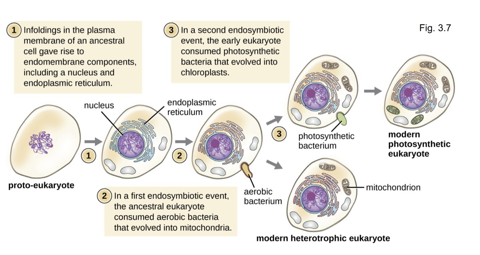
How did the eukaryotic cell arise?
Endosymbiotic theory: An ancient endosymbiosis gave rise to organelles such as the mitochondrion and chloroplast and possibly other over time
Organellogenesis
Formation of organs during embryotic development
Evidence that mitochondria and chloroplasts are derived
size
single circular chromosome with similar genes
70s ribosomes & Methionine (formyl-met)
Division by binary fission using homologous machinery( FtsZ rings)
Classifying bacteria and archaea
def: group of individuals that can reproduce naturally and produce fertile offspring
→ But this doesn’t work for prokaryotes because their asexual reproduction
So, classification is based on metabolism or physiology is possible for organisms that can be cultures
Prokaryotic Cells Structure
Cell envelope: Membrane + layers leading to environment
External Structure
Internal Structures
Nucleoid
Inclusions
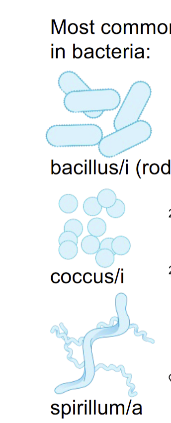
3 Common Morphology of Bacteria
Bacillus | Coccus | Spirllium
Bacterial and Archaeal Cell - Envelope
capsule, slime layer
s-layer
( other membrane with lipopolysaccharides)
( PG- associated lipoprotein)
(teichoic acid)
cell wall( peptidoglycan, PG)
Cell membrane
() means its only for some
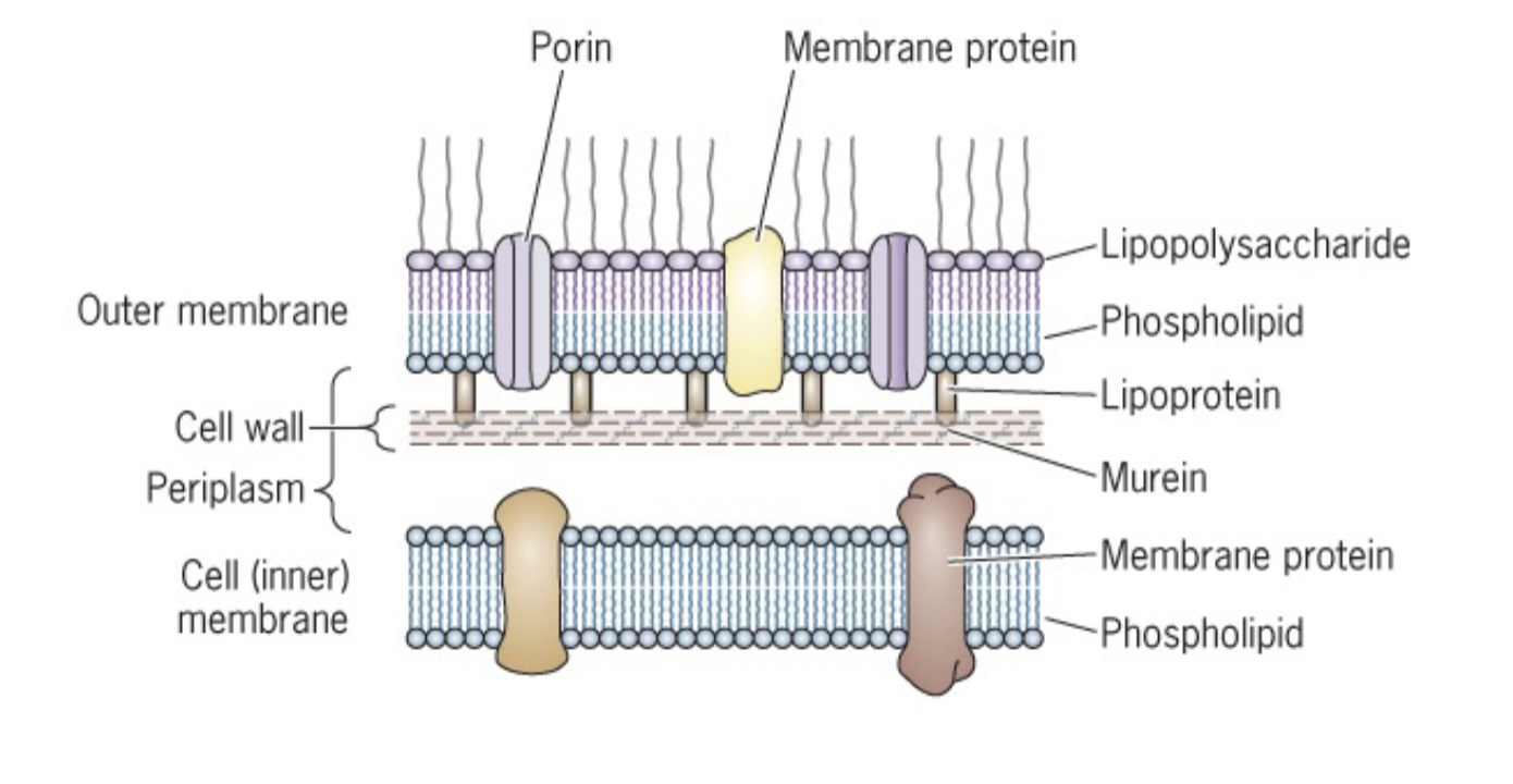
Gram- Negative Bacterial Envelope
Cell membrane (CM)
Peptidoglycan( PG)→ Cell Wall Material, mesh of peptide and glycan( sugars) → thin
Outer membrane
lipopolysaccharides(LPS)
PG- associated Lipoprotein (LP)→ Covalently binds to peptidoglycan and outer membrane which binds them together
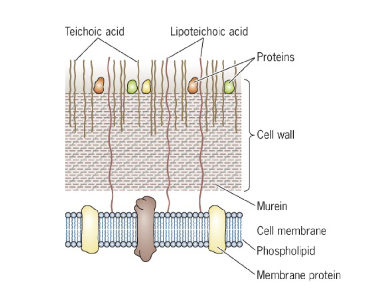
Gram- Postiive Bacterial Enevelope
Cell Membrane( CM)
Peptidoglycan( PG)
Teichoic Acid ( TA): Stabilizes layers or anchors weaving in and out of peptidoglycans due to x10 layers of it
doesn’t have an outer membrane, so it doesn’t need L,P which binds PG and Outer Membrane
Two solutes that would not cross a phospholipid bilayer by simple diffusion
Proton: Charged
Monosaccharides: Large + Polar which is repelled by nonpolar phosolipid tails which are nonpolar as well
Osmosis
Net movement of water from an area of higher concentration to an area of lower water concentration
Turgor Pressure
Pressure inside cell that pushes on the membrane
Osomotic Pressure
Pressure that must be applied to the solution side to stop fluid movement across a semi-permeable membrane
Hypertonic environment
Loss of cytoplasm volume (Plasmolysis)
Excess solubility outside of cell . water moving out / Shirvling inside
Hypotonic environment
Higher concertation inside cell
Water moving inside
Fills up until cell membrane constrains it
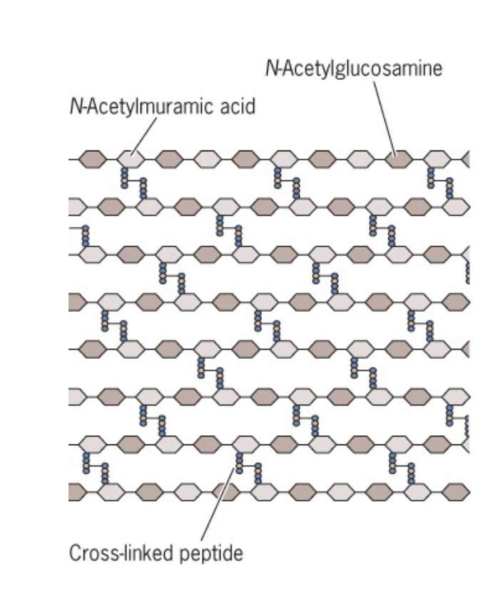
Peptidoglycan cell wall(Peptidoglycan = murein) Components - gram neg
N-Acetylglucosamine (NAG)
N-Acetylmuramic acid (NAM)
linked by transglucosylases to form glycan strands
Short peptides attach to NAMs lined by transpeptidases
this creates the multiple layers of peptidoglycan
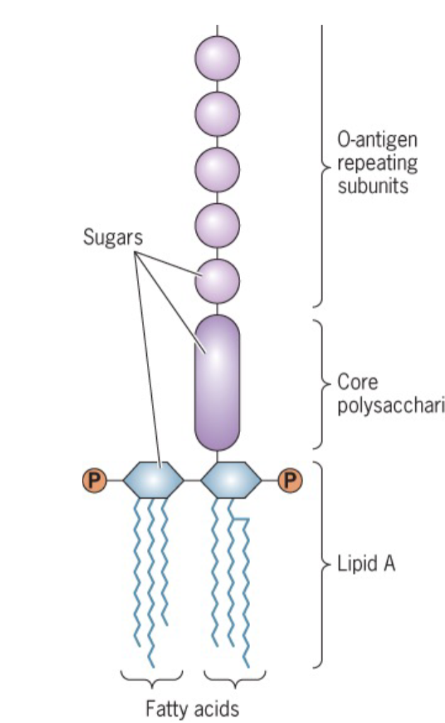
Lipopolysaccharides- gram neg
fatty acids: If person is infected with gram negative, then this is being released and at high concentration leads to shock: also known as endotoxin
o-antigen: immune system good at detecting this
Surface layer( S-layer)
a crystalline layer of protein; in many bacteria and nearly all archaea and it is a reinforcement of the cell and structural stability to cell wall
Glycocalyx : Sugar(glyco) ; Coat( calyx)
Slime layer and capsule; made by many species, as needed
Archaeal Envelope
Inner lipid membrane of isoprenoids linked to glycerol via more stable ether linkage( not ester) this creates more stability and resistance against extreme environmental conditions for archaea
External of Bacteria Cell Structures
Appendages
flagella
pili/fimbriae
Flagella
Movement for bacteria towards better environments/ bacteria can keep moving as long as there is enough energy
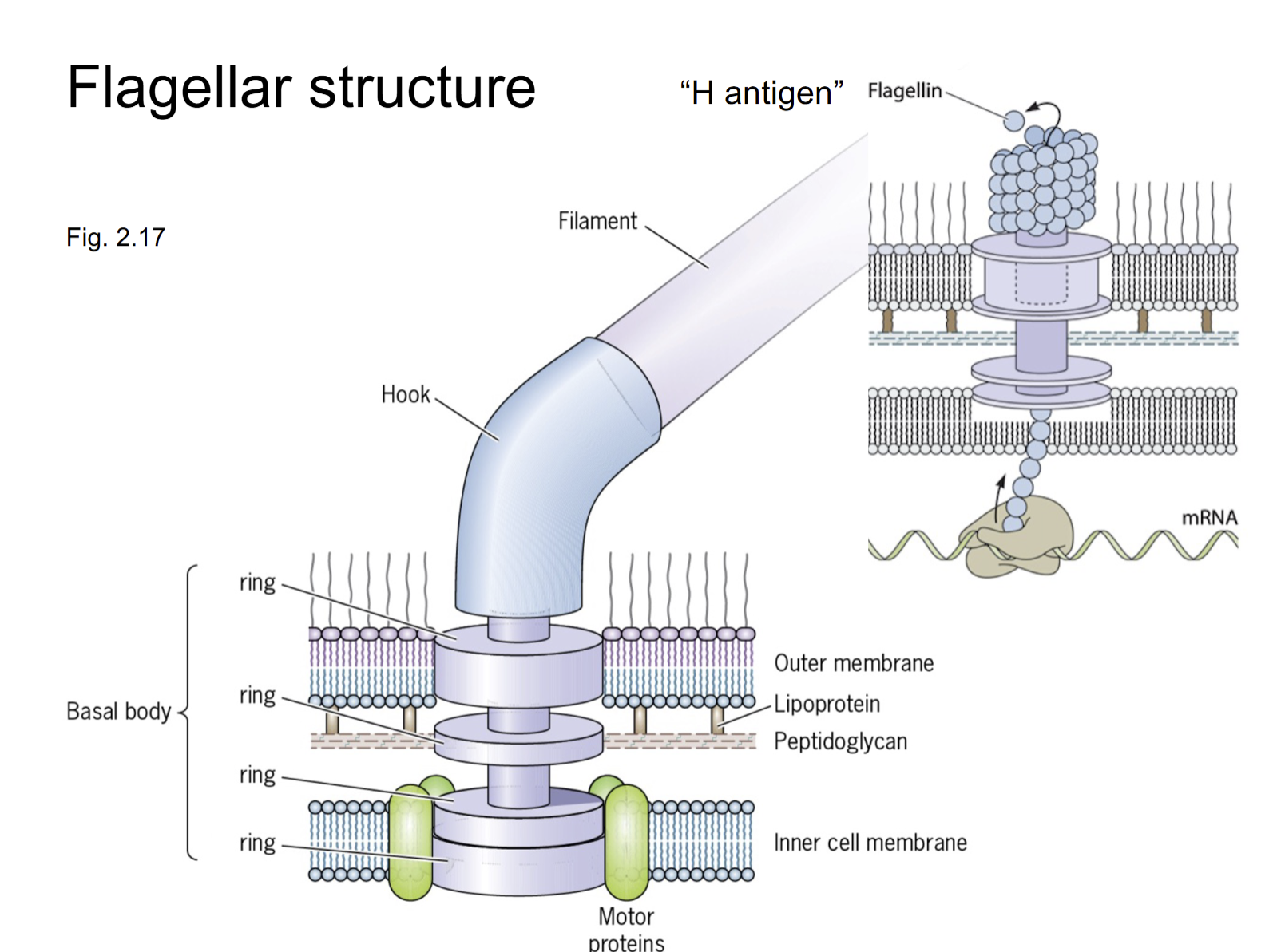
Flagellar structure
Anchored to envelop embedded in every layer (inner cell membrane, peptidoglycan, etc). to stabilize. Hook + Filament(10x length of cell) and is made from inside to outside( envelope to tip of filament)
Bacterial Pilus
Greek for hair
Pilin monomers assembled into a helical polymer
Key functions
attachment (fimbriae)
conjugation(sex pilus) → exchanging DNA
Twitching motility( type 4 pili)
Attachment via fimbrae
mostly on gram negative species
fimbriae allow attachment to surface via adhesins at their tips which binds to the structures or cell
reach out and drag cell if it adheres to surface
secures bacteria in gut if it doesnt want to leave
Which structures of a virus or immune cell could access from the outside of an intact gram-negative bacterial cell
Capsule, Liposaccharides, S-layer, maybe not cell membrane or peptidoglycan → nuance on environment
What about Gram-positive cells?
out membrane
Internal Bacterial Cell structures
Cytoplasm
cytoskeleton
nucleoid/ chromosome
ribosomes
inclusions
( Endospore):
Do bacteria, archaea, and eukaryotes have cytoskelentons?
yes
Bacteria and Diffusion
Limited by diffusion and has not active processes , has to be small to be able to diffuse everything
Eukaryotic Cell
x 10 larger than bacteria and has a smaller surface area to volume ratio
Have an active process since they cannot diffuse over a larger body
3 types of major cytoskeletal proteins
Actin, Tubulin, Creatinine
Organization of bacterial genome
Chromosome
singular, circular
millions of base pairs
condensed, organizes (different than histones since they don’t have histones)
May have plasmids
circular
thousands of base pairs
can be high or low copy number
Inclusions(microcompartments)
Carboxysomes: enclose carbon dioxide fixation machinery inside protein shell to increase efficiency
Thylakoids: membrane stacks that increase surface area for photosynthesis light harvesting and reactions
Inclusion: Gas vesicles
gas permeable protein shells that exclude water and provide buoyancy to non-swimming organisms in a water column
Inclusion: Magnetosomes
Magnetic crystals formed inside invaginations of inner membrane allowing magnetostatic bacteria to orient to the earth’s magnetic field and swim toward the N or S pole
How do bacteria grow
By binary fission which is the division of one cell doubling in the next generation 1→ 2→ 4→ 8 → 12
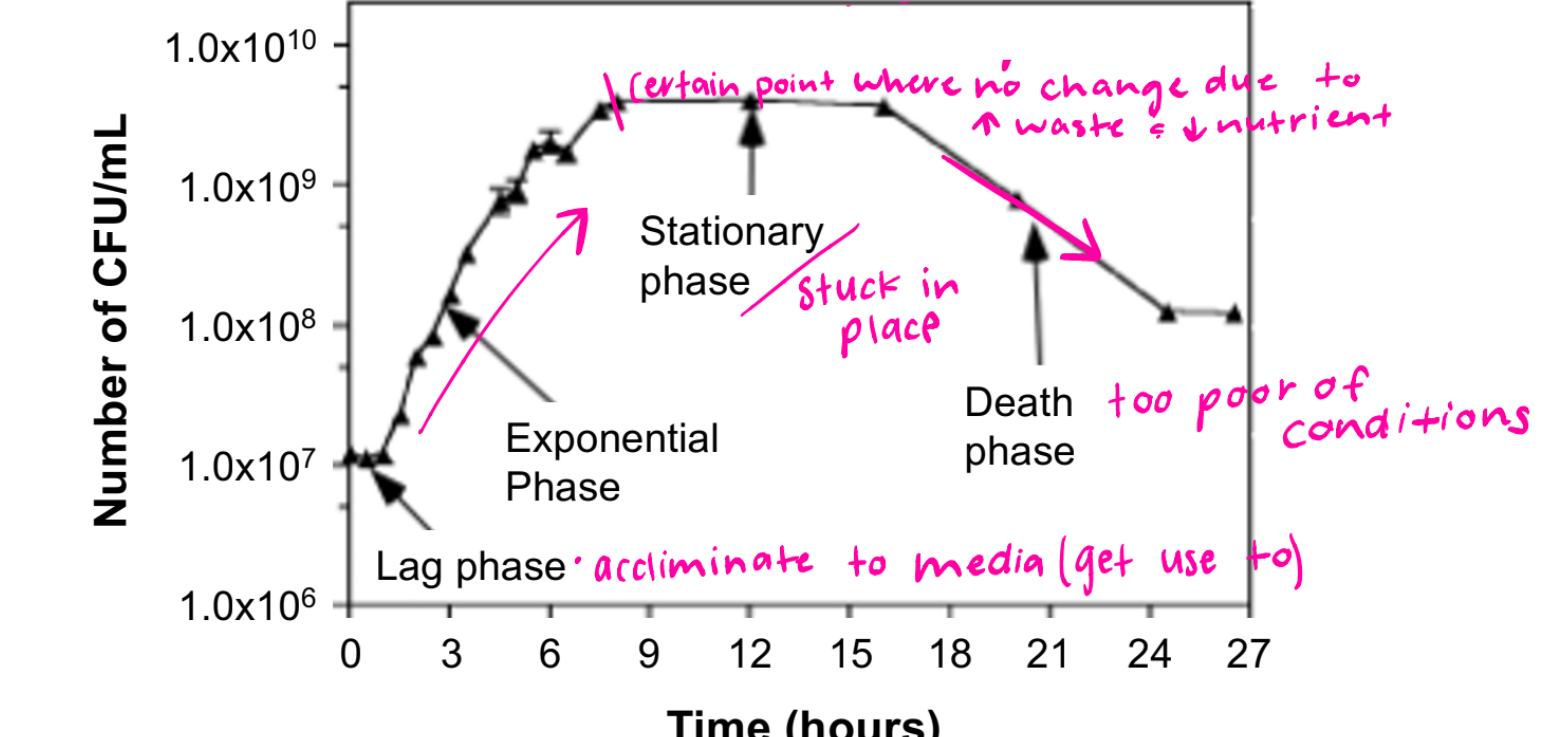
batch culture growth curve
Lag phase: acclimating to media/ metabolizing but not dividing
Exponential Phase: rapid growth/ division
Stationary Phase: certain point where no change due to decreasing in division bc food is becoming limited and waste is increasing/ trying to find balance
Death Phase: too poor of conditions, too much waste
growth and death are in logs, x10
Calculating population size
Nt= No x 2n
Nt: population size at time t
No: initial population size
n= number of generations
Calculating # of generations (n)
1) n=log10Nt-log10No/.301
2)g=t/n
Culture Organisms Nutrients
Macronutrients: CHONPS
Culture Organisms Needs
Incubation Conditions
Light or dark
atmosphere
Temperature
pH
Solute Concentration
Hydrostatic Pressure
Extremophiles are adapted to environments most organisms find inhabitable - examples
thermophiles: heat loving
halophiles: salt loving
amido/alkaliphiles: acid / basic loving
barophiles: pressure loving
Optimal temperature for Extremophiles
Psychrophiles: 10 C, best at low conditions
Mesophiles: 37 C, mild conditions
Thermophiles: 50+ C
Hyperthermophiles: 80 C +
Why does growth increase as the temperature goes up from the minimum?
For every 10C increase the reaction will increase of 2-3 because it allows for my energy to be used/ made
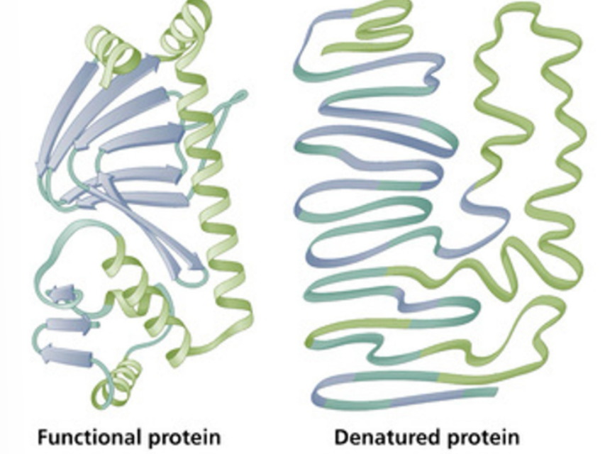
Why does growth decrease at temperatures above optimum?
Because of the denaturation of macromolecules and cell structures
How are thermophiles adapted to high temperatures
Challenges with solutions
DNA strands denature(separate)→ additional twist (positive supercoiling) and compaction
Proteins denature (unfold)→ stabilize with more intramolecular bonds
Membranes melt (too fluid/ permeable), glycerophosphate head of phospholipid monomers is hydrolyzed →archaea only, isoprenoid acyl side chains, monolayer membrane, ETHER linkage between acyl group and phosphoglycerol
Unsaturated v Saturated
Unsaturated cis fatty acid: kinks in membrane
saturated: no kinks
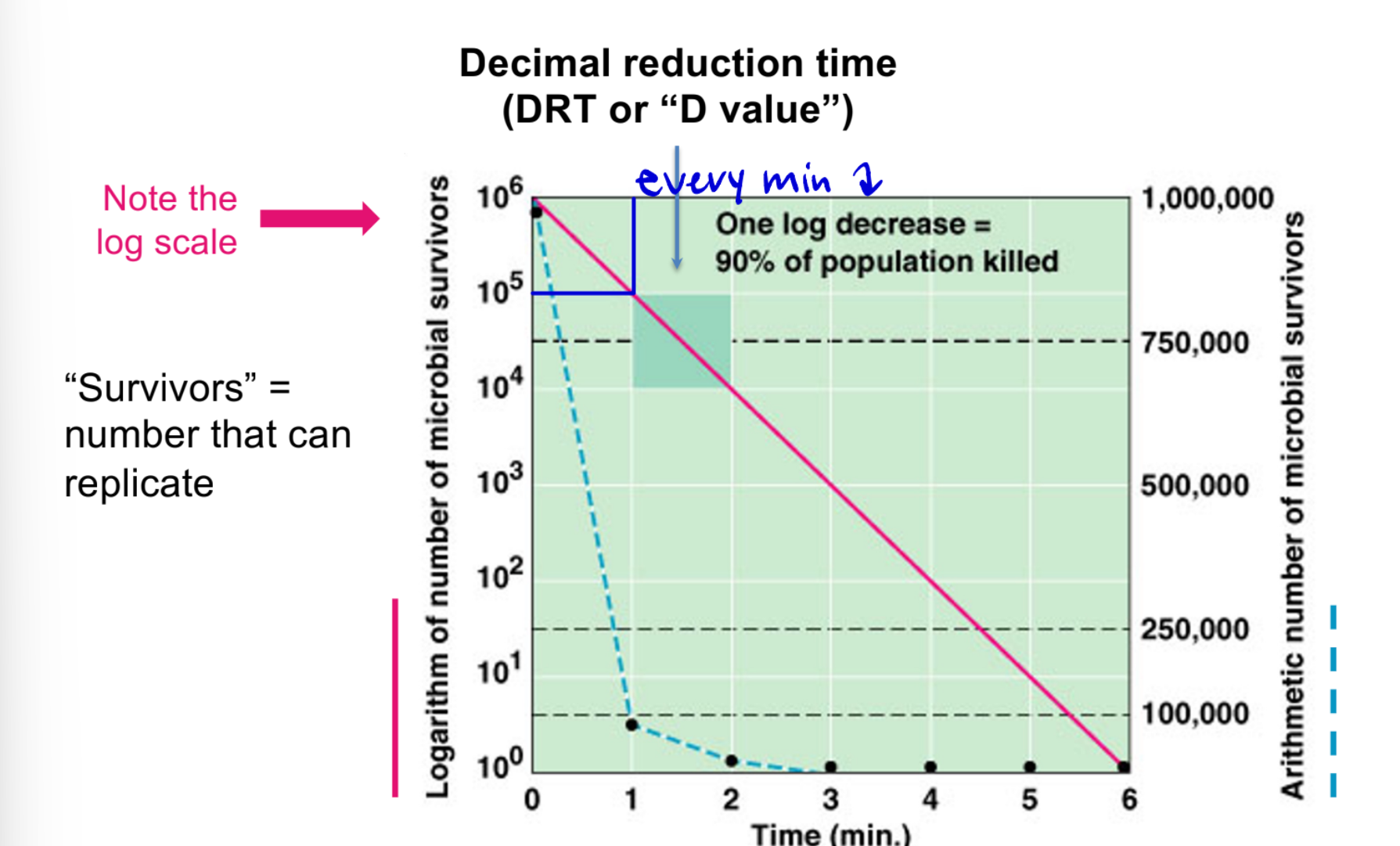
Pattern of microbial death: log reductions over time
90% of population killed per log scale
Catabolism- breaking it down
Energy, electron, and carbon sources
Entry, feeder pathways
Fueling (making of precursor metabolites, reductant, ATP)
Anabolism( build it up)
Autotrophy
Photosynthesis
Biosynthesis (of building blocks)
What is Metabolism?
Series of chemical reactions performed by living to make energy, build cell material, and maintain homeostatic
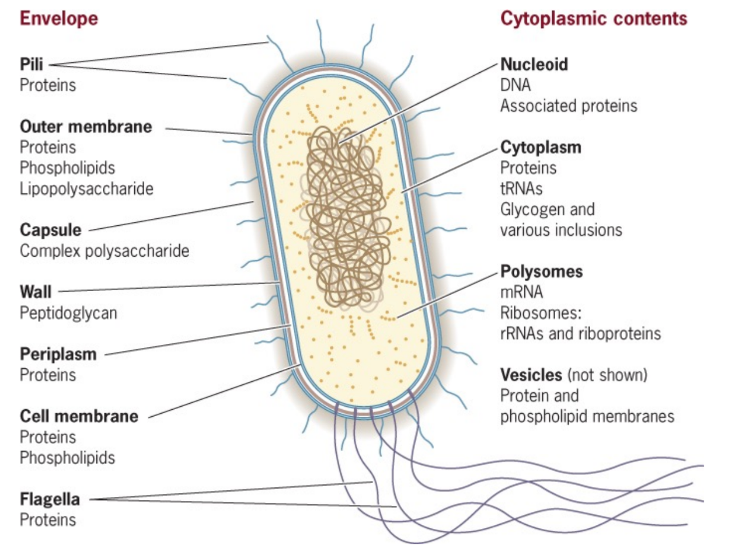
Image of Cell- gram negative bacteria
11 Components - Pili, outer membrane, capsule, wall, periplasm, cell membrane, flagella, nucleoid, cytoplasm, polysomes, vesicles
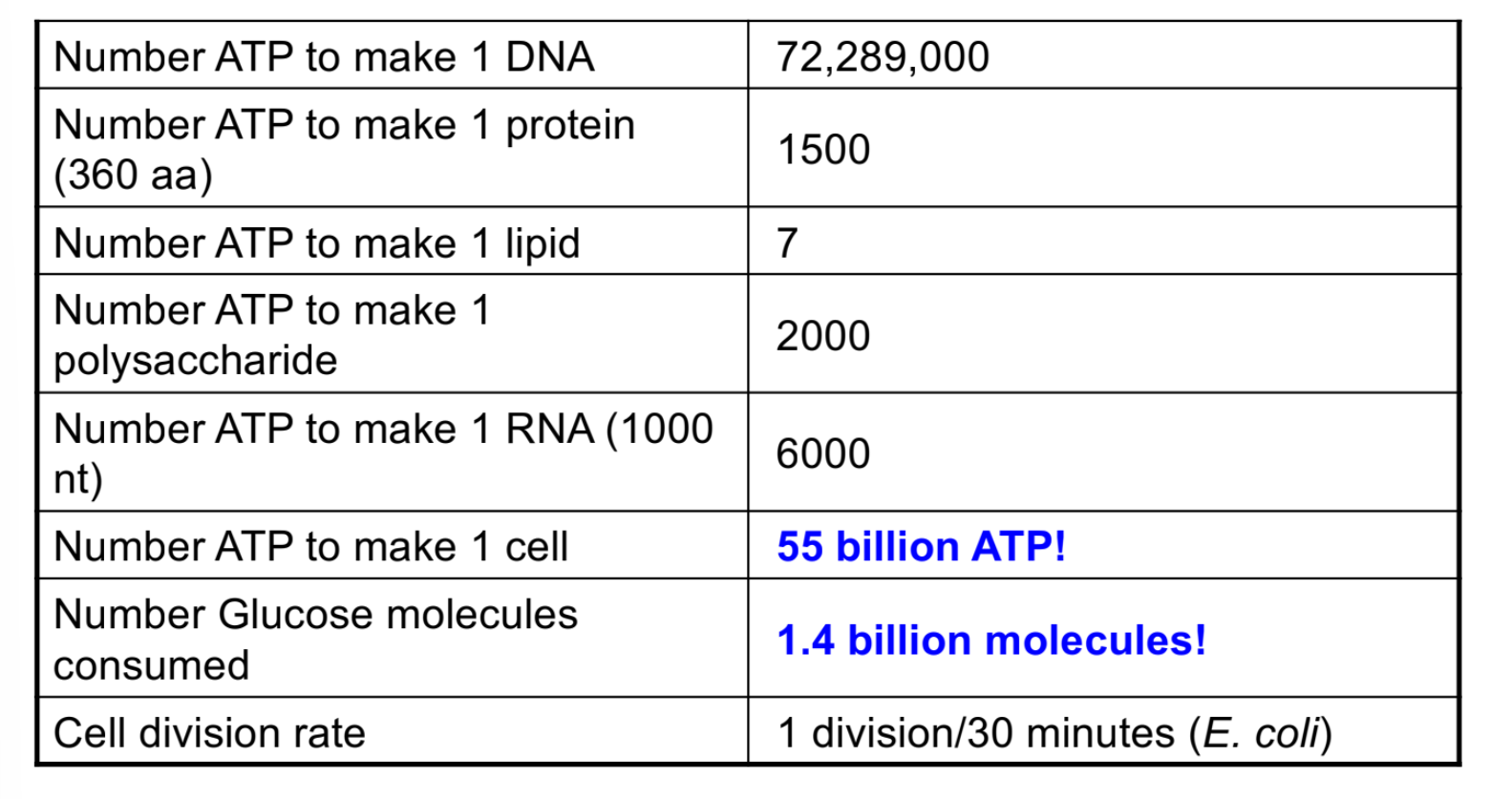
How expensive is it to make a cell
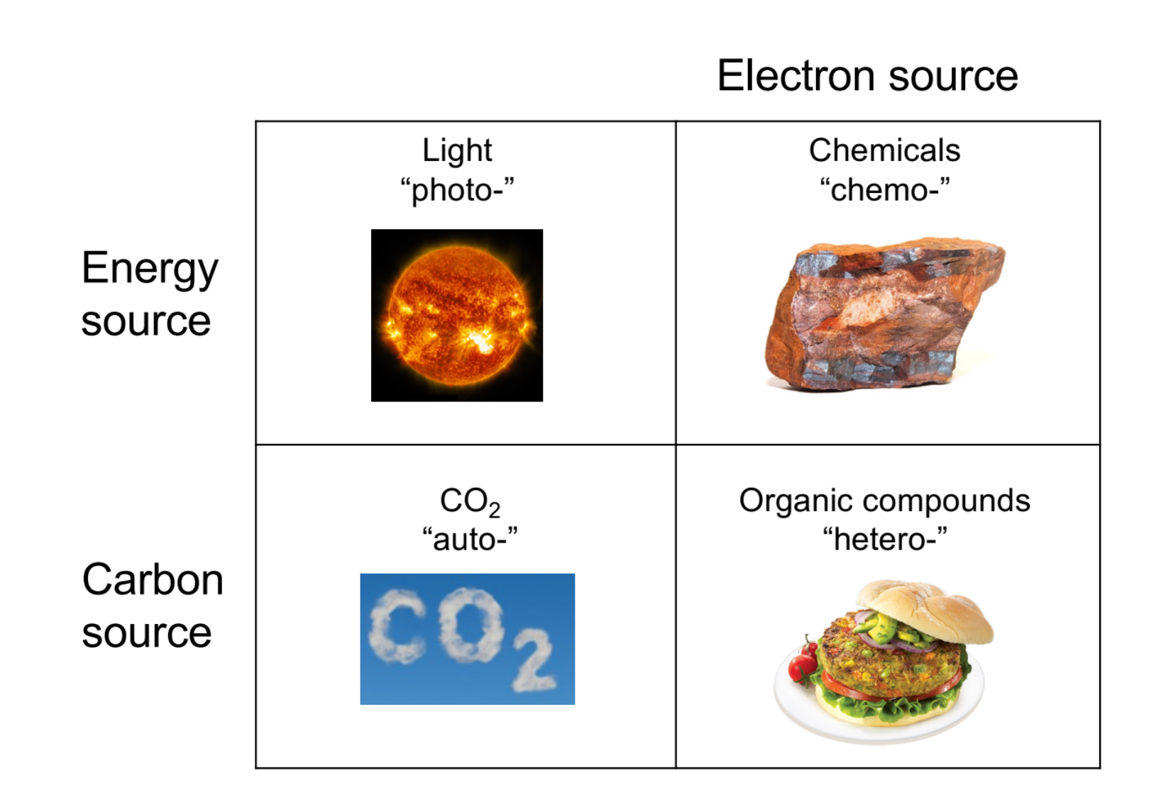
Type of -Troph
Energy source: Photo, Chemo
Carbon: Auto, Hetero
Electron: Chemo, Hetero
Metabolism Overview
CO2 (autotrophs/ carbon fixation) →or Organic macromolecules (feeder pathways for heterotrophs)→ Org C→ Fueling products Building Blocks→ Macromolecules → Cell
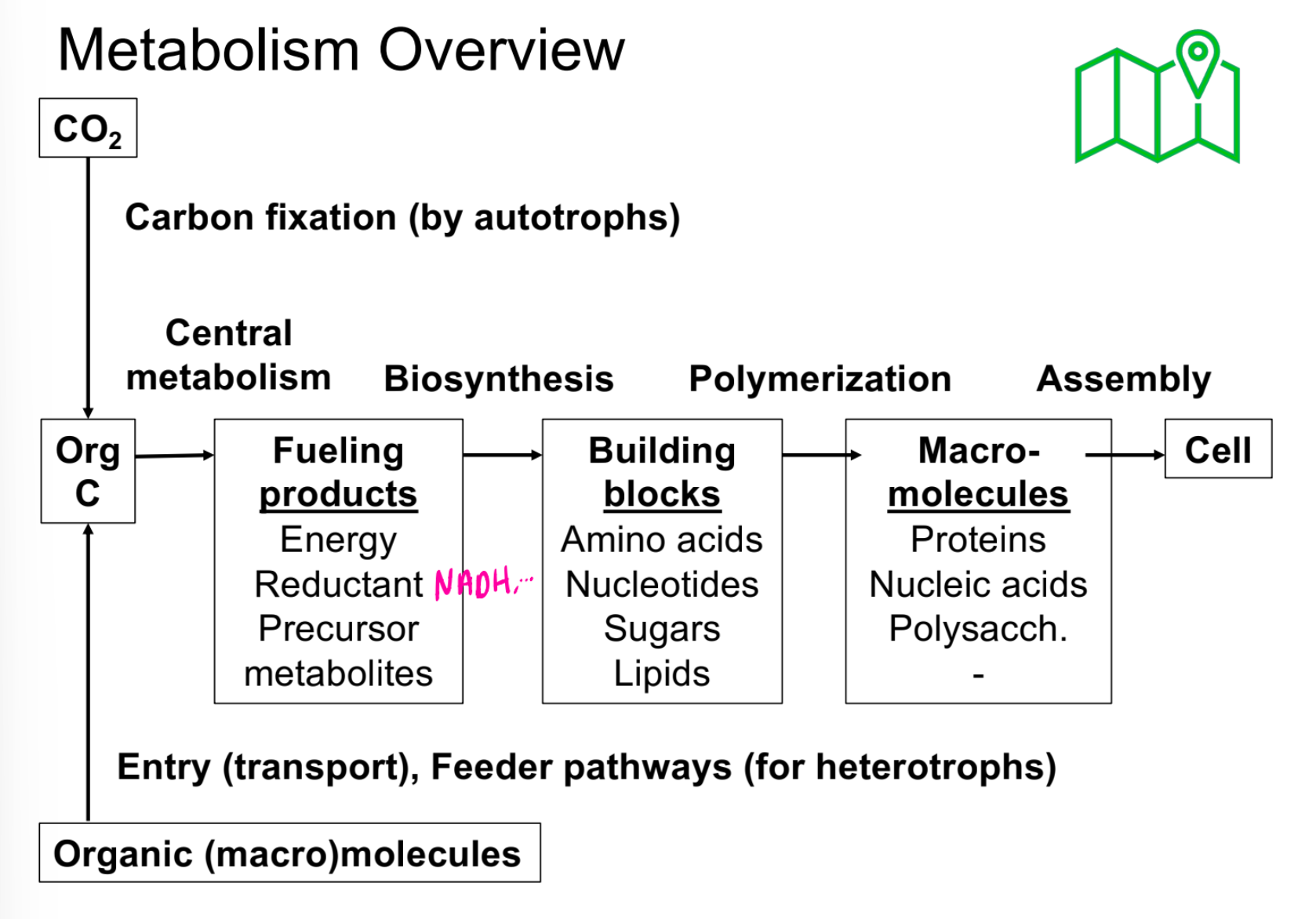
Fueling Products
Energy
Reductant
Precursor
Metabolites
Building Blocks
Animo Acids
Nucleotides
Sugars
Lipids
Macromolecules
Proteins
Nucleic Acids
Polysaccharides
Entry Barrier: Semi Permeable membranes
extracellular enzymes to break down larger macromolecules
Porins, transporters
Proteins?
High concentrations of solutes inside the cell
Hypotonic: Molecules at a higher concentration outside the cell can diffuse down their concentration gradient (passive transport)
Hypertonic: Molecules at higher concentrations inside the cell require energy to be transported against them concentration gradient (active transport)
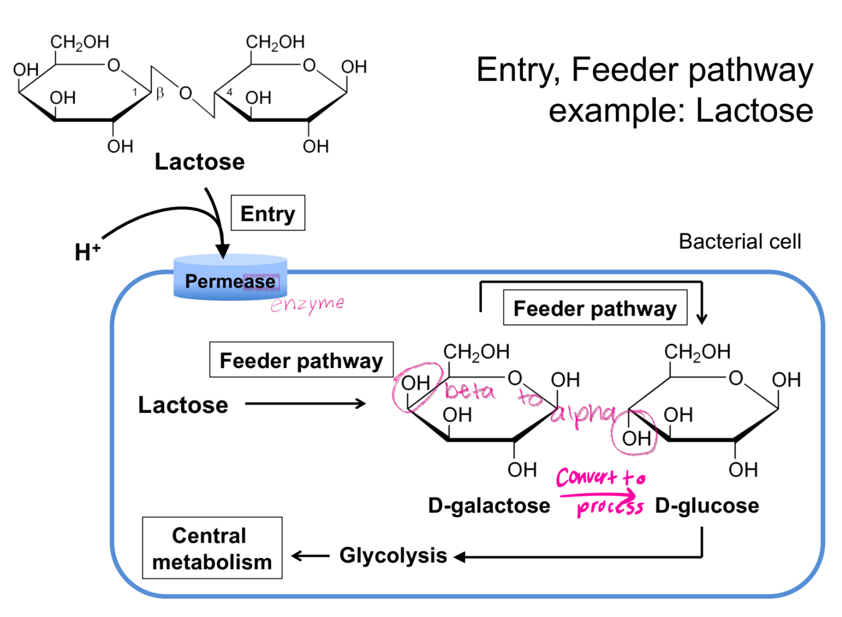
Entry, Feeder pathway example: Lactose
Lactose→ (permease)feeder pathway→ glycolysis→ Central metabolism
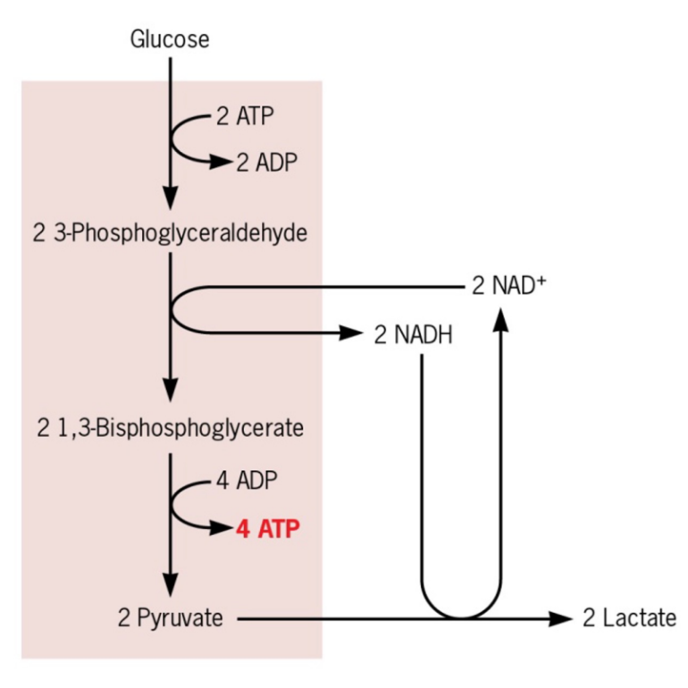
Fermentation
Produces energy during glycolysis
uses organic molecules
Regenerates NAD+
does not require oxygen, does not use the TCA cycle or electron transport chain