Chapter 2 - general anatomy/
 Call Kai
 Learn
Learn Practice Test
 Spaced Repetition
 Match
Match Flashcards
 Knowt Play
1/65

Earn XP
Description and Tags
Vocabulary flashcards covering body planes, cavities, abdominal divisions, body habitus, skeletal divisions, bone classifications, joint types, and bone markings from the General Anatomy lecture notes.
Name | Mastery | Learn | Test | Matching | Spaced | Call with Kai | Chat |
|---|
No analytics yet
Send a link to your students to track their progress
66 Terms
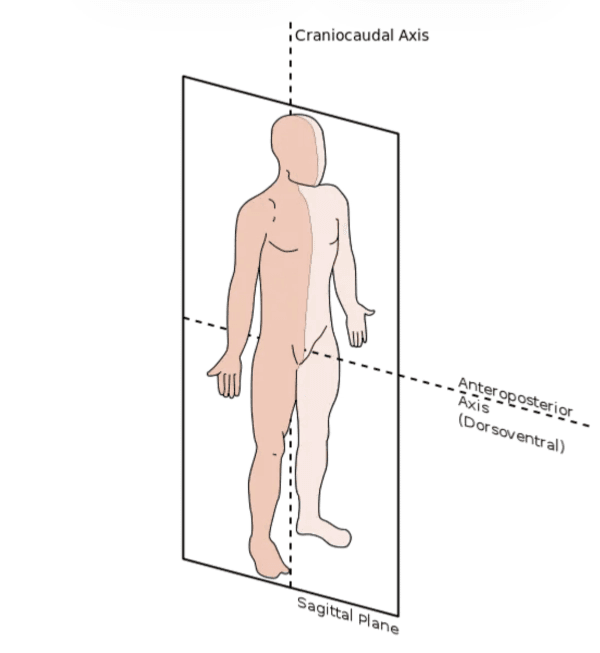
Sagittal plane
A body plane that divides the body into left and right portions.

Midsagittal plane
A body plane that passes through the midline and divides the body into equal right and left halves.
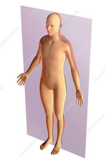
Coronal plane
A body plane that divides the body into anterior and posterior portions (front and back).
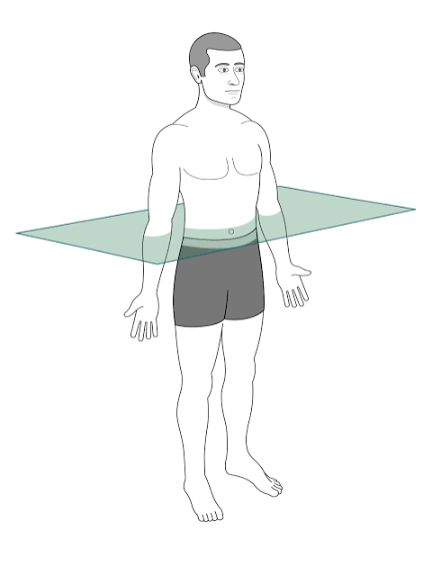
Horizontal plane (aka transverse)
A body plane that passes crosswise through the body or body part at right angles to the longitudinal axis (divides body into superior and inferior parts)
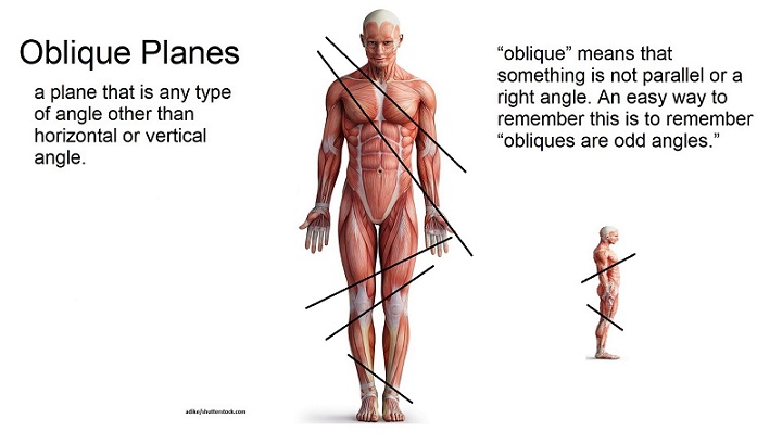
Oblique plane
A body plane that passes through a body part at any angle between the sagittal, coronal, and horizontal planes.

Diaphragm
The structure that divides the thoracic and abdominal cavities

Thoracic cavity
Body cavity containing pleural membranes, lungs, trachea, esophagus, pericardium, heart, and great vessels.

Abdominal cavity
Body cavity containing the peritoneum, liver, gallbladder, pancreas, spleen, stomach, intestines, kidneys, ureters, and major blood vessels.

Spleen
Abdominal organ located in the upper left side of the abdomen, posterior to the stomach.

Stomach
Abdominal organ located on the left side, anterior to the spleen
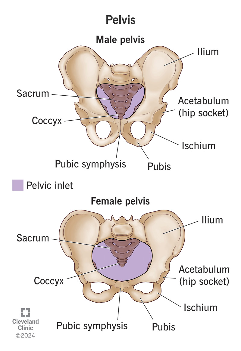
Pelvic cavity
Body cavity containing the rectum, urinary bladder, and part of the reproductive system.

Superior regions of the abdomen
The three upper abdominal regions: Right hypochondrium, Epigastrium, and Left hypochondrium.

Middle regions of the abdomen
The three central abdominal regions: Right lumbar/lateral, umbilical, and Left lumbar/lateral

Inferior regions of the abdomen
The three lower abdominal regions: Right inguinal/iliac, hypogastrium, and Left inguinal/iliac

T7 landmark
Spinal level landmark corresponding to the inferior angle of the scapula
T9, T10 landmark
Spinal level landmarks corresponding to the level of the xiphoid process.
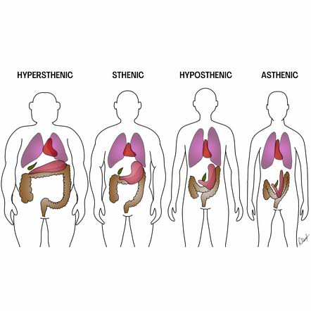
Hyposthenic (35% of people)
Body habitus type defined as a slender physical build that falls halfway between the average (sthenic) and very thin (asthenic)

Asthenic (10% of people)
Body habitus type defined as very skinny and tall.

Sthenic (50% of people)
Body habitus type defined as the average body type, with a standard physical build and typical organ anatomical positioning

Hypersthenic (5% of people)
Body habitus type defined as a massive, broad, and heavy build, with a short/wide thorax

Axial skeleton
Skeletal division consisting of 80 bones, including the vertebrae, skull, and ribs.

Appendicular skeleton
Skeletal division consisting of 126 bones, including the hips (which join vertebrae) and the upper and lower extremities

Epiphyseal plate
thin layer of hyaline cartilage located near the ends of long bones that allows the bone to grow longer

Red bone marrow
Marrow that produces red and white blood cells in the body

Yellow marrow
Marrow that stores fat cells and provides energy

Long bones
Bones found only in limbs, including the femur, humerus, tibia, fibula, and phalanges. Provide support
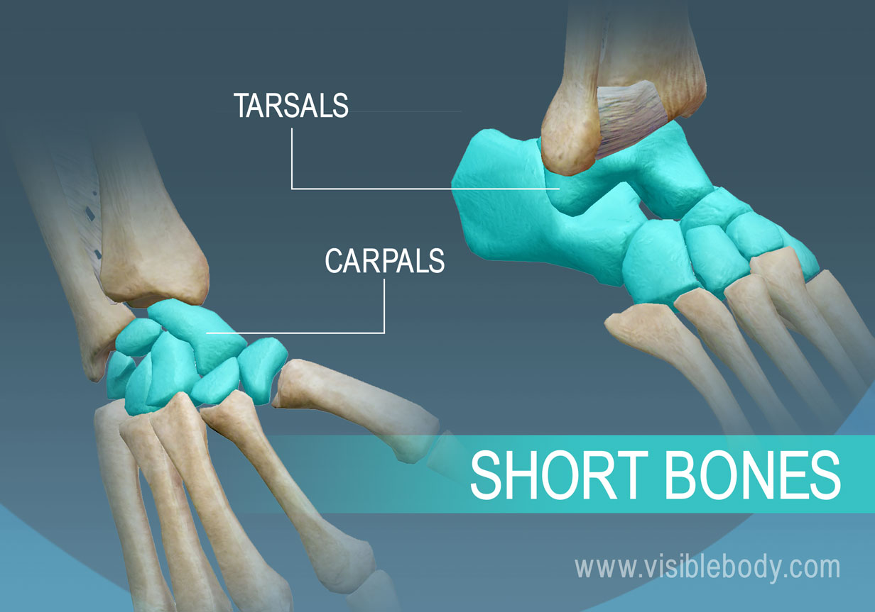
Short bones
Bones containing red marrow and have a thin outer layer of compact bone. Ex: wrist carpals, ankle tarsals. Allow minimum flexibility of motion in a short distance
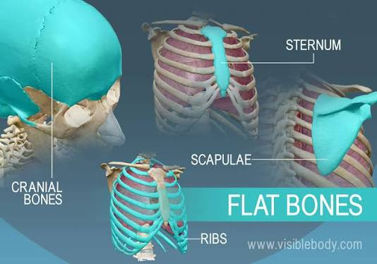
Flat bones
Bones with a hard part on top and bottom and a soft part in the middle. Ex: sternum, cranium, scapula, and ribs. Provide protection, and their broad surfaces allow for muscle attachment

Irregular bones
Bones with complex shapes, such as vertebrae and bones in the pelvis

Sesamoid bones
Small and oval bones that develop inside and beside tendons, such as knee caps (patella) and under the thumbs.

Functional Joint Classification
When joints are classified as functional, they are based on the mobility of the joint (ended in “throses”)

Synarthroses
immovable joints
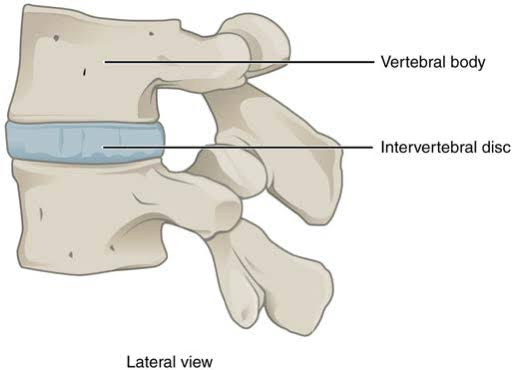
Amphiarthroses
slightly movable joints

Diarthroses
freely movable joints
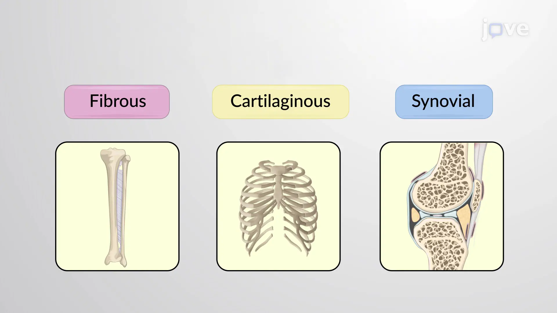
Structural Joint Classification
3 main groups based on the types of tissues that unite or bind the articulating bones

Fibrous joints (3 types)
Structural joint type with no joint cavity; they are the strongest joints in the body and include syndesmosis, suture, and gomphosis.

Suture
Immovable fibrous joint found only in the skull
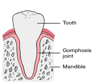
Gomphosis
Immovable fibrous joint found only in the roots of the teeth

Syndesmosis
Immovable/slightly movable fibrous joint united by sheets of fibrous tissue
Cartilaginous joints (2 types)
Structural joint type with no joint cavity, including symphysis and synchondrosis
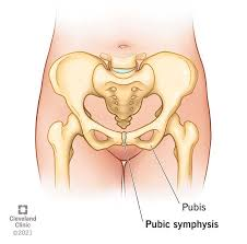
Symphysis
slightly movable cartilaginous joint. They are designed for strength and shock absorbency
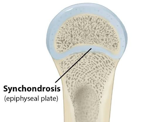
Synchondrosis
An immovable cartilaginous joint. This joint contains a rigid cartilage that unites two bones
Synovial joints
Structural joint type that is freely movable, comprising 6 specific subtypes

Gliding joint
Synovial joint type found in the intercarpal joints of the wrist/intertarsals of the foot

Hinge joint
Synovial joint found in the elbow and knee joint that opens and closes 90 degrees (flexion and extension)
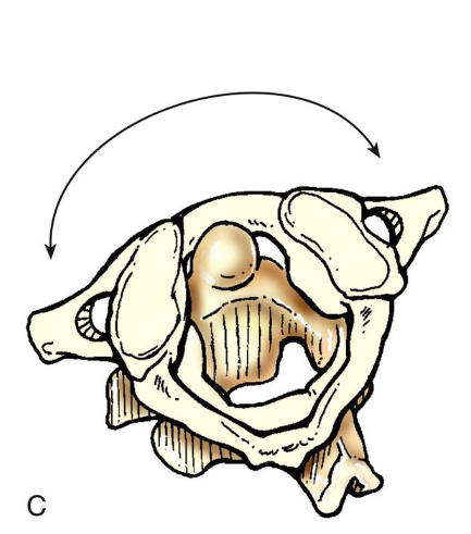
Pivot joint
Synovial joint found at the atlas and axis of the cervical spine
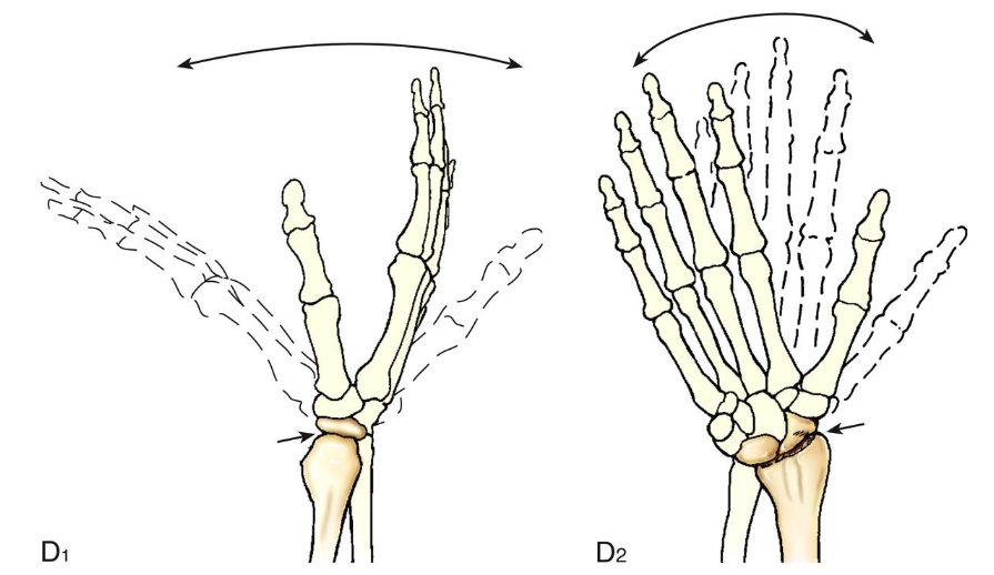
Ellipsoid joint
Synovial joint found at the radiocarpal joint of the wrist. Flexion and extension occur along with abduction and adduction
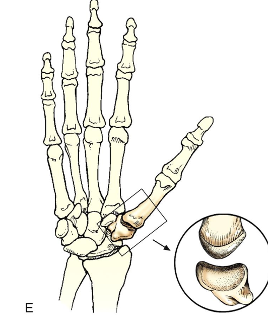
Saddle joint
Synovial joint found at the carpometacarpal joint

Ball and socket joint
Synovial joint found at the hip joint that permits the widest range of motion
Processes or projections
Extensions of the bone that go beyond the normal area

Condyle
Rounded process at an articular end of a bone

Coracoid/coronoid
Beaklike/crownlike process on a bone
Crest
Ridgelike process on a bone.

Epicondyle
Projection located above a condyle.
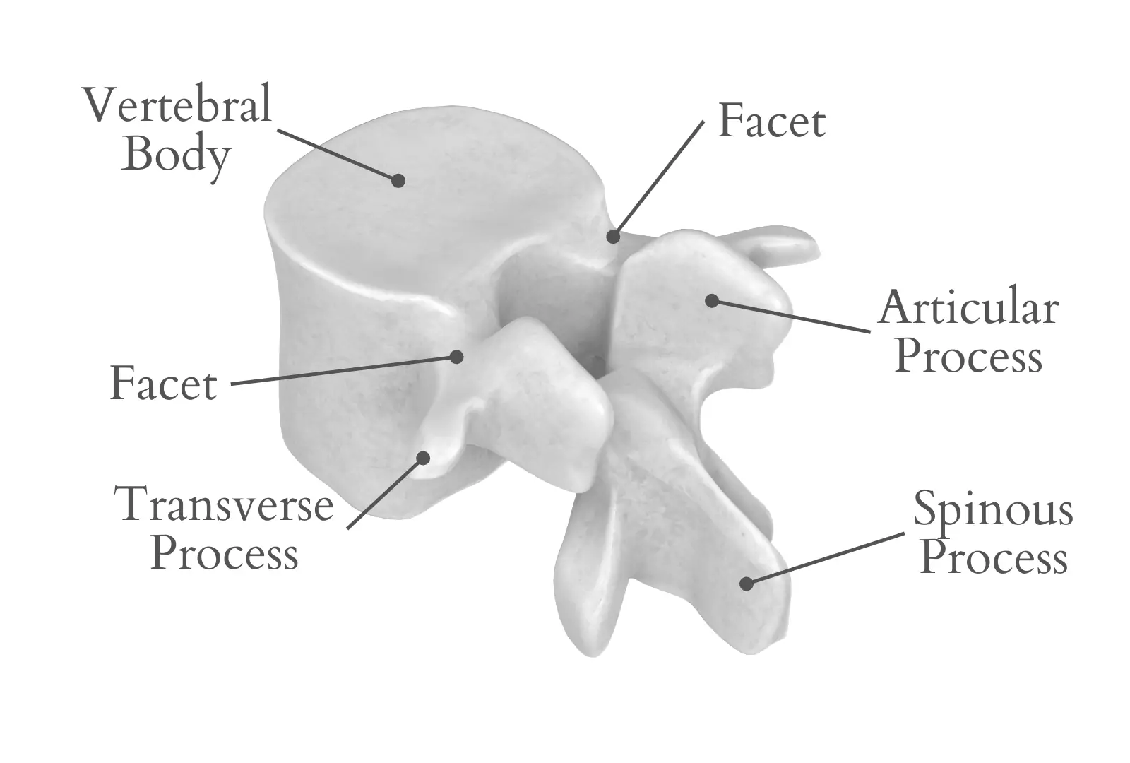
Facet
Small, smooth-surfaced articular process on a bone.

Head (bone marking)
Expanded end of a long bone

Horn
Hornlike process on a bone

Line (bone marking)
Linear elevation on a bone

Malleolus
Club-shaped process (ball of the ankle)

Protuberance
Projecting prominence on a bone

Styloid
Long, pointed process on a bone

Trochanter
Either of the 2 large, rounded, and elevated processes located at junction of neck and shaft of femur

Tubercle
Small, rounded, and elevated process on a bone

Tuberosity
Large, rounded, and elevated process on a bone.
Depression
Hollow or depressed areas of a bone
Fractures
A break in bone.