Lect 6: Thorax and Abdomen
 Call Kai
 Learn
Learn Practice Test
 Spaced Repetition
 Match
Match Flashcards
 Knowt Play
1/64

There's no tags or description
Looks like no tags are added yet.
Name | Mastery | Learn | Test | Matching | Spaced | Call with Kai | Chat |
|---|
No analytics yet
Send a link to your students to track their progress
65 Terms
What are the key functions of the thorax?
PROTECT VITAL ORGANS
resist neg internal pressure from breathing
support upper limb weight
provide muscle attachment points for back / extremities

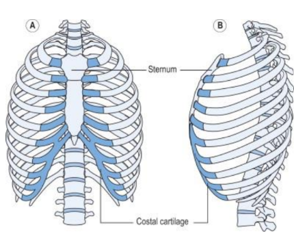
Thoracic Cage Osteology Overview
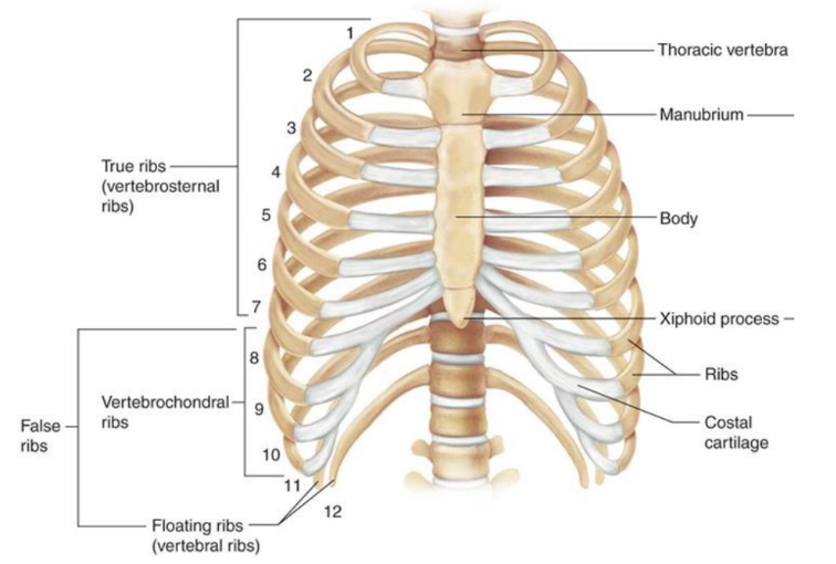
True vs False ribs (which #s are which)
1-7 true
8-10 false
11-12 float

What forms the anterior wall of the thoracic cage?
the sternum


Where is the manubrium located? (level)
T3-4
What ia the body of the sternum level with?
T5-9
What ribs articulate with the body of the sternum?
(T2- manubrium and body)
T3-6
(T7 xiphoid process)

How many costal notches are on the body of the sternum?
6 pairs
2nd-7th costal cartilage
(2 & 7 partial)

Are there any costal notches on the xyphoid process?
yes 1 pair for 7th costal cartilage
Xyphoid process development
initially cartilagenous > ossifies in adulthood
*meaning can vary in shape: point, bifid, or deflected to one side
What type of bone are the ribs?
curved flat bones
What are the true ribs?
ribs 1-7
costal cartilage articulates directly with sternum (costochondral joints)

What are the false ribs?
ribs 8-10
attatch to sternum indirectly via cartilage to the rib superior to it
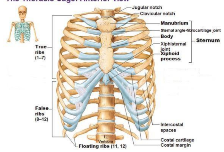
Where do floating ribs attach?
What are the floating ribs?
ribs 11-12
don’t attatch to sternum at all
end in posterior abdominal musculature

What are the (anatomically) typical ribs?
ribs 3-9

Shaft: outward convexity, costal groove- intercostal arteries/nerves
Posterior angle: bend in shaft 2” from tubercle
costal groove: protect intercostal nerve vessels
tubercle: junction of neck / shaft, has articular + non-articular portions
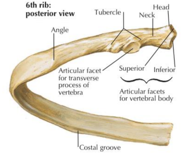
What does the head of a typical rib articulate with?
(2 facets with transverse crest between)
upper facet — body of vertebra above
Lower facet —body of cooresponding vertebra
*sandwhichh sup art proc T5 > up Rib 5 > rib 5 upper facet > T4
Tubercle of the Rib
junction of neck—shaft
articular potion: facet for numerically cooresponding TP
non-articular portion: roughened for ligaments
Which part of the rib articulates with the TP of vertebra?
articular portion of the tubercle
(to cooresponding vert)
What are the anatomically atypical ribs?
1-2
10-12
What is costal cartilage?
bars of hyaline cartilage
forward/medial from ant rib ends
mobility/elasticity to thorax wall
(no movement at costochondral joints)

ow many pairs of costal cartilage are there?
12
Where do each pair of costal cartilage connect?
1-7 — directly to sternum (costochondral)
8-10 — connect to costal cartilage of one above
11-12 —just cap floating rib and end in abdomical muscle wall
What type of joints are costovertebral + costotransverse joints?
what do they enable?
plane gliding synovial
*enable elevation + depression
(inhale / exhale)
*strong ligaments so movement is minimal

What parts of the rib do T vertebrae articulate with?
(rib sits on top of numerically cooresponding vert)
body T7 > rib 7 inf facet
TP T7 > tubercle of rib
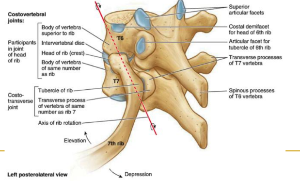
What joints connect vertebra > > ribs?
vertebra > (costovertebral/transverse joints) > costal cartilage (sternocostal) > sternum
What movement do sternocostal joints allow?
slight gliding / “give” for respiration
ribs move as unit with sternum
Movement of ribs 1-2
minimal in quiet respiration
inspiration: move up / laterally (mainly pump handle)
What are the 2 ways ribs 3-10 move?
pump handle
bucket handle
Which movement increases ribs transverse diameter?
bucket handle
(elevation up + lateral)
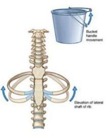
Which movement increases the thorax’s anteroposterior diameter?
pump handle

Describe the movement of the floating ribs?
ribs 11-12
small movement permissed in all directions
*depressed in inspiration! by quadratus lumborum to form fixed point for diaphragm
Which way do the fibers of each intercostal muscle run?
1) external- hand in pocket (grabbing something out pulling up/out)
2) internal- opposite of ^ (originate inf Insert sup pull down/in)
3) innermost- vertical (origin infer, insertion superior- pull down- depress ribs)

What is the action of each intercostal muscle?
1) external- elevation / inspiration
2) internal - depression / exhalation
3) innermost - depression / exhilation

What muscles form the “anterior thoracic wall”?
intercostals (3)
Transversus Thoracis
Transversus Thoracis
(OIA)
(spiderman muscle ‘picturing him pulling ship’)
O: post sruface lower sternum + xyphoid process (thats where spidy stand)
I: (‘webs’) to inner surface of costal cartilage 2-6
rib depression (pull in/down) —expiration
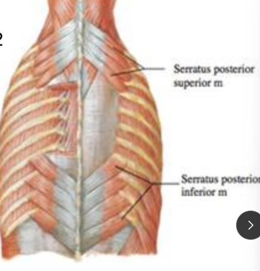
Wat makes up te posterior toracic wall?
serratus posterior (superior + inferior) *the diamond

Serratus posterior superior
(OIA)
O: nuchal lig & SP C7-T3
I: ribs 2-5 lateral to posterior angles
rib elevation (inspiration)
Serratus Posterior inferior
(OIA)
O: sp T11-12
I: inf boarder ribs 8-12 (lateral to post angles)
rib depression (expiration)
The innermost intercostals assist with what?
rib depression
The transversus thoracis is deep to the sternum.
A. True
B. False
true
What are the walls of the abdomen?
“cube”
anterior
lateral
posterior
superior / inferior

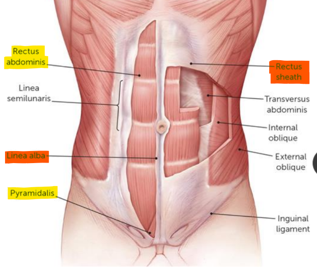
What muscles make up the anterior wall of the abdomen?
rectus abdominus
rectus sheath
pyramidalis (within sheath, triangle at bottom tense linea alba)
What innervates most of the abdomen?
ventral rami of thoracic nerves
Rectus Abdominis
(OIAN)
O: pubic crest + symphysis
I: xiphoid process + costal cartilage ribs 5-7
N: ventral rami thoracic nerves
flex vertebral column
*tendinous structures: linea alba + transverse
What tendinous structures appear in the rectus abdominis?
linea alba: xiphoid process > pubic symphysis
(seperate rectus abdom vertically)
tendinous intersections: 3 zig zags adherent to ant layer of rectus sheath
What points does the linea alba run between?
xiphoid process > pubic symphysis
What do the 3 tendinous intersections of the rectus abdominis adhere to?
anterior layaer of the rectus sheath
Rectus Sheath
(composition, runs-)
aponeurotic sheath
enclose rectus abdom + pyramidalis
anterior + posterior layer
ends blended into linea alba (blending with rectus sheath of opp side)

Pyramidalis
(location, action)
small triangular at base of abdominal sheath
pubis > linea alba
anterior to rect abdom
tenses linea alba
What muscles make up the lateral wall of the abdomen?
transversus abdominis
internal + external obliques
iliacus
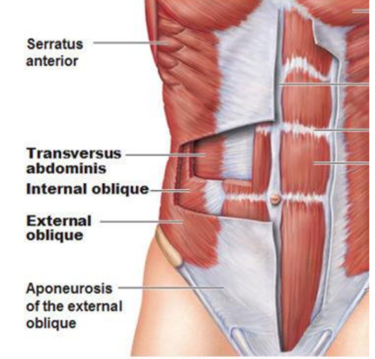
Transversus Abdominis
(OIAN)
“deep core”
Origin:
costal cart 7-12
thoracolumbar fascia
iliac crest
connective tissue under inguinal ligament
Insertion: linea alba
(with combined aponeurosis of intern obliq + pubic crest + pubis)
ACTION:
compress abdominal viscera
assist in forced expiration
role in Lumbar stabilization
dorsal rami thoracic n

Internal Oblique
(OIA)
(Insert superior - pull down)
O:
lat ½ inguinal lig
iliac crest
thoracolumbar fascia
I: (*A)
inf boarder rib 10-12
linea alba
pubic crest
pectineal line
ACTION:
support/compress abdominal viscera
assist force exp
bilat: flex
unilat: ipsi lat trunk flex + rot

External Oblique
(OIA)
(put hand in orientation use fingers to pull)
O: ext surface rib 5-12
I: (*V)
ant ½ iliac crest
anoneurosis from 9th cost cart to ASIS, pubic tubercle + linea alba
ACTION:
support / compress abdom viscera
assist forced exp
bilat: flex spinal column
unilat: ipsilat flex, contralat rot
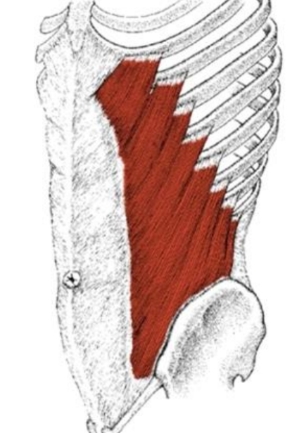
Unilaterally, which muscle is responsible for ipsilateral
rotation:
A. External obliques
B. Internal obliques
C. Transversus abdominis
D. Rectus abdominis
internal obliques
If lying supine and using the rectus abdominis to lift the legs and pelvis off the floor, this movement would be considered what type of chain?
closed chain
(open- lifting feet off floor)
What muscles make up the posterior abdominal wall?
quadratus lumborum
psoas major + minor
iliacus
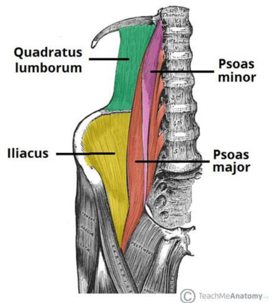
What muscle fixes rib 12 with inspiration?
quadratus lumborum (working bilaterally)
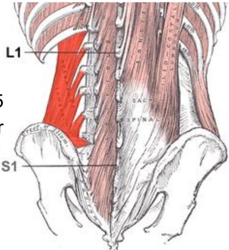
Quadratus Lumborum
(OIA)
O: iliolumbar ligament + iliac crest
I: lower boarder rib 12 + TP L1-5
bilat: ext lumbar vert + fix rib 12 with inspiration
unilat: ipsilat trunk sidebending
Closed vs Open Chain
closed: extremities fixed, body part in middle move
open: middle body fixed, extremity move
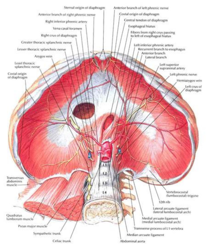
Diaphragm
(OIAN)
O: circumference of thorax + fibers from xiphoid, cost cart 7-12, L1-3
I: central tendon
draw central tendon down / forward in inspiration
relax up in expiration
N: (Phrenic nerve- C3, 4, 5 keep the diaphram alive)
3 parts

What are the 3 parts of the diaphragm?
And where to they each arise from?
1) Sternal - back of xiphoid process
2) costal- internal surface cost cart
3) lumbar- med/lat arcuate ligaments + crura fibers => central tendon
What muscles make up the inferior abdominal wall?
pelvic floor muscles
(unspecified for this class)

What nerves contribute to moving the diaphragm?
Phrenic Nerve
C3, 4, 5 (keep the diaphragm alive)