Kidney and Lower Urinary Tract
 Call Kai
 Learn
Learn Practice Test
 Spaced Repetition
 Match
Match Flashcards
 Knowt Play
1/98

There's no tags or description
Looks like no tags are added yet.
Name | Mastery | Learn | Test | Matching | Spaced | Call with Kai | Chat |
|---|
No analytics yet
Send a link to your students to track their progress
99 Terms
What is the renal capsule?
Thin fibrous layer overlaying cortex
May adhere to perinephric fat with tumor invasion
Why is the renal capsule important during gross examination?
It’s relationship to tumor
Tumor may invade through the capsule and into surrounding perinephric tissue
What is the orientation of the renal cortex and medulla?
Cortex extends inward as renal columns
Medulla forms renal pyramids that terminate at papillae → Minor calyces
Why is the renal sinus an important area when evaluating a renal tumor?
Due to lack of capsule, it is an important pathway for tumor extension and spread
What is the renal sinus?
Fat-filled space between pelvis mucosa and parenchyma
No capsule here
What structures are enclosed by Gerota’s fascia?
Kidney
Perinephric fat
Adrenal gland
It is typically located anterolaterally on radical nephrectomy specimens
What is the anterior-to-posterior arrangement of the hilar structures of the kidney?
Vein → Artery → Ureter
NOTE: Tumors may distort normal anatomy
(VAU)
How can a nephrectomy specimen be oriented using the hilar structures and adrenal gland?
Renal vein — Anterior to the artery
Ureter — Extends inferiorly
Adrenal gland — Superior and slightly medial
What is typically included in a partial nephrectomy specimen?
Tumor (confined to a pole)
Encapsulated tumor
Renal parenchyma
Renal capsule
± Perinephric fat
What is typically included in a radical nephrectomy specimen?
Kidney
Segment of ureter
Perinephric fat
Gerota’s fascia
± Adrenal gland
What is typically included in a simple nephrectomy specimen?
Kidney
Ureteric portions
(Generally removed for non-neoplastic/non-tumorous kidneys)
What is the typical gross examination for radical nephrectomy specimen?
Weigh and measure
Describe envelope
Ink margins
Hilar structures
Bivalve kidney
Measure and describe tumor
Sample margins and interfaces
What should be documented when examining the outer envelope of a radical nephrectomy?
Presence/absence of perinephric fat
Presence/absence of Gerota's fascia
Presence/absence of adrenal gland
Any defects or disruptions of the specimen envelope
What surfaces should be inked in a radical nephrectomy containing a tumor?
Perinephric fat overlying the tumor
Gerota’s fascia near the tumor
(Ink outer surface and bivalve (section along ureter and into kidney if TCC))
How should the hilar structures be handled in a radical nephrectomy?
Measure the length and diameter of the:
Renal vein
Renal artery
Ureter
Take cross-sections of each margin
What measurements should be obtained for a radical nephrectomy specimen?
Weight of the entire specimen
Overall specimen dimensions
Dimensions of the kidney proper
Measurements of associated structures when present, such as the adrenal gland and hilar structures
Why is the ureter probed before bivalving a kidney?
Helps guide sectioning along the collecting system
Allows exposure of the collecting system and tumor–renal sinus interface
Allows bivalve specimen if fixing overnight
What tumor features should be documented in a renal tumor?
Size in 3D
IMPORTANT FOR STAGING THRESHOLD
Location
Color/Appearance
Percentage of necrosis
Hemorrhage
Cystic change
What important tumor interfaces should be sampled in a radical nephrectomy?
Sample tumor in relation to:
Renal capsule
Renal sinus fat
Renal vein
Renal pelvis
Renal parenchyma
Also sample uninvolved parenchyma and adrenal gland when present
What are the most important structures to evaluate for extension of a renal tumor (radical nephrectomy specimen)?
Evaluate involvement of:
Renal sinus
Capsule
Perirenal fat
Gerota's fascia
Calyces
Renal vein and its branches
Inferior vena cava, when applicable
After bivalving a kidney with a tumor, how should the kidney be further sectioned?
Bread-loaf the kidney at approximately 0.5 cm intervals
Section extensively to identify:
Additional tumors
Tumor extension into adjacent structures

What is the classic gross appearance of clear cell (conventional) RCC?
Golden-yellow
Due to its lipid-rich nature
It may also show:
Cystic change
Hemorrhage
Necrosis
Calcification
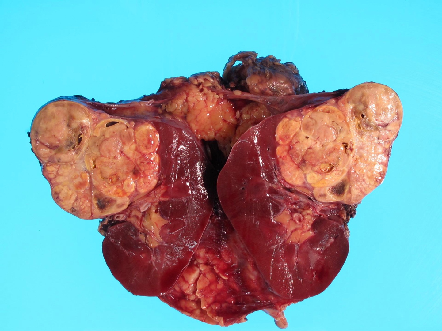
What is the typical gross appearance of papillary RCC?
Tan-yellow to dark brown
Variegated
Often surrounded by a fibrous pseudocapsule
May show extensive necrosis

What is the typical gross appearance of chromophobe RCC?
Solitary
Well-circumscribed
Lobulated
Homogeneous pale tan to dark brown
May have central scarring

What is the typical gross appearance of collecting duct carcinoma?
Unifocal
Poorly circumscribed
Centered in the medulla
Solid
Tan-white
Firm

How is gross tumor necrosis described, and why should the percentage be documented?
Gross necrosis may appear:
Geographic
Yellow-white
Soft
Granular/friable
Dry on the cut surface
The percentage of necrosis should be recorded because it has prognostic importance
What gross features may suggest sarcomatoid change in a renal tumor?
Solid gray-white appearance
Invasive margins
Firm, fleshy-to-fibrous cut surface
Often associated with hemorrhage and necrosis
How should a partial nephrectomy specimen be inked?
Ink the cauterized parenchymal margin one color
Ink the perinephric/Gerota's fascia surface a different color
How should a partial nephrectomy be sectioned?
Serially section the specimen perpendicular to the resection (cauterized) margin
What tumor features should be assessed in a partial nephrectomy?
Determine:
Whether the tumor is contained within the capsule
Tumor size + distance to each margin
Relationship to capsule and perinephric tissue
Features of uninvolved renal parenchyma and corticomedullary junction
What should be submitted from a partial nephrectomy with tumor?
Submit sections demonstrating:
Tumor to all margins
Tumor to capsule
Tumor to perinephric fat/fascia
Representative uninvolved renal parenchyma
What type of specimen is this cross section?

Partial nephrectomy
What is missing from this partial nephrectomy?

No perinephric fat attached
MAKE NOTE OF THAT IN DICTATION
How is Wilms tumor grossed differently from RCC?
Wilms tumor is grossed generally like an RCC
An important additional step is mapping the tumor
Its staging system is also NOT TNM-based
What ancillary studies may be requested for Wilms tumor?
Depending on available tissue:
Snap freezing for molecular studies
Cytogenetics
FISH (formalin fixed paraffin sections)
IHC, including markers such as:
p53
WT1
CD56
What is this?

Wilms tumor
What is this?

Adult polycystic kidney disease
What is this?

Pediatric polycystic kidney disease (autosomal recessive)
What is meant by the continuous urothelial unit?
The urothelial-lined system extends continuously from the minor calyces to the ureterovesical junction (UVJ)
What are the major wall layers of the upper urinary tract?
Urothelium
Lamina propria
Muscularis propria
The walls of minor calyces overlying the papillae are particularly thin
Should be sampled generously to avoid overstaging
What are the approximate dimensions of the ureter?
~30 cm long
~0.5 cm in diameter
What is the bladder cuff in a nephroureterectomy specimen?
The distal-most ureter at the ureterovesical junction (UVJ)
Is included in a nephroureterectomy specimen
Why is it important to distinguish intrarenal from extrarenal renal pelvis?
Intrarenal pelvis — Surrounded by renal parenchyma
pT3 invasion involves renal parenchyma
Extrarenal pelvis — Surrounded by peripelvic fat
pT3 invasion involves peripelvic fat
What are the regional LNs associated with the renal pelvis?
Renal hilar
Paracaval
Para-aortic
Retroperitoneal
What are the regional LNs associated with the ureters
Renal hilar
Paracaval
Iliac (common/internal/external)
Periureteral
Pelvic
What gross appearance is typical of an upper tract urothelial carcinoma?
Soft
Friable
Gray-pink
Glistening or granular
Polypoid, papillary, or sessile

What gross appearance is typical of more invasive upper tract urothelial carcinoma?
Tend to be:
Firm
Solid
Infiltrative
May cause stricture, hydroureter, or hydronephrosis
(NOT soft or friable anymore)
What should be measured in a ureterectomy specimen?
Length and diameter
Also document:
Disruption
Dilatation
Visible tumor
What is the typical gross examination for ureterectomy specimen?
Measure
Ink periureteral surface
Palpate for tumor
Section around tumor
Measure invasion depth
Assess uninvolved urothelium
How should the external surface of a ureterectomy specimen be inked?
Ink the periureteral surface
The proximal, middle, and distal thirds → Use three colors to preserve orientation
How should a ureter with a tumor be sectioned?
Serially cross-section through the tumor
Open the remaining ureter longitudinally with scissors
Avoid cutting directly through the tumor when opening the uninvolved portion
What tumor measurements should be recorded for a ureteral tumor?
3-dimensional size when possible
Or 2 dimensions plus depth if flattened
Depth of invasion
Distance to the inked margin
What additional findings should be evaluated in the uninvolved ureter?
Additional lesions
Hemorrhage
Discoloration
Dilatation/Stenosis
What is the typical gross examination for radical nephroureterectomy with bladder cuff specimen?
Weigh and measure
Bladder cuff
Ink margins
Bivalve kidney
Localize tumor
Assess invasion depth
Document extension
Sample uninvolved tissue
What structures and measurements should be documented in a radical nephroureterectomy with bladder cuff?
Entire specimen weight and 3D dimensions
Kidney proper
Ureter length and diameter
Renal vein and artery
Bladder cuff dimensions
How should the bladder cuff be handled?
Measured in 3 dimensions
Inked
Amputated if the tumor is not adjacent
Radially sectioned from the ureteral orifice
Submitted entirely
What margins or surfaces are inked in a radical nephroureterectomy?
Hilar soft tissue
Periureteral soft tissue
Bladder cuff
(cross-section hilar vascular margins)
How should the kidney be opened in a radical nephroureterectomy with a pelvic tumor?
Bivalve from the lateral/convex surface through the pelvis toward the hilum
Keep the hilum intact
Adequate fixation is important because pelvic tumors may be friable
What tumor information should be documented for an upper tract urothelial carcinoma?
Location: calyces, renal pelvis, UPJ, or ureter
Unifocal vs multifocal
Size in 3 dimensions
Depth of invasion
Extension into adjacent structures
Why should the tumor-to-wall interface be sampled extensively in upper tract tumors?
Portions of the upper urinary tract can make depth of invasion difficult to assess
Especially the thin subepithelial areas overlying papillae
Thorough sampling helps avoid overstaging or understaging
What sites of extension should be evaluated in a renal pelvis/ureter tumor?
Pelvic wall
Hilar/peripelvic fat
Renal parenchyma
Renal sinus fat
Ureter
Capsule/perinephric fat
Gerota's fascia
Adrenal gland
What uninvolved tissues should be sampled in a radical nephroureterectomy?
Pelvic and ureteral urothelium
Renal parenchyma with corticomedullary junction
Adrenal gland
Hilar lymph nodes, if present
What are the layers of the urinary bladder wall?
Urothelium (stratified “transitional” epithelium)
3 layers when distended; 5-7 when relaxed
Lamina propria
Muscularis propria (detrusor muscle)
Serosa or adventitia, depending on location
Serosa → Dome
Adventitia → Remainder
(inner to outer)
What is the difference between bladder serosa and adventitia?
Serosa — Covers only the bladder dome and is continuous with the peritoneum
NOTE: Perivesical fat is the true surgical margin, NOT serosa
Elsewhere, the bladder has adventitia
What is the muscularis propria of the bladder?
Detrusor muscle
Consists of three smooth muscle layers:
Inner longitudinal
Middle circular
Outer longitudinal
Why is identifying muscularis propria important in bladder tumor specimens?
Relationship of tumor to the bladder wall and depth of invasion into or through the muscularis propria is critical to pathologic assessment and staging
What is the typical gross examination for bladder specimen?
Size (including ureters, prostate etc…)
Probe ureters and ink margins
Fill with formalin and fix if possible
Open anteriorly with a Y-shaped cut (scissors work well) avoiding mass
Measure mass, report location and distance from margins (need closest at least)
Section and report depth/structures involved
Open ureters longitudinally to look for lesions
How should a bladder biopsy be grossed?
Measure the aggregate tissue in 3 dimensions
Submit the specimen entirely
How should a TURBT specimen be grossed?
Weigh and measure aggregate fragments in 3 dimensions
Fragments are often pink-tan and cauterized, with or without blood
Submit the specimen entirely
What is TURBT?
Transurethral Resection of Bladder Tumor
What is the gross appearance of papillary urothelial carcinoma?
Large
Exophytic
Friable mass
Pattern is usually low grade and arises from hyperplasia
What is the gross appearance of flat urothelial carcinoma/CIS-associated lesions?
Hemorrhagic
Granular
Flattened areas
These contrast with the normal mucosal folds and arise from dysplasia/CIS
Associated pattern with higher-grade disease
What gross appearance may be seen in a cystectomy after a prior TURBT?
Rather than a discrete mass, there may be a flat, granular tumor bed or biopsy site
When is a partial cystectomies commonly used for?
Used for bladder dome tumors
What is the typical gross examination for partial cystectomy specimen?
Measure specimen (3D)
Note serosal integrity/attached perivesical ST
Any orientation markers?
Ink transmural resection margin
Section through mass
Measure tumor distance to adjacent structures
Describe remaining mucosa/average wall thickness
How should a partial cystectomy specimen be oriented?
Look for surgeon-placed orienting sutures
Many partial cystectomy specimens have no inherent orientation
What margin is inked in a partial cystectomy?
Ink the transmural resection margin:
One color if unoriented
Multiple colors if orientation is provided
What should be assessed when sectioning through a bladder tumor in a partial cystectomy?
Third dimension of the tumor
Depth of invasion into or through muscularis propria
Distance to serosa
Distance to perivesical margin
Distance to the transmural excision margin
How can a radical cystectomy specimen be oriented?
Identify the ureters entering posterolaterally
Assess the integrity and amount of perivesical soft tissue/fat
What measurements should be obtained for a radical cystectomy?
Measure:
Bladder in 3 dimensions
Urethra in 2 dimensions
Ureters in 2 dimensions
How should a radical cystectomy be opened?
Cut anteriorly through the urethra and into the bladder
Extend the cuts right and left in a Y-shaped fashion
Reflect anterior wall upwards
Avoid cutting through the tumor
What should be documented for a bladder tumor in a radical cystectomy?
Location
Size
Gross appearance
Depth of invasion into or through muscularis propria
Distance to perivesical soft tissue margin
Distance to urethral margin
Distance to each ureteral margin
How are ureteral margins handled in a radical cystectomy?
The ureteral margins are shaved and submitted separately
The ureters are opened longitudinally to evaluate for additional lesions
Where should lymph nodes be searched for in a radical cystectomy?
The perivesical fat should be sectioned and examined for lymph nodes
What additional structures are present in a cystoprostatectomy compared with a radical cystectomy?
In addition to the radical cystectomy specimen, a cystoprostatectomy includes:
Prostate
Seminal vesicles
Vas deferentia
What is the typical gross examination of a cystoprostatectomy specimen?
Identify and ink
Prostate (R/L), anterior staple margin, bladder margins (A/P)
Serially section prostate
Seminal vesicles and vas deferens?
Assess prostatic invasion
Apex margin
Incidental findings
How should the prostate be sectioned in a cystoprostatectomy according to the lecture?
Serially section the prostate from:
Posterior aspect
Base to apex
At approximately 5 mm intervals
What important distinction must be made when assessing prostatic involvement by bladder carcinoma?
Distinguish
Transmural invasion into prostatic stroma
from
Transurethral mucosal spread
How should the apical prostate margin be handled?
Shave the apical prostate margin
Bisect it into right and left portions
Section it perpendicularly
Submit it entirely
What margins from the seminal vesicles and vas deferens should be submitted?
Submit base of the:
Seminal vesicles
Vas deferens
margins en face
What is the key principle when selecting sections from a genitourinary tumor specimen?
Representative sections should demonstrate:
Tumor
Maximum depth of invasion
Tumor-to-margin relationship
Tumor-to-important anatomic interfaces
Uninvolved tissue
Adjacent structures
Lymph nodes, when present
What are the most important interfaces to demonstrate in a renal tumor?
Sample tumor in relation to:
Renal capsule
Renal sinus fat
Renal vein
Renal pelvis
Renal parenchyma
Perinephric fat
Gerota's fascia
Adrenal gland
(Depending on the specimen)
What are the important margins in bladder cancer specimens?
Ureteral margins
Distal urethral margin
Deep/perivesical soft tissue margin
Transmural excision margin in partial cystectomy
Distance of carcinoma from the closest margin when margins are uninvolved
(Depending on the procedure)
What should always be included when documenting a urinary tract tumor grossly?
When documenting a UTT, one should include:
Tumor location + size
Gross appearance
Focality, when relevant
Depth/extent of invasion
Relationship to adjacent structures
Relationship to surgical margins
Presence and extent of necrosis or other significant changes when applicable
(At the minimum)
What is the key pathway for renal tumor spread between the pelvis mucosa and renal parenchyma?
The renal sinus
It is fat-filled and lacks a capsule
What are the list of bladder regions that should be selected if applicable tumor sites?
Trigone
R. lateral wall
L. lateral wall
Anterior wall
Posterior wall
Dome
Other (specify)
Not specified
What surrounds the abdominal vs the distal ureter?
Abdominal → Adventitia (Gerota’s fascia)
Distal → Pelvic fat
What are the two growth patterns of urothelial carcinoma?
Papillary
Flat
How should an intact radical cystectomy vs a pre-opened radical cystectomy be fixed?
Intact → Perfuse w/ formalin overnight via urethra
Pre-opened → Pin out on paraffin/cork board, fix overnight